210 Korean J Radiol 5(3), September 2004
Fish Bone as a Nidus for Stone Formation in the Common Bile Duct:
Report of Two Cases
We report two cases of common bile duct stone formed around a fish bone which migrated from the intestinal tract, along with their characteristic imaging findings. Two patients who had no history of previous operation were admitted because of cholangitis. Percutaneous transhepatic biliary drainage (PTBD) was performed and the cholangiogram showed filling defects with an unusually elon- gated shape in the common bile duct. After improvement of the cholangitic symp- toms, the stones were removed through the PTBD tract under fluoroscopic guid- ance. A nidus consisting of a 1.5 cm sized fish bone was found in each stone removed.
igment stones in the bile duct are thought to form as a result of the deconjugation of bilirubin by bacterial beta-glucuronidase, which results in the precipitation of calcium bilirubinate (1). Numerous factors are thought to influence their origin. However, there have only been sporadic reports about foreign bodies, especially fish bones, as the cause of lithiasis (2, 3). Herein, we report two cases of common bile duct stones caused by the migration of a fish bone from the intestinal tract and present their characteristic imaging findings.
CASE REPORTS
Case 1
A 75-year-old woman who had no history of previous abdominal surgery was admitted to the emergency room for the treatment of cholangitis. Ultrasonography (US) showed the presence of a common bile duct (CBD) stone with mild dilatation of the bile duct (Fig. 1A). Computed tomography (CT) also revealed the presence of a CBD stone, containing a characteristic bright dot corresponding to the density of bone (Fig. 1B). Percutaneous transhepatic biliary drainage (PTBD) was performed to control for biliary sepsis. The percutaneous transhepatic cholangiogram (PTC) showed an elongated filling defect in the CBD (Fig. 1C). After 5 days, sepsis was successfully controlled and the infected bile containing pus changed to a clear yellow color. After obtaining informed consent from the patient, we extracted the stone through a previously created PTBD tract under fluoroscopic guidance. After the patient was prepared and draped on the fluoroscopic table, pethidine 50 mg was given intramus- cularly prior to the procedure. 0.035-inch Radifocus guidewire M, of the angled stiff type (Terumo, Tokyo, Japan) was inserted and the PTBD catheter was replaced by a 12-F sheath catheter (Cook, Bloomington, IN). A cholangiogram was obtained to evaluate the contour of the bile ducts and the location of the stones within the CBD.
After obtaining the cholangiogram, a Wittich nitinol stone basket (Cook Bloomington, Young Hwan Kim, MD1
Yong Joo Kim, MD2 Won Kyu Park, MD3 Sang Kwon Lee, MD1 Jung Hyeok Kwon, MD1 Seong Ku Woo, MD1
Index terms : Biliary duct stones Foreign bodies Fish bone
Korean J Radiol 2004;5:210-213 Received March 15, 2004; accepted after revision June 17, 2004.
1Department of Diagnostic Radiology, Dongsan Medical Center, Keimyung University College of Medicine;
2Department of Diagnostic Radiology, Andong General Hospital; 3Department of Diagnostic Radiology, Youngnam University Hospital
Address reprint requests to : Young Hwan Kim, MD, Department of Diagnostic Radiology, Dongsan Medical Center, Keimyung University College of Medicine, 194 Dongsan-dong, Jung-gu, Taegu 700-712, Korea.
Tel. (8253) 250-7770 Fax. (8253) 250-7766 e-mail: [email protected]
P
IN) was inserted through the sheath catheter beyond the stone. The stone was trapped by rotating the basket and was then extracted through the sheath catheter. There was no evidence of residual stone or choledochoenteric fistula on the completion cholangiogram. The postoperative course was uneventful. The stone was friable, brittle, elongated and pigmented, containing a nidus of fish bone 1.5 cm in length (Fig. 1D).
Case 2
A 67-year-old man who had no previous history of abdominal surgery was admitted to our institution because of epigastric pain and fever. Serum bilirubin was 10 mg%, and was mostly conjugated. CT showed mild dilatation of the intra- and extra-hepatic ducts, but there was no demonstrable obstructive cause. US demonstrated the
presence of a CBD stone. PTC showed an elongated filling defect in the CBD (Fig. 2A). After improvement of the cholangitic symptoms, stone removal was performed through a PTBD tract under fluoroscopic guidance. A 1.5 cm sized nidus consisting of fish bone was found in the removed stone (Fig. 2B).
DISCUSSION
The pathogenesis of stone formation in the biliary duct remains unknown. With regard to the formation of stones in the biliary duct, there are a variety of predisposing factors, including benign or malignant strictures, bacterial infection or parasites, metabolic changes and unusual dietary habits, as well as anatomical conditions in the bilioduodenal region. Foreign bodies in the CBD are rarely Fish Bone as a Nidus for Common Bile Duct Stone Formation
Korean J Radiol 5(3), September 2004 211
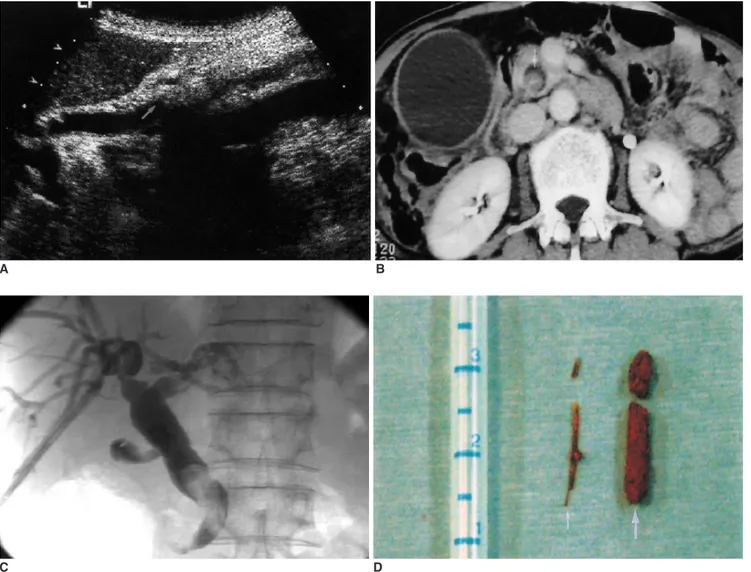
Fig. 1. A 75-year-old woman with common bile duct stone.
A. Ultrasonography demonstrates a hyperechoic stone with acoustic shadowing (arrow) in the common bile duct.
B. Computed tomography shows a common bile duct stone containing a bright dot of bone density (arrow).
C. Cholangiogram shows an elongated filling defect in the common bile duct.
D. Photograph of specimen extracted from common bile duct using a stone basket shows a friable pigment stone (thick arrow), that was formed around a fish bone (thin arrow).
C D
A B
the cause of biliary stone. The commonest cause of foreign bodies in the CBD is residual objects from a previous operation, like portions of tubes, suture material or surgical clips, which act as a nidus for stone formation. Another cause is ingested foreign bodies such as food materials.
Ingested foreign bodies in the biliary tree are usually associated with a previous biliary tree operation. However, there have been very few case of ingested foreign bodies, such as the fish bones in our patients, acting as a nidus for stone formation in the CBD without any previous history of operation (2, 3).
Most ingested foreign bodies pass through the gastroin- testinal tract uneventfully within 1 week. Gastrointestinal perforation following the ingestion of a foreign body usually occurs in the case of a material with a sharp, pointed end. Perforation may occur at any site in the gastrointestinal tract, but the ileocecal or rectosigmoid regions are the most commonly affected areas (4). There have been many reports of ingested fish bones penetrating the aerodigestive tract and migrating into various parts of the thoracic cage and abdominal cavity, resulting in retropharyngeal abscess, mediastinitis, pleural empyema, liver abscess and perivesical abscess (5 9).
Orda et al. (3) reported one case of CBD stone caused by a fish bone, which occurred in a patient who had no history of previous abdominal surgery. In their case, the presence of a choledochoduodenal fistula, which acted as the route of migration for a fish bone originating from the intestinal tract, was demonstrated by endoscopic
retrograde cholangiopancreatography (ERCP). In contrast to their case, no choledochoenteric fistula was observed on the cholangiograms obtained either before or after stone
removal in our cases. Prochazka et al. (2) reported two cases of choledocholithiasis caused by foreign material in patients who had not undergone any prior operation. They mentioned manometric studies of the sphincter of Oddi in patients with choledocholithiasis, and in a comparative study they found that the patients with choledocholithiasis had sequences of antegrade phasic waves in 18 5% and retrograde waves in 53 9%, while the control group of patients without choledocholithiasis had antegrade waves in 60 4% and retrograde waves only in 26 3%. They concluded that the presence of foreign material within the stones in patients without a history of previous operation could be explained by a possible reflux from the
duodenum. Because there was no demonstrable choledo- choenteric fistula on the cholangiogram in our cases, we retrospectively speculated that the fish bone in the CBD might be the result of reflux from the duodenum through the ampulla of Vater, although we did not perform a manometric study.
Although their visibility varies depending on the fish species, location and orientation, ingested fish bones are poorly identified by plain radiography. CT is useful for diagnosing fish bone impaction (10). One of our cases demonstrated a unique bright dot of bone density in the stone on CT. In this particular case, the cholangiograms of the stone formed from the nidus of fish bone showed filling defects with an unusually elongated shape, because the stone was formed over a linear fish bone nidus. When CT shows a stone containing a bright dot of bone density or the cholangiogram shows an unusually elongated filling defect in the biliary tree, the possibility of a stone formed over fish bone should be considered.
Kim et al.
212 Korean J Radiol 5(3), September 2004
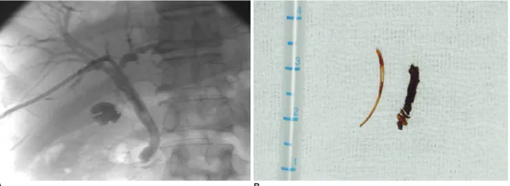
Fig. 2. A 67-year-old man with common bile duct stone.
A. Cholangiogram shows an elongated filling defect in the common bile duct.
B. Photograph of specimen extracted from common bile duct using a stone basket shows a friable pigment stone, that was formed around a fish bone.
A B
Fish Bone as a Nidus for Common Bile Duct Stone Formation
Korean J Radiol 5(3), September 2004 213
References
1. Stewart L, Ponce R, Oesterle AL, Griffiss M, Way LW. Pigment gallstone pathogenesis: slime production by biliary bacteria is more important than beta-glucuronidase production. J Gastrointest Surg 2000;4:547-553
2. Prochazka V, Krausova D, Kod’ousek R, Zamecnikova P.
Foreign material as a cause of choledocholithiasis. Endoscopy 1999;31:383-385
3. Orda R, Leviav A, Ratan I, Stadler J, Wiznitzer T. Common bile duct stone caused by foreign body. J Clin Gastroenterol 1986;8:
466-468
4. McCanse DE, Kurchin A, Hinshaw JR. Gastrointestinal foreign bodies. Am J Surg 1981;142:335-337
5. Singh B, Kantu M, Har-El G, Lucente FE. Complications associ- ated with 327 foreign bodies of the pharynx, larynx, and esophagus. Ann Otol Rhino Laryngol 1997;106:301-304 6. Nazoe T, Kitamura M, Adachi Y, et al. Successful conservative
treatment for esophageal perforation by a fish bone associated with mediastinitis. Hepatogastroenterology 1998;45:2190-2192 7. Solomonov A, Best LA, Goralnik L, Rubin AE, Yigla M. Plerual empyema: an unusual presentation of esophageal perforation.
Respiration 1999;66:366-368
8. Horii K, Yamazaki O, Matsuyama M, Higaki I, Kawai S, Sakaue Y. Successful treatment of a hepatic abscess that formed secondary to fish bone penetration by percutaneous transhep- atic removal of the foreign body: report of a case. Jpn J Surg 1999;29:922-926
9. Imamoto T, Tobe T, Mizoguchi K, Ueda T, Igarashi T, Ito H.
Perivesical abscess caused by migration of a fish bone from the intestinal tract. International J Urology 2002;9:405-406 10. Lue AJ, Fang WD, Manolidis S. Use of plain radiography and
computed tomography to identified fish bone foreign bodies.
Otolaryngol Head Neck Surg 2000;123:435-438