분화된 3T3-L1 세포에서 잣송이 초임계 추출물의 지방분해 효과
이민희1․남다은1․김옥경1․허석현2․이정민1,3
1경희대학교 의학영양학과
2한국건강기능식품협회
3경희대학교 임상영양연구소
Lipolytic Effect of Supercritical Extraction from Pine Cone (Pinus koraiensis) in Mature 3T3-L1 Adipocytes
Minhee Lee1, Da-Eun Nam1, Ok Kyung Kim1, Seok Hyun Heo2, and Jeongmin Lee1,3
1Department of Medical Nutrition and 3Research Institute of Medical Nutrition, Kyung Hee University
2Korea Health Supplements Association
ABSTRACT Seeds of Korean pine cone (Pinus koraiensis) have long been consumed as an edible food in countries located in North-East Asia, On the other hand, Korean pine cone, containing various polyphenols, is discarded as a useless garbage after removing seeds. This study investigated the lipolytic effects of pine cone extract in differentiated 3T3-L1 adipocytes. Intracellular lipid accumulation was measured by Oil red O staining, free glycerol release by colori- metric reaction, and expression of genes related to lipid metabolism by real-time PCR. Compared to control, pine cone extract reduced intracellular lipid accumulation by 8.8% and increased free glycerol release by 8.2% a concentration of 5 μg/mL in differentiated 3T3-L1 adipocytes. mRNA levels of fatty acid synthesis were not significantly different between control and pine cone extract, but mRNA levels of lipoprotein lipase (LPL) and hormone-sensitive lipase (HSL) significantly increased by 38.7% and 94.1% at a concentration of 5 μg/mL, respectively. Thus, pine cone extract is suggested to have lipolytic effects through induction of LPL and HSL gene expression.
Key words: pine cone, Pinus koraiensis, 3T3-L1, lipolysis
Received 16 May 2014; Accepted 6 August 2014
Corresponding author: Jeongmin Lee, Department of Medical Nutrition, Kyung Hee University, Yongin, Gyeonggi 446-701, Korea E-mail: [email protected], Phone: +82-31-201-3779
서 론
2012년 국민건강조사에 따르면 최근 생활환경의 변화 및 식생활의 서구화 등으로 인하여 국내 비만 인구가 1998년 26.0%에서 2012년 32.4%로 급증하였다(1). 비만은 환경 적, 유전적 및 사회적 요인 등 다양한 원인들이 관여하는 복합적 증후군으로 섭취한 에너지 중 소비되고 남은 것이 체내에 지방조직으로 과다하게 축적된 상태를 의미하며, 고 혈압, 심혈관 질환, 고지혈증, 당뇨와 같은 성인병을 일으키 는 주된 원인으로 밝혀지고 있다(2,3). 이러한 비만의 예방 및 치료를 위한 방법으로는 음식 섭취의 감소, 지방 또는 단백질 대사 또는 지방과 단백질의 저장에 대한 조절, 영양 소 흡수의 억제, thermogenesis 증가 등이 있다. Adip- ogenesis 억제, lipogenesis 억제, 지방분해 증가 등이 지방 저장의 조절에 속한다(4-7). 지방조직의 축적은 지방세포의 과형성과 비대를 초래하고, 지방세포의 과형성은 전지방세 포(preadipocyte)의 활발한 증식과 활성화된 지방세포의
분화과정으로 인해 발행한다. 반면 지방세포의 비대는 중성 지방의 축적에 의해 유발된다(8,9).
현재까지 개발된 비만치료제로는 지방 흡수를 억제하거 나 식욕을 억제한다. 미국 FDA(U.S. Food and Drug Administration)에서 승인된 제니칼(orlistat)은 lipase 활 성을 감소시켜 지방 흡수를 억제하고 지방을 배설시키지만 장기 복용 시 지방변 및 위장 질환 등의 문제점이 있고, phentermine, diethylpropion, mazindol은 중추신경계에 서의 교감신경 유사작용을 통해 식욕 억제효과가 있으나 입 마름과 수면장애 등과 같은 부작용이 있다(10-13). 따라서 보다 안전하고 효과적인 항비만 물질을 개발하기 위해 천연 소재를 이용한 연구가 요구되고 있다.
잣나무(Pinus koraiensis)는 소나무과에 속하는 상록목 으로 해발고도 1,000 m 이상에서 높이 30 m, 지름 1 m 정도 까지 자라고, 우리나라 중부 이북과 경남지역 등에서 널리 생장하고 있으며 만주 동부, 시베리아 및 일본 일부에까지 분포하고 있다(14,15). 잣나무에는 영양물질 이외의 항균, 살충 효과가 있고(16,17), 잣나무 진에는 phenol계 성분, terpenoid계 성분, 탄닌 성분이 함유되어 있으며, 잣나무의 terpinolene과 bomeol 성분은 담즙 분비를 촉진하여 콜레 스테롤 수치를 낮추는 작용을 하는 것으로 알려져 있다(18,
19). 뿐만 아니라 잣나무로부터 추출한 정유 성분(essential oil)은 식물의 휘발성 이차대사산물로서 항산화(20), 항염 증, 항알레르기(21), 항암 활성(22), 항균 활성(23,24)을 가 지고 있다. 잣나무를 이용한 연구들은 활발히 이루어져 있으 나 잣송이를 이용한 연구로는 정유성분 추출 및 항균 활성 (25)만이 보고되어 있는 실정이다. 농림축산부 통계에 의하 면 2008년 국내에서는 3,042톤의 잣이 생산되고 있으며, 잣 은 수확 후 잣 알갱이만을 식용으로 사용하고 그 외의 부산 물은 그대로 버려지며 이는 환경 오염원으로 방치되고 있다 (26). 최근 부산물을 이용하여 환경 오염원의 배출량을 감소 시키고 건강기능성식품의 원료로 이용하고자 하는 연구가 활발히 진행되고 있다. 잣 껍질에는 여러 생리활성 물질들이 함유되어 있고 항비만 효과가 있을 것으로 사료된다(27).
따라서 본 연구에서는 잣 알맹이와 잣피를 제외한 나머지 부산물인 잣송이의 항비만 효과를 평가하기 위하여 3T3- L1 지방세포를 이용하여 잣송이 추출물의 지방분해 효과와 지방분해 효소인 lipoprotein lipase(LPL)와 hormone- sensitive lipase(HSL) 유전자 발현에 미치는 영향을 알아 보고자 하였다.
재료 및 방법
재료 및 시약
3T3-L1 preadipocyte는 American Type Cultured Collection(ATCC; Rockville, MD, USA)에서 분주 받아 실 험하였다. High-glucose Dulbeco's modified Eagle's medium(DMEM), newborn calf serum(NCS), fetal bo- vine serum(FBS), PBS, penicillin-streptomycin, L-glu- tamin, Sodium pyruvate, hepes, NEAA mixture은 Ther- mo Scientific(Hyclone, Thermo Fisher Scientific, Wal- tham, MA, USA)에서 구입하였고, gentamicin reagent solution은 Gibco-BRL(Grand Island, NY, USA)에서 구입 하였다. Thiazolyl blue tetrazolium bromide(MTT), di- methyl sulfoixde(DMSO), insulin, dexamethasone(DEX), 3-isobutyl-1-methylxanthine(IBMX), Oil red O, free glycerol reagent는 Sigma-Aldrich Co.(St. Louis, MO, USA)에서 구입하였다. 그 외의 모든 시약 및 용매는 일급 또는 특급 이상의 등급을 사용하였다.
잣송이 초임계 추출물의 제조
1 L의 초임계 추출장비에 분쇄된 잣송이 310 g을 투입한 후, energized seal로 밀봉하고 추출기의 head 부분과 본체 는 clamp 체결 방식을 사용하였다. 초임계 유체로 사용된 CO2는 cylinder에서 가스 상태로 나와서 원하는 압력에 도 달할 때까지 Haskel gas booster(No. 30/75, Holiday Inn., Burbank, CA, USA)를 이용하여 압력을 높이고 150 bar에 도달한 후, 추출기 내부가 일정한 압력을 유지하도록 nee- dle valve를 조작하면서 추출물을 500 mL 가지 달린 플라
스크에 포집하였다. 추출기의 온도는 자동 온도 조절이 가능 한 전기히터를 사용하여 40°C로 일정하게 유지하였다. 추출 종료 시점은 추출물의 무게가 증가될 때까지 최대한 진행하 였다. 초임계 추출을 통해 얻어진 잣송이의 수득율은 약 4.2%로 나타났다.
3T3-L1 세포배양과 분화
3T3-L1 preadipocyte를 10% newborn calf serum (NCS)과 1% penicillin-streptomycin, 1% L-glutamin, 1% sodium pyruvate, 1% hepes, 1% NEAA mixture를 함유한 high-glucose DMEM을 사용하여 37°C, 5% CO2가 유지되는 배양기에서 배양하였다. 2일마다 배양액을 교환하 였고 세포가 단일 층으로 플라스크 바닥에 70% 이상 부착하 면 PBS 용액을 사용하여 세포표면을 세척하였으며, 0.25%
trypsin-EDTA를 첨가한 뒤 배양기에서 3분간 방치하여 세 포를 분리하였다. 그 후 원심분리기(GYROZEN 416G, BMS, Seoul, Korea)를 사용하여 1,600 rpm에서 5분간 원 심분리 하여 세포를 모았다. 이들 세포를 6 well plate(TPP) 에 1×10⁵ cells/well이 되게끔 균등하게 분주하였고, 100%
confluent 상태인 4일 후 10% FBS와 1% sodium pyr- uvate, 1% hepes, 1% NEAA mixture를 함유한 high-glu- cose DMEM에 adipogenic cocktail[MDI solution인 3- IBMX(0.5 mM), insulin(5 μg/mL), DEX(0.25 μM)]을 혼합 하여 분화 유도를 시작하였다. 분화기간은 총 9일간 지속되 었으며 분화 초기 3일 동안 매일 같은 배양액을 교환해 주었 다. 분화 중기 3일 동안 배양액을 insulin(5 μg/mL)만을 포 함하는 10% FBS를 함유한 DMEM으로 매일 교환해 주었 고, 분화 후기 3일 동안은 10% FBS를 함유한 DMEM 배양 액으로 매일 교환해 주었다.
잣송이 추출물의 세포 독성
본 실험에 사용한 잣송이 초임계 추출물이 지방세포에 독 성을 나타내는지를 측정하기 위하여 3-(4,5-dimethyl- thiazol-2-yl)-2,5 diphenyl tetrazolium bromide(MTT) assay(28)를 수행하였다. MTT solution(5 mg/mL)은 PBS 에 녹인 뒤 여과하여 사용하였다. 분화하기 직전의 상태인 3T3-L1 preadipocyte를 96 well plate에 1×10⁴ cells/
well로 총 용적을 100 μL가 되도록 분주하였으며, 배양액은 10% NCS를 함유한 DMEM을 사용하였다. 12시간 동안 배 양한 후 잣송이 추출물을 5, 10, 20 μg/mL로 처리하여 24시 간 동안 배양하였다. 그 후 MTT reagent 20 μL를 첨가하여 빛에 의한 MTT 환원을 최소화하기 위해서 호일로 빛을 차 단하고 4시간 동안 반응시켰다. 배양액과 MTT reagent를 suction 하고 PBS 용액으로 2회 세척한 후, 각 well당 DMSO 200 μL를 첨가하여 배양기에서 용해시켰으며, 20분 후 iMARKTM Microplate Reader(Bio-Rad Laboratories, Hercules, CA, USA)를 이용하여 560 nm 파장에서 optical density를 측정하였다.
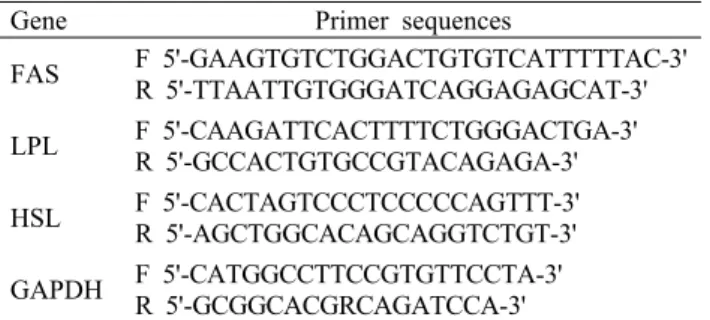
Table 1. Primer sequences used in real time PCR quantification of mRNA
Gene Primer sequences
FAS F 5'-GAAGTGTCTGGACTGTGTCATTTTTAC-3' R 5'-TTAATTGTGGGATCAGGAGAGCAT-3' LPL F 5'-CAAGATTCACTTTTCTGGGACTGA-3'
R 5'-GCCACTGTGCCGTACAGAGA-3' HSL F 5'-CACTAGTCCCTCCCCCAGTTT-3' R 5'-AGCTGGCACAGCAGGTCTGT-3' GAPDH F 5'-CATGGCCTTCCGTGTTCCTA-3'
R 5'-GCGGCACGRCAGATCCA-3'
FAS: fatty acid synthase, LPL: lipoprotein lipase, HSL: hor- mone-sensitive lipase, GAPDH: glyceraldehyde-3-phosphate de- hydrogenase.
Oil red O 염색
60 mm cultured dish에서 분화된 3T3-L1 세포를 1, 5 μg/mL 농도로 처리하여 48시간 동안 배양한 후 배양액을 suction 하고 PBS 용액으로 2회 세척한 뒤 PBS 용액을 완 전히 suction 하였으며, 10% formalin을 넣고 5분간 실온에 서 고정시킨 후 10% formalin을 suction 하고 다시 새로운 10% formalin을 넣어 2시간 이상 실온에서 고정시켰다. 그 후 formalin을 suction 하고 60% isopropanol을 넣어 바로 suction 한 후 flask를 완전히 건조시켰으며, Oil red O sol- ution 용액을 첨가하여 60분 동안 지방구를 염색하였다. 염 색 후 증류수로 4번 세척하여 지방구의 세포 내 축적을 현미 경을 이용하여 관찰하였으며 사진촬영을 통해 육안으로 잣 송이 추출물이 지방세포 분화에 미치는 영향을 관찰하였다 (29). 지방 정량을 위해 잘 건조된 상태에서 100% iso- propanol을 첨가하여 Oil red O dye를 용출한 뒤 iMARKTM Microplate Reader(Bio-Rad Laboratories)를 이용하여 560 nm 파장에서 optical density를 측정하였다. 이때 100
% isopropanol을 blank로 사용하였다.
Free glycerol의 측정
잣송이 추출물이 지방세포의 분해과정에서 glycerol 방 출량에 미치는 영향은 glycerol phosphate oxidase- TRINDER 효소반응법(30)을 적용한 free glycerol re- agent를 이용하여 배양액 내 glycerol 함량을 측정하였다.
지방세포의 분해과정에서의 free glycerol을 측정하기 위해 분화 후기(11일째)에 배양액을 모아 사용하였다. Free glycerol reagent 800 μL와 배양액 10 μL를 혼합하여 37°C에서 5분간 반응시킨 후, iMARKTM Microplate Reader (Bio-Rad Laboratories)를 이용하여 560 nm 파장에서 optical density를 측정하였다. Glycerol 함량은 free glyc- erol을 표준시약으로 사용한 표준곡선을 작성함으로써 측정 하였다.
RNA 추출 및 real-time polymerase chain reaction (real-time PCR)
잣송이 추출물 처리 시 지방 대사 관여 효소의 발현을 확 인하기 위해 real-time PCR을 실시하였다. 지방조직을 적 출하여 RNeasyⓇ Lipid Tissue Mini kit(Qiagen Sciences Inc., Gaithersburg, MD, USA)로 제조사의 protocol에 따 라 RNA 추출을 실시하였다. iScript cDNA synthesis kit (Bio-Rad Laboratories)를 사용하여 cDNA를 합성하였다.
유전자들의 발현을 측정하기 위하여 SYBR Green(iQ SYBR Green Supermix, Bio-Rad Laboratories)을 이용한 실시 간 정량 PCR을 실시하였고, 기기는 Real-Time PCR(Applied Biosystems, Foster City, CA, USA)을 사용하였다. 각각 의 유전자에 대한 PCR primer의 염기서열은 Table 1에 제 시하였다. Real-time PCR 반응은 총 20 μL 내에 cDNA 2 μL와 2X SYBR mix 10 μL, forward, reverse primer는
각각 100 pmol/μL를 1 μL씩 첨가하였고, 나머지는 H2O로 채워주었다. PCR 증폭 단계는 다음과 같고 증폭 cycle은 40 cycle을 실시하였다. Hot start를 위해 95°C에서 8분, 증폭 단계의 denaturation을 95°C에서 15초, annealing을 52°C에서 30초, extension을 72°C에서 30초간 반복하며, 각 cycle의 extension 후에 값이 기록되었다. 모든 cycle이 완료된 후 primer의 특이성을 확인하기 위해 melting curve 분석을 실시하였다. 결과의 분석은 Applied Biosystems에 서 제공하는 One step system software v2.1로 분석하였다.
통계처리
본 실험 결과는 SPSS(Statistical Package for the Social Science, SPSS Inc., Chicago, IL, USA) version 20 프로 그램을 이용하여 분석하였다. 모든 측정항목의 결과는 평균 (mean)±표준편차(standard deviation, SD)로 표시하였고 실험군간 평균의 차이는 one-way ANOVA로 유의성을 확 인한 후 Duncan's multiple range test를 이용하여 사후 검 증하였으며 P<0.05 수준에서 유의성의 여부를 검증하였다.
결과 및 고찰
세포 생존율
MTT 분석법은 살아있는 세포의 미토콘드리아 탈수소 효 소를 이용하여 MTT를 환원시켜서 분광 광도법으로 측정이 가능한 청색의 formazan 결정을 형성하는 특성을 이용한 방법이다(31,32).
잣송이 초임계 추출물의 3T3-L1에 대한 세포 독성을 알 아보기 위하여 MTT assay를 실시하였다. 3T3-L1 세포에 대한 잣송이 초임계 추출물의 세포 생존율은 Fig. 1에 나타 내었다. 5, 10 μg/mL 농도에서는 세포 생존율이 100%, 95%
로 세포독성은 나타나지 않았으나, 20 μg/mL 농도에서 세 포 생존율이 74.5% 유의적으로 감소하였다(P<0.05). 이상 의 결과를 토대로 추후 실험에는 세포가 사멸하지 않고 독성 이 일어나지 않은 5 μg/mL 농도를 최대로 하여 실험에 사용 하였다.
*
0 20 40 60 80 100 120
0 5 10 20
Concentration (μg/mL)
Cell viability (%) .
Fig. 1. Effect of pine cone extracts on cell survival percentage in mature 3T3-L1 adipocytes based on MTT assay. Significant differences were determined by Student's t-test compare with 0 μg/mL at P<0.05.
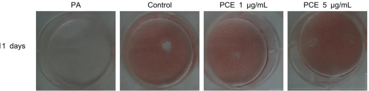
11 days
PA Control PCE 1 μg/mL PCE 5 μg/mL
Fig. 2. Oil red O staining of neutral lipids in pine cone extracts treated 3T3-L1. Cell were differentiated with medium containing adipogenic cocktail for 9 days. After 9 days for differentiation, pine con extract treated 3T3-L1 for 2 days. Oil red O staining was performed at 11 day. PA: preadipocyte, PCE: pine cone extract.
b a a
c
0 100 200 300 400 500 600 700
PA Control PCE (1 μg/mL) PCE (5 μg/mL)
Lipid accumulation (%of control) .
Fig. 3. Lipid accumulation in pine cone extracts treated 3T3-L1.
Cell were differentiated with medium containing adipogenic cocktail for 9 days. After 9 days for differentiation, pine con extract treated 3T3-L1 for 2 days. The lipid accumulation was evaluated by Oil red O staining at 11 day. All data are presented as mean±standard deviation. Statistical analyses were performed by Duncan's multiple range tests after one-way ANOVA using SPSS software. Differences were considered statistically sig- nificant at P<0.05. PA: preadipocyte, PCE: pine cone extract.
Oil red O 염색의 의한 세포 내 지방구 함량 측정 항비만 소재를 탐색하기 위한 in vitro 연구에서는 주로 3T3-L1 세포주를 이용하여 지방세포의 분화를 억제하거나 지방세포의 분해를 촉진하는 소재의 효능 및 기전을 연구한 다. 3T3-L1 세포주는 성장이 시작되면 성장기간 동안 100
% 융합이 일어나게 되는데, 이때 배양액에 insulin, DEX, IBMX와 같은 유도물질을 첨가하면 성장이 멈추고 지방세포 로의 분화가 시작된다. 분화 시 생화학적, 형태학적으로 포 유류의 지방조직 성장과 유사한 것으로 알려져 비만에 대한 효과검증에 사용된다(33-36).
분화된 3T3-L1 지방세포에 잣송이 초임계 추출물을 1, 5 μg/mL 농도로 처리한 후 생성되었던 지방구에 미치는 영 향을 확인하기 위해 중성지방만을 붉은색으로 염색하는 Oil red O 염색법을 통해 3T3-L1 지방세포 내 생성된 중성지방 의 양을 측정한 결과를 Fig. 2, Fig. 3에 나타내었다. 지방 축적 함량을 측정한 결과, 전지방세포군에 비해 지방 분화 유도군이 84.7% 유의적으로 증가하였고, 잣송이 초임계 추 출물을 1 μg/mL 농도로 처리한 군은 지방 분화 유도군에 비해 0.8% 감소하였으나 유의성은 나타나지 않았다. 잣송이 초임계 추출물 5 μg/mL 농도로 처리한 군은 지방 분화 유도 군에 비해 8.8% 유의적으로 감소하였다(P<0.05). 지방구에 존재하는 중성지방은 3T3-L1 지방전세포로부터 지방세포 로 분화하는 단계에서 생성된다. 세포 내에 생성된 중성지방
은 분화 마지막 단계까지 계속적으로 지방구를 형성하게 되 는데 시간이 지날수록 지방구끼리 결합하여 그 크기가 점점 증가하게 된다. 본 연구에서는 잣송이 초임계 추출물의 지방 구 감소 효과를 검증하기 위하여 형성된 지방구를 분해 또는 감소시킬 수 있는지를 측정한 것이다.
Glycerol release 측정
인체의 경우 지방세포 내 축적된 중성지방은 유리지방산 과 glycerol로 가수분해 되어 혈액 속으로 방출되어 간으로 이동하게 된다(37). 3T3-L1 지방세포에서는 유리된 glyc- erol의 함량이 지방구 내 중성지방의 분해 정도를 간접적으 로 나타내는 측도가 된다.
잣송이 초임계 추출물을 1, 5 μg/mL 농도로 처리한 후 유리된 glycerol의 함량을 측정한 결과를 Fig. 4에 나타내었 다. Free glycerol 함량을 측정한 결과, 전지방세포군에 비 해 지방 분화 유도군이 68.0% 유의적으로 증가하였고, 잣송 이 초임계 추출물을 1 μg/mL 농도로 처리한 군은 지방 분화 유도군에 비해 35.0% 유의적으로 증가하였다(P<0.05). 잣 송이 초임계 추출물을 5 μg/mL 농도로 처리한 군은 지방
b a
c
d
0 2 4 6 8 10 12 14 16
PA Control PCE (1 μg/mL) PCE (5 μg/mL)
Free glycerol (μg/mL) .
Fig. 4. The effect of pine cone extracts on glycerol release in 3T3-L1 cells. Cell were treated pine cone extract for 2 days after inducing differentiation with medium containing adipo- genic cocktail for 9 days. Increase of lipolytic effect was eval- uated by the release of glycerol to the cultured medium at 11 day. All data are presented as mean±standard deviation. Statisti- cal analyses were performed by Duncan's multiple range tests after one-way ANOVA using SPSS software. Differences were considered statistically significant at P<0.05. PA: preadipocyte, PCE: pine cone extract.
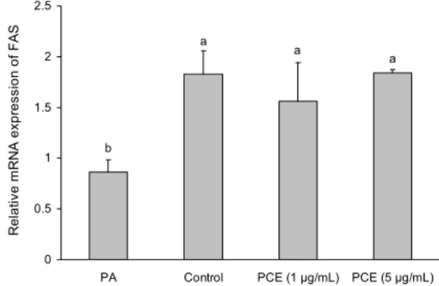
a a a
b
0 0.5 1 1.5 2 2.5
PA Control PCE (1 μg/mL) PCE (5 μg/mL)
Relative mRNA expression of FAS
Fig. 5. Effect of pine cone extracts on expression of fatty acid synthase in 3T3-L1. Cell were treated pine cone extract for 2 days after inducing differentiation with medium containing adi- pogenic cocktail for 9 days. All data are presented as mean±
standard deviation. Statistical analyses were performed by Dun- can's multiple range tests after one-way ANOVA using SPSS software. Differences were considered statistically significant at P<0.05. PA: preadipocyte, PCE: pine cone extract.
분화 유도군에 비해 8.2% 유의적으로 증가하였다(P<0.05).
저농도에서 유의적인 증가를 보인 것은 실험적인 오차에 의 한 것으로 판단된다. 그 이유는 Fig. 5, Fig. 6, Fig. 7에서 농도에 따른 변화가 나타나지 않았기 때문이다. 이와 같은 결과는 잣송이 초임계 추출물이 지방세포에 저장되어 있던 중성지방을 유리지방산과 glycerol로 가수분해 시켜 세포 밖으로 방출시켰음을 시사한다.
지방분해 효과를 측정하기 위해 세포로부터 배양액으로 방출된 glycerol 농도를 분석한 다른 연구 결과를 보면 Kim 등(38)이 tannic acid를 처리하였을 때 3T3-L1 지방세포로 부터 배양액으로 방출된 glycerol 함량이 유의적으로 증가 하였다고 보고하였다. 잣나무 진에도 tannic acid가 함유되 어 있는 것으로 알려져 있는데 잣송이에도 tannic acid가 함유되어 있을 것으로 예측되고 이 tannic acid에 의해 중성 지방을 가수분해 시켜 세포 밖으로 glycerol이 방출되었을 것으로 사료된다.
지방합성 효소의 mRNA 발현
지방대사에 관여하는 지방합성 효소인 fatty acid syn- thase(FAS)는 세포질에서 acetyl-CoA와 malonyl-CoA의 축합반응을 통해 긴 사슬 지방산의 합성반응을 촉매하며, 식이 및 영양상태, 호르몬 등에 의해 활성이 변화한다(39).
본 연구에서 잣송이 초임계 추출물을 1, 5 μg/mL 농도로 처리한 후 FAS mRNA의 발현을 측정한 결과, 잣송이 초임 계 추출물의 처리에 따른 효과는 나타나지 않았다(Fig. 5).
지방분해 효소의 mRNA 발현
지방분해는 중성지방이 지방분해과정을 통해 지방산과 glycerol로 가수분해 되는 과정을 일컫는 것으로, 지방세포
에서의 지방분해 과정은 신체의 항상성 조절을 위한 중요한 작용 중의 하나이다(40). 지방분해 효소로는 LPL, HSL이 대표적으로 알려져 있다. LPL은 혈중 지방산을 세포 내로 전달하는 효소로서 장에서 흡수되어 대사되는 중성지방과 간에서 흡수되어 대사되는 중성지방을 분해하여 다른 장기 에서 지방산을 이용할 수 있게 하거나 지방조직에서 지방 저장에 관여한다(41,42). HSL은 cAMP의 영향을 받아 지방 조직에서 지방을 분해할 때 중성지방을 유리지방산과 glyc- erol로 분해하는 지방분해 효소로 알려져 있다(43,44).
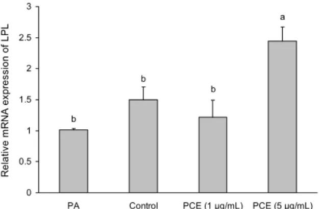
잣송이 초임계 추출물이 지방분해 효소의 mRNA 발현에 미치는 영향을 알아보기 위해 real-time PCR을 실시한 결 과를 Fig. 6과 Fig. 7에 나타내었다. LPL mRNA의 발현을 확인한 결과, 전지방세포군에 비해 지방 분화 유도군이 32.6
% 증가하였으나 유의성은 나타나지 않았고, 잣송이 초임계 추출물을 1 μg/mL 농도로 처리한 군은 지방 분화 유도군에 비해 18.9% 감소하였으나 유의성이 나타나지 않았다. 잣송 이 초임계 추출물을 5 μg/mL 농도로 처리한 군은 지방 분화 유도군에 비해 38.7% 유의적으로 증가하였다(P<0.05). HSL mRNA의 발현을 확인한 결과, 전지방세포군에 비해 지방 분화 유도군이 90.5% 유의적으로 감소하였고, 잣송이 초임 계 추출물을 1 μg/mL 농도로 처리한 군은 지방 분화 유도군 에 비해 69.8% 유의적으로 증가하였다(P<0.05). 잣송이 초 임계 추출물을 5 μg/mL 농도로 처리한 군은 지방 분화 유도 군에 비해 94.1% 유의적으로 증가하였다(P<0.05).
Kim과 Jang(45)은 epigallocatechin gallate(EGCG)와 glucosamine-6-phosphate를 3T3-L1 세포에 처리하였 을 때 HSL 유전자 발현이 증가하여 지방분해 효과가 있을 것이라고 보고하였고, Choi 등(46)은 alkaloid 성분이 풍부 한 berberine을 3T3-L1 세포에 처리하였을 때 HSL 유전 자 발현이 증가하여 지방분해 효과가 있을 것이라고 보고하
a
b b
b
0 0.5 1 1.5 2 2.5 3
PA Control PCE (1 μg/mL) PCE (5 μg/mL)
Relative mRNA expression of LPL .
Fig. 6. Effect of pine cone extracts on expression of lipoprotein lipase in 3T3-L1. Cell were treated pine cone extract for 2 days after inducing differentiation with medium containing adipogenic cocktail for 9 days. All data are presented as mean±standard deviation. Statistical analyses were performed by Duncan's mul- tiple range tests after one-way ANOVA using SPSS software.
Differences were considered statistically significant at P<0.05.
PA: preadipocyte, PCE: pine cone extract.
a
c d
b
0 0.5 1 1.5 2 2.5
PA Control PCE (1 μg/mL) PCE (5 μg/mL)
Relative mRNA expression of HSL .
Fig. 7. Effect of pine cone extracts on expression of hormone- sensitive lipase in 3T3-L1. Cell were treated pine cone extract for 2days after inducing differentiation with medium containing adipogenic cocktail for 9 days. All data are presented as mean±
standard deviation. Statistical analyses were performed by Dun- can's multiple range tests after one-way ANOVA using SPSS software. Differences were considered statistically significant at P<0.05. PA: preadipocyte, PCE: pine cone extract.
였다. 잣송이의 성분 분석이 이루어지지 않아 정확한 생리활 성 물질이 무엇인지 알 수는 없으나 잣나무 진에 함유되어 있는 phenol과 alkaloid 성분이 잣송이에도 포함되어 있을 것으로 생각된다. 이러한 성분들이 HSL과 LPL 유전자를 증가시켜 지방분해 효과를 나타내는 것으로 사료된다. 결론 적으로 분화된 3T3-L1 지방세포에서 잣송이 초임계 추출 물의 지방분해 효과는 LPL과 HSL 지방분해 효소의 발현 증가에 의한 것으로 사료된다.
요 약
건강에 대한 관심의 증가로 천연식품뿐만 아니라 산업 부산 물을 이용한 기능성 소재 탐색에 대한 연구들이 활발하게
진행되고 있다. 본 연구에서는 산업 부산물인 잣송이를 이용 하여 초임계 방법으로 추출을 하여 항비만 식품 소재로서의 기능성을 알아보고자 하였다. 분화된 3T3-L1 지방세포에 서 잣송이 초임계 추출물이 지방의 축적 및 지방분해에 미치 는 영향을 확인한 결과, 잣송이 초임계 추출물을 1, 5 μg/mL 농도로 처리 시 세포 내 지방 축적이 억제되었고 중성지방 분해 산물인 glycerol의 함량은 증가하였다. 잣송이 초임계 추출물이 지방합성 효소 및 지방분해 효소에 미치는 영향을 알아보기 위해 real-time PCR을 실시한 결과, 잣송이 초임 계 추출물은 지방합성 효소인 FAS에 아무런 영향이 나타나 지 않았고, 지방분해 효소인 LPL과 HSL의 유전자 발현이 증가됨에 따라 세포 내 중성지방이 지방산과 glycerol로 분 해되었음을 확인하였다. 따라서 이 결과들은 잣송이 초임계 추출물이 지방조직에서의 LPL과 HSL 유전자 발현 증가를 통한 지방분해로 항비만 효과를 나타냄을 보여주고 있다.
잣송이 초임계 추출물이 항비만 생리활성을 나타내는 기능 성 식품 신소재로서 개발 가능성이 있을 것으로 사료된다.
감사의 글
본 연구는 2013년도 농림수산식품부 고부가가치사업 연구 비 지원에 의해 수행된 결과의 일부이며, 이에 감사를 드립 니다.
REFERENCES
1. Jin JA, Oh KS, Hyun YN, Choi SK, Kwon YH, Kim HJ, Woo B. 2013. Antiadipogenic effect of Vitis amurensis root methanol extract and its solvent fraction in 3T3-L1 pre- adipocytes. J Life Sci 23: 69-78.
2. Spiegelman BM, Flier JS. 1996. Adipogenesis and obesity;
rounding out the big picture. Cell 87: 377-389.
3. Visscher TL, Seidell JC. 2001. The public health impact of obesity. Annu Rev Public Health 22: 355-375.
4. Bray GA, Tartaglia LA. 2000. Medicinal strategies in the treatment of obesity. Nature 404: 672-677.
5. Spiegelman BM, Flier JS. 2001. Obesity and the regulation of energy balance. Cell 104: 531-543.
6. Ardévol A, Bladé C, Salvadó MJ, Arola L. 2000. Changes in lipolysis and hormone-sensitive lipase expression caused by procyanidins in 3T3-L1 adipocytes. Int J Obes Relat Metab Disord 24: 319-324.
7. Balkau B, Valensi P, Eschwège E, Slama G. 2007. A review of the metabolic syndrome. Diabetes Metab 33: 405-413.
8. Holst D, Grimaldi PA. 2002. New factors in the regulation of adipose differentiation and metabolism. Curr Opin Lipidol 13: 241-245.
9. De Ferranti S, Mozaffarian D. 2008. The perfect storm: obe- sity, adipocyte dyfunction, and metabolic consequences.
Clin Chem 54: 945-955.
10. Padwal RS, Majumar SR. 2007. Drug treatments for obesity:
orlistat, sibutramine, and rimonabant. Lancet 369: 71-77.
11. Zhi J, Moore R, Kanitra L, Mulligan TE. 2003. Effects of orlistat, a lipase inhibitor, on the pharmacokinetics of three highly lipophilic drugs (amiodarone, fluoxetine, and simvas- tatin) in healthy volunteers. J Clin Pharmacol 43: 428-435.
12. Kim KK, Cho HJ, Kang HC, Youn BB, Lee KR. 2006.
Effects on weight reduction and safety of short-term phen- termine administration in Korean obese people. Yonsei Med J 47: 614-625.
13. Cercato C, Roizenblatt VA, Leanca CC, Segal A, Lopes Filho AP, Mancini MC, Halpern A. 2009. A randomized double- blind placebo-controlled study of the long-term efficacy and safety of diethylpropion in the treatment of obese subjects.
Int J Obes 33: 857-865.
14. Critchfield WB, Little EL Jr. 1966. Geographic Distribution of the Pines of the World. US Department Agriculture, Forest Service, Washington, DC, USA. No 991, p 4.
15. Lee HJ, Choe YJ, Choe DH, Hong IP. 2003. Extractives of Pinus koraiensis wood. J Korean Wood Sci Technol 31:
49-56.
16. Cimanga K, Kambu K, Tona L, Apers S, De Bruyne T, Hermans N, Totte J, Pieters L, Vlietinck AJ. 2002. Correla- tion between chemical composition and antibacterial activity of essential oils of some aromatic medicinal plants growing in the Democratic Republic of Congo. J Ethopharmacol 79:
213-220.
17. Baricevic D, Milevoj L, Borstnik J. 2001. Insecticidal effect of oregano Origanum vulgare L. ssp. hirtum Ietswaart) on bean weevil (Acanthoscelides obtectus Say). Inter J Horti- cult Sci 7: 84-88.
18. Hong WT, Ko KM, Lee JG, Jang HJ, Kwak JJ. 2002. Volatile compounds of pine needle (Pinus rigida miller) extracts.
J Korea Soc Tabacco Sci 24: 53-59.
19. Kim YK, Chung KN, Hirosh I, Shigeru M. 1986. Volatile components of pinenut. Korean J Food Sci Technol 18: 105- 109.
20. Caldefie-Chézet F, Guerry M, Chalchat JC, Fusillier C, Vasson MP, Guillot J. 2004. Anti-inflammatory effects of Melaleuca alternifolia essential oil on human polymorpho- nuclear neutrophils and monocyte. Free Radic Res 38:
805-811.
21. Medeiros R, Passos GF, Vito CE, Koepp J, Mazzuco TL, Pianowski LF. 2007. Effect of two active compounds ob- tained from the essential oil of Cordia verbenacea on the acute inflammatory responses elicited by LPS in the rat paw.
Br J Pharmacol 151: 618-627.
22. Sylvestre M, Pichette A, Longtin A, Nagau F, Legault J.
2006. Essential oil analysis and anticancer activity of leaf essential oil of Croton flavens L. from Guadeloupe. J Ethno- pharmacol 103: 99-102.
23. Lee EK, Kim HS, Chung SJ. 2011. Antimicrobial activity of chitosan solution with PKS (Pinus koraiensis S. et Z) and PRM (Pinus rigida Mill) extract. J Chitin Chitosan 16:
117-122.
24. Hwang HJ, Yu JS, Lee HY, Kwon DJ, Han W, Heo SI, Kim SY. 2014. Evaluations on deodorization effect and anti-oral microbial activity of essential oil from Pinus koraiensis.
Korean J Plant Res 27: 1-10.
25. Kim CH, Lee SY, Pak JI. 2008. The extraction of essential oil from by-product of pine nut cone and its antibacterial activity. Annal Animal Resource Sci 19: 63-70.
26. Ministry of Agriculture, Food and Rural Affairs. 2010. Stat- istical yearbook of forestry. Vol 36, p 296.
27. Kim JE, Kim WY, Kim JW, Park HS, Lee SH, Lee SY, Kim MJ, Kim AR, Park SN. 2010. Antibacterial, antioxida- tive activity and component analysis of Pinus koraiensis leaf extracts. J Soc Cosmet Scientists Korea 36: 303-314.
28. Berridge MV, Tan AS. 1993. Characterization of the cellular reduction of 3-(4,5-dimethylthiazol-2yl)-2,5-diphenyltetra- zolium bromide (MTT): subcellular localization, substrate dependence, and involvement of mitochondrial electron
transport in MTT reduction. Arch Biochem Biophys 303:
474-482.
29. Green H, Kehinde O. 1975. An established preadipose cell line and its differentiation in culture. Ⅱ. Factors affecting the adipose conversion. Cell 5: 19-27.
30. Trinder P. 1969. Determination of blood glucose using an oxidase-peroxidase system with a non-carcinogenic chrom- ogen. J Clin Pathol 22: 158-161.
31. Carmichael J, DeGraff WG, Gazdar AF, Minna JD, Mitchell JB. 1987. Evaluation of a tetrazolium-based semiautomated colorimetric assay: assessment of radiosensitivity. Cancer Res 47: 943-946.
32. Carmichael J, DeGraff WG, Gazdar AF, Minna JD, Mitchell JB. 1987. Evaluation of a tetrazolium-based semiautomated colorimetric assay: assessment of chemosensitivity testing.
Cancer Res 47: 936-942.
33. Ailhaud G, Grimaldi P, Negrel R. 1992. Cellular and molec- ular aspects of adipose tissue development. Annu Rev Nutr 12: 207-233.
34. Haugen F, Zahid N, Dalen KT, Hollung K, Nebb HI, Drevon CA. 2005. Resistin expression in 3T3-L1 adipocytes is re- duced by arachidonic acid. Lipid Res 46: 143-153.
35. Tenney R, Stansfield K, Oekala PH. 2005. Interleukin 11 signaling in 3T3-L1 adipocytes. J Cell Physiol 202: 160- 166.
36. MacDougald OA, Hwang CS, Fan H, Lane MD. 1995. Re- gulated expression of the obese gene product (leptin) in white adipose tissue and 3T3-L1 adipocytes. Proc Natl Acad Sci USA 92: 9034-9037.
37. Frayn KN, Coppack SW, Fielding BA, Humphreys SM.
1995. Coordinated regulation of hormone sensitive lipase and lipoprotein lipase in human adipose tissue in vivo: im- plications for the control of fat storage and fat mobilization.
Adv Enzyme Regul 35: 163-178.
38. Kim HJ, Yun YR, Song YB, Song YO. 2008. Ani-lipogenic effects of tannic acid in 3T3-L1 adipocytes and in high fat diet-fed rats. Food Sci Biotechnol 17: 219-445.
39. Ronnett GV, Kim EK, Landree LE, Tu Y. 2005. Fatty acid metabolism as a target for obesity treatment. Physiol Behav 85: 23-35.
40. Jeon T, Hwang SG, Hirai S, Matsui T, Yano H, Kawada T, Lim BO, Park DK. 2004. Red yeast rice extracts suppress adipogenesis by downregulating adipogenic transcription factors and gene expression in 3T3-L1 cells. Life Sci 75:
3195-3203.
41. Rea S, James DE. 1997. Moving GLUT4: the biogenesis and trafficking of GLUT4 storage vesicles. Diabetes 46: 1667- 1677.
42. Mead JR, Irvine SA, Ramji DP. 2002. Lipoprotein lipase:
structure, function, regulation, and role in disease. J Mol Med 80: 753-769.
43. Langin D, Holm C, Lafontan M. 1996. Adipocyte hormone sensitive lipase: a major regulator of lipid metabolism. Proc Nutr Soc 55: 93-109.
44. Morimoto C, Kameda K, Tsujita T, Okuda H. 2001. Rela- tionships between lipolysis induced by various lipolytic agents and hormone sensitive lipase in rat fat cells. J Lipid Res 42: 120-127.
45. Kim KB, Jang S. 2014. Anti-obesity effect of EGCG and glucosamine-6-phosphate through decreased expression of genes related to adipogenesis and cell cycle arrest in 3T3-L1 adipocytes. J Nutr Health 47: 1-11.
46. Choi BH, Ahn IS, Kim YH, Park JW, Lee SY, Hyun CK, Do MS. 2006. Berberine reduces the expression of adipo- genic enzymes and inflammatory molecules of 3T3-L1 adi- pocyte. Exp Mol Med 38: 599-605.