Analysis of corneal real astigmatism and high order aberration changes that cause visual disturbances after lower eyelid epiblepharon repair surgery
Dong cheol Lee
this retrospective, cross-sectional study investigated changes in corneal low and high order aberrations (LOAs and HOAs) after lower eyelid epiblepharon repair surgery. In total, 108 eyes from 54 patients were evaluated. Wavefront analyses for calibrated LOAs and HOAs were performed using a Galilei G4 Dual Scheimpflug Analyzer before surgery and during the first and second follow-ups (f/u), adjusting for several risk factors. Flat keratometry (K) and axis values decreased significantly from baseline at the first f/u. At the second f/u, mean K and axis values decreased. Coma and trefoil increased from baseline at the first f/u and normalized by the second f/u. Spherical aberrations (SA) only decreased at the second f/u. After correction for risk factors, cylinder, coma, trefoil, and SA significantly increased at the first f/u; axis and flat K values decreased. At the second f/u, cylinder increased while axis and mean K values significantly decreased. Epiblepharon repair surgery may result in a shift from ‘with-the-rule’
to ‘against-the-rule’ axis change. Flat K, coma, and trefoil may be affected by mechanical force changes immediately post-surgery, while mean K values and SA may also change with corneal state changes including corneal erosion healing after the second f/u during the postoperative period.
Epiblepharon is a congenital anomaly of eyelids that occurs more frequently among Asian children than among children of other demographic backgrounds
1. The prevalence of epiblepharon in the Japanese population was reported to be 9.9% in children aged 3 months to 18 years
2. In addition, epiblepharon comprised 9.5% of the oculoplastic surgery clinical cases in a tertiary care hospital setting in Singapore
3. Epiblepharon is defined as a horizontal fold in the hypertrophied pretarsal orbicularis muscle and redundant skin below the margin of the eyelid, which cause the eyelashes to project inwards towards the cornea while the eyelids with tarsus remains in normal positions. Epiblepharon is different from congenital entropion, which is an inward rotation of the eyelid margin with the tarsus
4,5.
Patients with epiblepharon experience photophobia, foreign body sensation, irritating tearing, and visual dis- turbances caused by corneal problems
6. In addition, astigmatisms are most frequently observed in patients with epiblepharon as compared with the normal population. For instance, Preechawai et al. reported a high prevalence of astigmatism in epiblepharon patients (52.2% had a 1 D or worse astigmatism)
7. Furthermore, several studies have reported a greater prevalence of astigmatism in patients with epiblepharon and changes in astigmatism after epiblepharon repair surgery
7–10.
Modern vision research has benefited from wavefront technology, which makes it possible to measure low and high order aberrations (LOA and HOA), including posterior corneal astigmatisms, with total cornea curva- ture via the Galilei G4 Dual Scheimpflug Analyzer (Ziemer, Port, Switzerland). Ray tracing using Snell’s law and pachymetry data with a reference plane in the posterior corneal surface are used
11. Furthermore, HOAs from all refractive errors, such as those that occur with astigmatisms, cannot be corrected by ordinary glasses or contact lenses. Recently, HOAs have been represented mathematically by calculating a Zernike coefficient
12–14. Moreover, corneal transplantation, intraocular lens (IOL) implantation, pterygium surgery, and scleral buckling result in
Department of Ophthalmology, Keimyung University School of Medicine, 1095 Dalgubeol-daero, Dalseo-gu, Daegu, 42601, Republic of Korea. e-mail: [email protected]
open
HOA changes
15–17. In HOAs, ‘spherical aberration’ changes are known to result in decreased contrast sensitivity, halos, and glare, and ‘coma’ changes are associated with tilt and double vision
18,19. These HOAs result in changes that may lead patients to experience a qualitative deterioration in sight
18–20.
Given that most epiblepharon patients are children, understanding the visual impairments caused by surgery is especially critical. The risk for astigmatism and HOAs due to surgery should be conveyed to patients, and eye- glass prescriptions should be considered. To our knowledge, although changes in anterior LOAs (sphere and cyl- inder) after surgery have been investigated, no studies on the changes in calibrated anterior and posterior LOAs together with HOAs, including their risk factors, have been conducted. The purpose of the present study was to evaluate the preoperative and postoperative calibration of LOA (anterior and posterior astigmatisms) and HOA changes in the postoperative period, while adjusting for potential risk factors such as age, sex, body mass index (BMI), and corneal keratitis status.
Methods
Approval for the retrospective review (IRB file No.: DSMC 2019-02-005) of our patients’ medical data was obtained from the Keimyoung University Dongsan Hospital Institutional Review Board (IRB), Daegu, Korea. All data and research collection procedures followed the tenets of the Declaration of Helsinki.
The medical and surgical records of 78 patients (156 eyes) diagnosed with epiblepharon who underwent epiblepharon repair surgery at Keimyoung University Dongsan Hospital from January 2016 to January 2019 were retrospectively reviewed. Due to the retrospective nature of this study, the Institutional Review Board of Keimyoung University Dongsan Hospital waived the requirement for patient consent. A single surgeon (LDC) participated in all surgeries and was involved in all clinical patient assessments. Severe corneal erosions and irri- tating symptoms such as epiphora, often eye blinking and/or rubbing, and photophobia, were included as surgical indications. Patients with post-surgery f/u of as long as 3 months were included. Patients who had congenital entropion, distichiasis, trichiasis, or a history of ocular or eyelid surgery were excluded. A total of 48 eyes of 24 patients were excluded from this study.
Preoperatively, all patients underwent ophthalmological examinations, including best-corrected visual acuity, slit-lamp examinations, cycloplegic refraction, and indirect fundus examinations. In addition, wavefront analyses for calibrated LOAs (anterior and posterior astigmatisms including keratometry were calibrated in ray-traced mode) and HOAs (root mean square [RMS], coma, three-piece aberrations [Trefoil], secondary astigmatisms [2
ndAstig], and spherical aberrations [SA]) were measured via a Galilei G4 Dual Scheimpflug Analyzer. These analyses were performed by a single technician preoperatively, and at the first and second f/u in group 1 (less than 45 days post-surgery), group 2 (from 45 to 75 days post-surgery), and group 3 (more than 75 days post-surgery).
Surgical technique. All surgeries were performed under general anaesthesia due to patient age. A mixture of lidocaine 2% and epinephrine (1:100,000) was injected subcutaneously into the lower eyelid. Excess lower eyelid skin was grasped using forceps to cause mild ectropion, and the lid was marked with a pen. The upper skin incision line was approximately 1–2 mm below the eyelash line, with a delineated ellipse more lateral than prox- imal to the punctum. The width of the ellipse was extended laterally, until reaching close to the lateral canthus to achieve a good contour.
The infra-eyelash skin incision and excision of the pretarsal orbicularis muscle was performed. Subsequently, the surgeon sutured subcutaneous tissue of the upper skin-muscle flap and exposed tarsal plate using three inter- rupted 6.0 monosyn sutures (medial, centre, and lateral) to achieve a good eyelash contour
21. Moreover, a Bovie monopolar needle cautery instrument (Colorado Biomedical Inc, Evergreen, CO) was used for thermal cauteri- zation of the septum to create adhesions between the preseptal orbicularis oculi muscle and the septum
22. These adhesions minimized vertical overriding of the orbicularis oculi muscle at the lower eyelid. If there was excessive skin, such as in the form of a dog-ear at the lateral ends of the incision, the surgeon removed them with a triangu- lar skin excision. If there were small amounts of pretarsal orbicularis oculi muscle and redundant skin overlying the lower eyelid margin, the tissue was also removed. At the end of surgery, the skin was then closed with a con- tinuous 6–0 fast-absorbing plain gut suture. Ocuflox antibiotic ointment was used on the skin wound for 4 weeks postoperatively and was then tapered weekly.
Statistical Methods. R language version 3.3.3 (R Foundation for Statistical Computing, Vienna, Austria)
23and T&F program version 2.9 (YooJin BioSoft, Korea) were used for all statistical analyses. Normally distrib- uted variables are expressed as means ± standard deviations (SD), and non-normally distributed variables are expressed as medians (interquartile range). For categorical variables, data are expressed as sample numbers and percentages (N [%]).
Paired sample t-tests were used to test for differences in outcomes from pre- to post-operation. Patients were divided into two subgroups based on age and a Student t-test was used to determine significant differences between the two subgroups. When variables were not normally distributed, Wilcoxon signed rank tests were per- formed. Kolmogorov-Smirnov normality tests and Shapiro-Wilk normality tests were used to check the normality of all continuous variables.
Univariable linear regression analyses were performed to analyse the independent effects of risk factors such as age, sex, BMI, and cornea keratitis presence on continuous outcomes after surgery. For multivariable analysis, outcomes measured before and after surgery and risk factors such as age, sex, BMI, and cornea keratitis presence were used as paired dependent variables and independent variables, respectively, in a linear mixed-effect model.
Time and all risk factors were used as fixed effect covariates with random intercepts across subjects. Statistical
significance was reached when two-tailed p-values were less than 0.05.
Results
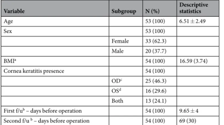
A total of 108 eyes of 54 patients were included in the study. Baseline patient characteristics, as well as the average f/u times, are shown in Table 1.
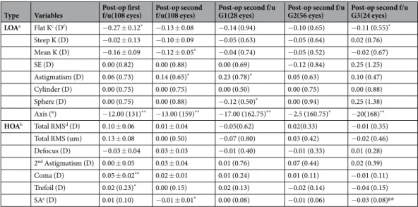
Baseline values for LOA and HOA variables are shown in Table 2. In terms of post-surgical changes for LOA variables, flat K was significantly lower at first f/u (p = 0.023) and G3 (p = 0.034) compared to the baseline (see Supplementary Fig. S1), and mean K values were significantly different only at the second f/u (p = 0.034; see Supplementary Fig. S2). Astigmatism values were significantly higher at second f/u (p = 0.026) and G1 (p = 0.026) compared to the baseline (see Supplementary Fig. S3), while sphere values were significantly lower at G1 (p = 0.041; see Supplementary Fig. S4). Axis values were significantly lower at all post-operative time points com- pared to baseline (p = < 0.001 and 0.015 at G2; see Supplementary Fig. S5). No significant changes across time points were observed for steep K (see Supplementary Fig. S6), SE (D) (see Supplementary Fig. S7), or cylinder values (see Supplementary Fig. S8, Table 3).
In terms of corneal HOAs, both coma (D) (p = 0.006; see Supplementary Fig. S9) and trefoil (D) (p = 0.037;
see Supplementary Fig. S10) values were significantly higher at first f/u compared to baseline while SA was signif- icantly lower at second f/u (p = 0.039) and G3 (p = 0.016) compared to baseline (see Supplementary Fig. S11). No significant differences at any of the post-operative time points, compared to baseline, were observed for total RMS (D) (see Supplementary Fig. S12), total RMS (μm) (see Supplementary Fig. S13), Defocus (see Supplementary Fig. S14), or 2
ndAstig values (see Supplementary Fig. S15, Table 3).
In this study, the age distribution of patients was from 3 to 12 years (mean ± SD, 6.51 ± 2.49 years). The median age was 6 years with 5 years in the 1
stquartile and 8.5 years in the 3
rdquartile. Given the wide age distri- bution, patients were divided into two subgroups based on the median age (6 years old). Patients in Number 1
Variable Subgroup N (%) Descriptive
statistics
Age 53 (100) 6.51 ± 2.49
Sex 53 (100)
Female 33 (62.3) Male 20 (37.7)
BMI
a54 (100) 16.59 (3.74)
Cornea keratitis presence 54 (100)
OD
c25 (46.3) OS
d16 (29.6) Both 13 (24.1)
First f/u
b– days before operation 54 (100) 9.65 ± 4 Second f/u
b– days before operation 54 (100) 69 (30)
Table 1. Baseline patient characteristics.
aBMI: body mass index,
bf/u: follow up,
cOD: Oculus Dexter (right eye),
dOS: Oculus Sinister (left eye).
Type Variable Mean ± SD
hor Median
(Interquartile range)
ia
LOA Flat K
b(D
g) 41.82 ± 1.49
Steep K (D) 43.41 ± 1.45 Mean K (D) 42.61 ± 1.30 SE
c(D) −0.25 (1.76) Astigmatism (D) 1.24 (1.14) Cylinder (D) −0.75 (1.25) Sphere (D) 0.25 (1.75)
Axis (°) 180 (90)
HOA
dTotal RMS
e(D) 1.23 ± 0.53
Total RMS
e(µm) 1.60 ± 0.69 Defocus (D) 0.09 ± 0.29 2
ndAstigmatism (D) 1.01 ± 0.61
Coma (D) 0.33 ± 0.15
Trefoil (D) 0.17 (0.16)
SA
f(D) −0.11 ± 0.06
Table 2. Baseline distribution of LOA and HOA variables.
aLOA: low order aberration,
bK: keratometry,
cSE:
spherical equivalent,
dHOA: high order aberration,
eRMS: root mean square,
fSA: spherical aberration,
gD:
Diopter,
hSD: standard deviation. Normally distributed variables are expressed as mean ± SD.
iNon-normally
distributed variables are expressed as median (interquartile range).
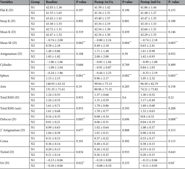
(N1) were 6 years of age and older (n = 29; mean ± SD, 8.28 ± 1.94 years old) and those in Number 2 (N2) were less than 6 years old (n = 24; mean ± SD, 4.38 ± 0.82 years old). Mean SE and sphere were statistically different between N1 and N2 at baseline, first f/u, and second f/u in LOA; defocus and SA were statistically different between N1 and N2 at baseline and second f/u in HOA (Table 4).
Univariable linear regression analyses were performed to study the effects of risk factors on postoperative outcomes. At the first and second f/u, sex had an effect on all LOA values except first f/u steep K. Additionally, sex had an effect on all values in G3. Age and BMI affected SE in G1, while sex, age, and BMI affected the first and second f/u times in G2. Sex had an effect on astigmatism values only in G2. The presence of cornea keratitis affected second f/u cylinder values in G3, while sex affected these in G2. Sex, age, and BMI further affected first and second f/u sphere values, while age and BMI affected G1 sphere values, and sex and age affected G2 sphere values. The risk factors assessed did not affect axis values at any time point.
In terms of HOAs, total RMS and RMS values were affected by the presence of cornea keratitis at the second f/u only. Age affected defocus values at the second f/u in G2 and G3. However, 2
ndastigmatism was not affected by any risk factors at any time point. Age only affected coma values at the second f/u and in G1, while sex, age, and BMI affected them in G2. Only sex had an effect on trefoil values in G3. Finally, age affected SA values in G2 alone.
Next, we analysed the significance of time as a fixed effect after correcting for the effects of the risk factors discussed above on the two outcomes (change from baseline values and univariable regression analysis) measured before and after surgery. To do this, a multivariable linear mixed-effect model was employed. At the first f/u, cyl- inder, coma, trefoil, and SA were significantly increased (p = 0.039, 0.008, 0.027, and 0.05, respectively), while axis and flat K decreased (p = < 0.001, 0.022) from baseline. At the second f/u, cylinder was increased (p = 0.05), while axis and mean K were significantly decreased (p = < 0.001 and 0.045, respectively) from baseline. In G1, sphere, axis, and flat K decreased from baseline (p = 0.041, <0.001, and 0.028, respectively), while astigmatism increased significantly (p = 0.028). In G2, axis decreased from baseline (p = 0.001), while coma increased significantly (p = 0.04). In G3, axis, flat K, and SA all significantly decreased from baseline p = < 0.001, 0.009, and 0.011, respectively).
Discussion
Given the current deficiencies in the understanding of visual impairments caused by surgery in patients with epi- blepharon, this study aimed to investigate post-operative changes in corneal LOAs and HOAs after lower eyelid epiblepharon repair in children. This surgical procedure was demonstrated to cause significant changes in axis, flat K, mean K, SA, coma, and trefoil values.
Many kinds of postoperative visual disturbances due to refractive power changes after ocular surgery have been reported previously. Specifically, severe or irregular astigmatism, changes in the astigmatic axis, myopic or hyperopic shifts, changes in axial length, and positional or dioptric errors in the implanted intraocular lenses have been attributed to visual disturbances and refractive changes in the postoperative period
24–29‘With-the-rule’
astigmatism often occurs in epiblepharon patients, as the present paper demonstrates.
The prevalence of ‘with-the-rule’ astigmatism, defined as an axis of astigmatism of 180 ± 15°, has been reported to range from 60.7% to 90.5%
7–9,30,31. Here, we report changes from ‘with-the-rule’ to ‘against-the-rule’
Type Variables Post-op first
f/u(108 eyes) Post-op second
f/u(108 eyes) Post-op second f/u
G1(28 eyes) Post-op second f/u
G2(56 eyes) Post-op second f/u G3(24 eyes) LOA
aFlat K
c(D
f) −0.27 ± 0.12
*−0.13 ± 0.08 −0.14 (0.94) −0.10 (0.65) −0.11 (0.55)
*Steep K (D) −0.02 ± 0.13 −0.10 ± 0.09 −0.05 (0.63) −0.05 (0.64) 0.02 (0.76) Mean K (D) −0.16 ± 0.09 −0.12 ± 0.05
*−0.04 (0.74) −0.05 (0.52) −0.02 (0.67)
SE (D) 0.00 (0.82) 0.00 (0.88) 0.00 (0.69) −0.12 (0.84) 0.25 (1.25)
Astigmatism (D) 0.06 (0.73) 0.14 (0.65)
*0.23 (0.78)
*0.05 (0.63) 0.10 (0.47)
Cylinder (D) 0.00 (0.75) 0.00 (0.75) 0.00 (0.50) 0.00 (0.75) 0.00 (0.88)
Sphere (D) 0.00 (0.75) 0.00 (0.88) −0.12 (0.50)
*0.00 (0.94) 0.25 (1.38) Axis (°) −12.00 (131)
**−13.00 (159)
**−17.00 (162.75)
**−2.5 (160.75)
*−20(168)
**HOA
bTotal RMS
d(D) 0.10 ± 0.06 0.01 ± 0.04 −0.05(0.62) 0.02(0.33) −0.01 (0.35) Total RMS (um) 0.13 ± 0.08 0.00 (0.50) −0.07 (0.80) 0.03 (0.42) −0.02 (0.46) Defocus (D) −0.03 ± 0.04 0.03 ± 0.03 −0.01 (0.40) −0.01 (0.33) 0.01 (0.28) 2
ndAstigmatism (D) 0.00 ± 0.05 0.03 ± 0.04 0.01 (0.76) 0.07 (0.44) 0.02 (0.39)
Coma (D) 0.05 ± 0.02
**0.02 ± 0.01 0.01 (0.24) 0.01 (0.11) −0.01 (0.11)
Trefoil (D) 0.02 (0.23)
*0.00 (0.15) 0.02 (0.13) −0.02 (0.14) −0.04 (0.15) SA
e(D) 0.01 (0.10) −0.01 ± 0.01
*0.00 (0.08) −0.01 (0.06) −0.03 (0.08)
g*
Table 3. Post-surgical changes in LOA and HOA variables compared to baseline. Values are presented as mean
± standard error or median (interquartile range).
aLOA: low order aberration,
bHOA: high order aberration,
c