Tuberc Respir Dis 2011;70:285-292
CopyrightⒸ2011. The Korean Academy of Tuberculosis and Respiratory Diseases. All rights reserved.
인플루엔자 연관 폐렴
중앙대학교 의과대학 호흡기내과학교실
김재열Influenza Associated Pneumonia
Jae Yeol Kim, M.D.
Department of Internal Medicine, Chung-Ang University College of Medicine, Seoul, Korea
After an outbreak of H1N1 influenza A virus infection in Mexico in late March 2009, the World Health Organization raised its pandemic alert level to phase 6, and to the highest level in June 2009. The pandemic H1N1/A influenza was caused by an H1N1 influenza A virus that represents a quadruple reassortment of two swine strains, one human strain, and one avian strain of influenza. After the first case report of H1N1/A infection in early May 2009, South Korea was overwhelmed by this new kind of influenza H1N1/A pandemic, which resulted in a total of 700,000 formally reported cases and 252 deaths. In this article, clinical characteristics of victims of H1N1/A influenza infection, especially those who developed pneumonia and those who were cared for in the intensive care unit, are described. In addition, guidelines for the treatment of H1N1/A influenza virus infection victims in the ICU, which was suggested by the Korean Society of Critical Care Medicine, are introduced.
Key Words: Influenza A Virus, H1N1 Subtype; Pneumonia; Intensive Care Units
Address for correspondence: Jae Yeol Kim, M.D.
Department of Internal Medicine, Chung-Ang University College of Medicine, 224-1, Heukseok-dong, Dongjak-gu, Seoul 156-755, Korea
Phone: 82-2-6299-1396, Fax: 82-2-825-7571 E-mail: [email protected]
Received: Dec. 3, 2010 Accepted: Dec. 3, 2010
역사 속에서의 Influenza 유행
언제부터 인간사회에서 독감이 유행하였는지 그 기원 을 정확히 밝히는 것은 어렵지만, 역사적으로 발한병 (sweat disease) 등으로 불리던 유행병이 독감에 의하였을 가능성이 있으며, 16세기에 유럽에 유행했던 전염병은 질 병의 특징과 전파양상을 고려할 때 독감에 의한 것일 가능 성이 높다. 과거에는 독감이 왜 발생하는지를 잘 몰랐기 때문에 나쁜 별의 영향(influence)이라고 여기던 시기가 있었으며, 여기에서 인플루엔자(influenza)라는 이름이 유 래되었다고 한다.
2009∼2010년 사이에 범세계적으로 유행한 H1N1 A형 인플루엔자의 역학
2009년 3월에 멕시코에서 호흡기 전염병이 대규모로 발생하였으며, 이것이 결국 H1N1 A형 인플루엔자에 의한 것으로 확인되었다. 멕시코에서 유래한 H1N1 A형 인플루 엔자는 빠른 속도로 전 세계적으로 전파되었으며, 검사를 통해 확인된 감염사례를 발표한 나라만 해도 214개 국가 에 달한다. 2009년 6월 11일에 WHO는 감염경보를 최고 단계인 6단계로 격상시켰다. 모델링기법에 의하면 미국에 서 H1N1 A형 인플루엔자에 감염된 사람은 6천 백만 명 정도일 것으로 추정된다1. 이를 연령별로 나눠서 살펴보 면 0세에서 17세까지 감염된 사람은 2천만 명, 18세에서 64세 사이에서 감염된 사람은 3천 5백만 명, 그리고 65세 이상에서 감염된 사람은 6백만 명일 것으로 추산된다. 이 렇게 나이에 따라 이환율에 차이가 나는 것은 노년층에서 인플루엔자에 대한 항체율이 높기 때문으로 해석된다.
2008년에 획득된 14,000명의 혈청을 이용하여 2009년형 H1N1 A형 인플루엔자에 대한 교차 항체반응 검사를 시행 하였을 때, 4세 이하의 어린이에서는 항체 양성률이 1.8%
Table 1. Multivariate analysis for survival predictors of pa- tients with H1N1-associated pneumonia
Variables Adjusted OR
(95% CI) p-value
Malignancy 12.0 (2.8∼51.5) 0.001
PSI score 1.03 (1.01∼1.04) 0.008
>50% increased extent of 4.00 (1.05∼15.2) 0.033 infiltrate on follow-up chest X ray
BUN>7 mmol/L 4.64 (0.93∼23.2) 0.062
Male gender 0.28 (0.07∼1.16) 0.080
CI: confidence interval; BUN: blood urea nitrogen.
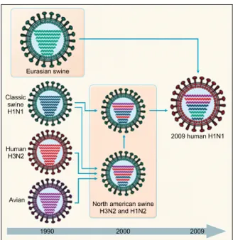
Figure 1. History of reassortment events in the evolution of the 2009 influenza A (H1N1) virus.
인 반면에 80세 이상인 사람에서의 항체 양성률은 31%였 다2. H1N1 A형 인플루엔자로 입원한 환자수는 274,000명 이고, 이로 인해 사망한 환자수는 12,470명이다3. 우리나 라에서는 약 70만 명의 환자가 발생하여 252명의 환자가 이로 인하여 사망한 것으로 공식 발표되었다.
H1N1 A형 인플루엔자의 유전적 특징
2009년과 2010년 사이에 걸쳐서 범세계적인 유행을 몰 고 온 H1N1 A형 인플루엔자 바이러스는 두 개의 돼지계 통 유전자(swine strain), 한 개의 사람계통 유전자(human strain), 그리고 한 개의 조류계통 유전자(avian strain)의 복합체로 과거에는 분리된 적이 없는 새로운 형태의 유전 적 구조를 가지고 있다(Figure 1)4. 이 때문에 신종 인플루 엔자라고 불렸다.
H1N1 A형 인플루엔자로 인한 사망률
H1N1 A형 인플루엔자에 의한 사망률은 0.5% 미만 (0.0004∼1.47%)인 것으로 추정된다5. 수학적 모델링에 의거하여 미국질병관리국(CDC)은 미국에서 H1N1 A형 인플루엔자로 인하여 사망한 숫자를 12,470명으로 추산하 였다3. 비록 H1N1 A형 인플루엔자에 의한 사망자수가 범
세계적인 유행이 없는 시기의 계절성 인플루엔자에 의한 사망자수보다는 적지만, H1N1 A형 인플루엔자에 의한 사 망이 주로 젊은 층에서 많이 일어났기 때문에 그 손실은 더 막대한 것으로 추산된다6. H1N1 A형 인플루엔자 감염 에 의한 사망의 위험을 높이는 위험인자로는 만성호흡기 질환, 면역억제질환, 심질환 등의 동반질환 존재7, 임산 부8, 신생아9, 고령7, 그리고 비만10 등이었다.
H1N1 A형 인플루엔자와 연관된 폐렴
국내의 14개 종합병원에서 2009년 6월부터 2010년 2월 까지 H1N1 A형 인플루엔자로 확진된 환자 중에서 폐렴이 발생한 환자 269명에 대하여 후향적 분석을 시행한 연구 에 따르면(unpublished data), 환자들의 평균 나이는 48세 (15∼93세)였으며, 남성이 143명으로 53.2%였다. 동반질 환은 천식이 36명(13.4%), 만성폐쇄성 폐질환이 14명 (5.2%), 기관지 확장증이 12명(4.5%), 그리고 악성 질환이 29명(10.8%)이었다. 사망한 환자는 19명으로 7.2%의 사 망률을 보였다. 사망한 환자의 연령대를 살펴보면 40대에 서 2명, 50대에서 5명, 60대에서 4명, 70대에서 5명, 그리 고 80대 이상에서 3명이 사망하였다. 다중회귀분석(multi- variate logistic regression analysis)에서 사망에 영향을 미 치는 인자는 악성 종양(odd ration [OR], 12.0; 95% con- fidence interval [CI], 2.8∼51.5; p=0.001), 추적한 가슴 X선 사진에서 병변의 범위가 50% 이상 증가하는 경우 (OR, 4.00; 95% CI, 1.05∼15.2; p=0.033), 그리고 폐렴 중증도 지표인 pneumonia severity index (OR, 1.03; 95%
CI, 1.01∼1.04; p=0.008)였다(Table 1).
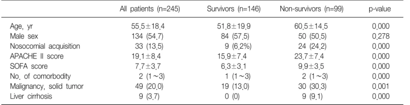
Table 2. Baseline characteristics of critically ill patients with pandemic influenza A/H1N1
All patients (n=245) Survivors (n=146) Non-survivors (n=99) p-value
Age, yr 55.5±18.4 51.8±19.9 60.5±14.5 0.000
Male sex 134 (54.7) 84 (57.5) 50 (50.5) 0.278
Nosocomial acquisition 33 (13.5) 9 (6.2%) 24 (24.2) 0.000
APACHE II score 19.1±8.4 15.9±7.4 23.7±7.4 0.000
SOFA score 7.7±3.7 6.3±3.1 9.9±3.5 0.000
No. of comorbodity 2 (1∼3) 1 (1∼3) 2 (1∼3) 0.000
Malignancy, solid tumor 49 (20.0) 19 (13.0) 30 (30.3) 0.001
Liver cirrhosis 9 (3.7) 0 (0) 9 (9.1) 0.000
Values are presented as number (%) or mean±SD unless otherwise indicated.
SD: standard deviation.
중환자실에 입원한 H1N1 A형 인플루엔자 환자의 특징
국내의 28개 종합병원에서 H1N1 A형 인플루엔자로 입 원치료를 받았던 환자들의 특징을 살펴보면(Hong et al, unpublished data), 총 245명의 환자 중에서 사망한 경우 는 99예(사망률, 40.4%)였다. 사망군에서 평균 나이, 질환 의 중증도(APACHE II, SOFA score)가 높았고, 동반 질환 의 수가 많았으며, 특히 악성 질환과 간경변의 빈도가 높 았다(Table 2).
중환자실에 입원한 H1N1 A형 인플루엔자 환자의 치료지침(대한중환자의학회 제공)
1. ICU admission criteria
1) Acute respiratory failure
(1) An arterial oxygen tension (PaO2) of <60 mm Hg with normal or low PaCO2 at FiO2 more than 0.5 (2) An arterial carbon dioxide tension (PaCO2) of > 50 mm Hg accompanied by a fall in pH<7.3 in addi- tion to hypoxaemia
(3) Respiratory rate >35 breaths/minute with ac- cessory muscle use
2) Hemodynamic instability
(1) Pulse <40 or >50 beats/minute
(2) Systolic arterial pressure <90 mm Hg or 40 mm Hg below the patient's usual pressure, or mean arterial pressure <60 mm Hg, or diastolic arterial pressure >120 mm Hg
2. ICU discharge criteria
When a patient's physiologic status has stabilized and the need for intensive patient monitoring is no longer necessary and the patient can be cared for on a general unit.
3. Treatment protocol
1) Antibacterial+antiviral treatment
(1) Initial antibiotics: 3rd generation cephalosporine+
respiratory quinolone
(2) Antiviral: tamiflu 150 mg po bid+amantidine 100 mg po bid+ribavirin 300 mg po tid
ㆍRibavirin-induced hemolysis and anemia should be monitored.
ㆍRibavirin is contraindicated in patients with CrCl
<50 mL/min. Ribavirin is not effectively re- moved by hemodialysis.
(3) Others: follow ATS/IDSA guideline11 2) Respiratory support
(1) General principles
① NPPV (noninvasive positive pressure ventilation) - generally not recommended, selected patients only ② Bronchodilator delivery: 6 puffs of metered dose inhaler of ventolin at the inspiratory limb of ventilatory tubing using a spacer or nebulizer
③ Moderate case - follow usual respiratory failure management
④ Severe case (PaO2/FiO2 100∼200) - mechanical
ventilation with or without NO inhalation, consider prone position or ARM (alveolar recruitment maneuver) ⑤ Very severe case (PaO2/FiO2 <100) - consider early ECMO
⑥ With unstable vital signs: heavy sedation with fen- tanyl or ketamine, and consider use neuromuscular blockades
(2) Ventilaror setting
① In case of FiO2 requirement less or equal than 0.6:
Lung protective ventilation (tidal volume 6 mL/pre- dicted body weight; if Pplat >30 cm H2O, consider re- duce tidal volume further, PEEP setting as ARDSnet PEEP table, I:E=1:1).
Consider permissive hypercapnea strategy (do not overventilate)
② In case of FiO2 >0.6
- if hemodynamic stable: decremental PEEP setting af- ter ARM
- if case of hemodynamic unstable: prone ventilation with or without NO inhalation (nitric oxide should be titrated the dose daily)
(3) Indication of early ECMO: Consider ECMO at early stage of ARDS if ARDS patients not be maintained appropriately (optional treatment)
① If FiO2 requirement is still higher 0.7 after decre- mental PEEP titration with alveolar recruitment maneu- ver and/or Prone ventilation & NO inhalation ② Refractory hypercapnea (pH <7.2) under me- chanical ventilation
③ Blood pressure could not be maintained with ap- propriate vasopressor use
④ Rapid progression of lung fibrosis (4) Criteria for Ventilator Weaning
① Stability/reversal of acute respiratory failure ② PaO2/FiO2 >150∼200, PEEP <5∼8 cm H2O, FIO2 <0.4∼0.5, pH >7.25
③ Hemodynamic stability (dop/dob <5) ④ Capable of reliable inspiratory efforts 3) Shock managements
Principle: follow early goal-directed therapy protocol If needed vasopressors,
(1) Norepinephrine (NE) with Dobutamine
(2) If the dose of NE >0.4μg/kg/min --- add low dose of vasopressin (0.01∼0.04 IU/hr) and low dose steroid (hydrocortisone 50 mg q 6 hr or continuous in- fusion 10 mg/hr)
(3) If the dose of NE >1μg/kg/min --- consider ear- ly ECMO
4) Renal replacement therapy: early CRRT or daily hemodialysis
Principle: follow early goal-directed therapy protocol (1) In case of urine output <0.5 mL/hour/kg even after intravenous Lasix 1 mg/kg injection over 20 min (2) In case of serum creatinine elevation more than 2.0∼3.0 times compared with the baseline value (3) Dialysis is better than high dose furosemide in- fusion in acute kidney injury
5) Steroid
(1) Generally not recommended
(2) It may be applied only in refractory shock and ARDS
6) Transmission prevention
(1) All health care workers; eye protection google, gown, gloves, N95 mask during aerosol-generating pro- cedure, handwahsing before and after patient's manage- ment or touching patient's related equipments
(2) Patient's bed; single mechanically or naturally ventilated room (12 air exchanges/hour), or negative pressure room if available
(3) Closed tracheal suction system
(4) In open spaced icu beds: the space between pa- tient's bed should be wider than 3 meter to prevent spread of H1N1 influenza via H1N1 influenza infected patient's droplet
7) Other general prophylaxis guideline (1) Deep vein thrombosis prophylaxis (2) Stress ulcer prophylaxis
(3) Bed head elevation (higher than 30 degree) to prevent aspiration
(4) Subglottic aspiration in case of intubated patients, if available
(5) Serum glucose control around 150 mg/dL
4. 참고 자료
1) 진단적 검사Laboratory of H1N1 RT-PCR in brocnoalveolar lavage (BAL) fluid or endotracheal aspiration : can be repeated twice for false negative
2) PEEP setting: Use PEEP table or decremental PEEP titration after ARM
(1) PEEP titraton
PEEP and FiO2 in ARDS Net study
FiO
20, 3 0, 3 0, 3 0, 3 0, 3 0, 4 0, 4 0, 5 0, 5 0, 5∼0, 8 0, 8 0, 9 1, 0 1, 0
PEEP 5 8 10 12 14 14 16
16 18 20 22 22 22 24
(2) Alveolar recruitment maneuver - an example Initial Lung Recruitment Procedure
① Before beginning recruitment maneuver, follow steps a∼f in order:
a. Ensure patient is ventilated on 100% O2. b. Sedate patient to produce apnea
c. Administer neuromuscular blockade if necessary d. Measure S O2, pulse pressure or CVP.
e. Determine if intravascular volume status is accept- able.
ㆍS O2 >70% or
ㆍPulse pressure variation <10% or ㆍCVP >12 mm Hg
f. Administer IV fluids for low S O2 or CVP and/or high pulse pressure
② Ventilator settings for the initial recruitment ma- neuver
a. Pressure A/C mode b. PEEP=25 cm H2O initially c. Pressure control (PC)=15 cm H2O
d. Peak inspiratory pressure (PIP)=40 cm H2O e. Inspiratory time=3.0 sec
f. Ventilator rate=10 breaths/min
g. FIO2=1.0 (Ventilator should already be set on 100%
O2)
③ After 5 breaths, increase PC to 20 cm H2O (PIP=45 cm H2O)
④ After 5 breaths, increase PEEP to 30 cm H2O
(PIP=0 cm H2O)
⑤ Continue for 20 breaths
⑥ Important: Abort lung recruitment procedure and immediately change ventilator to pre-recruitment set- tings if any of the following occur
ㆍMean arterial pressure <60 mm Hg or decreases by >20 mm Hg from baseline
ㆍSpO2 <88%
ㆍHeart rate >130 or <60/min ㆍNew onset of cardiac arrhythmias
ㆍSvO2 <65% or decreases by >20% or more ⑦ Change ventilator to Post-Recruitment settings ㆍVolume A/C mode
ㆍFIO2=1.0
ㆍPEEP=25 cm H2O ㆍVT=6 mL/kg ㆍPplat<45 cm H2O
ㆍDecrease VT to 5 or 4 mL/kg if needed to keep Pplat<45 cm H2O
ㆍInspiratory time=0.6 seconds
ㆍVentilator rate ≤40 breaths/min (highest possible without auto-PEEP)
⑧ Begin Decremental PEEP Trial Procedure immedi- ately
3) Prone positioning (복와위) (1) 복와위 시행 방법
① 복와위를 함께 수행할 사람들에게 체위 변경의 목적 과 절차를 설명한다.
② 손을 씻는다.
③ 필요한 물품(electrode, air ring, 욕창방지용 pad, 베개 등)을 준비한다.
④ 필요 시 기관절개관은 유연하고 긴 인공기도관으로 교체한다.
⑤ 욕창 호발 부위(앞가슴, 어깨)에 욕창방지용 pad를 붙인다.
⑥ 인공호흡기 tube, 말초동맥관, 중심정맥관, Swan- ganz catheter. IV-line, chest-tube 및 L-tube 등 환자가 가지고 있는 모든 line이 빠지지 않도록 충분히 길게 정리 한다.
⑦ 주치의와 간호직원에게 알리고 도움을 요청한다.
⑧ 상지부분 2명, 하지부분 2명, 모두 4명이 침대 양쪽 에 마주보며 선다.
⑨ 환자를 침대 가장자리로 옮긴 후 체위를 측위로 돌 린다.
⑩ Foley catheter, chest-tube는 복와위 자세에 맞게 이 동한다.
⑪ 가슴 위의 electrode를 제거한다.
⑫ 환자를 침상에서 10 cm 정도 들어올린 상태에서 동 시 구호를 외치면서 신속하게 돌린다.
ㆍ머리쪽 의사/간호사(I)는 기도유지와 인공호흡기를 담당하는데 한 손으로 인공기도를 지지하고 다른 한 손으로 목 뒷부분(경추)을 지지한다.
ㆍ상지쪽 간호사(II)는 상지부분의 모든 line을 지지하 고 양팔의 관절에 무리가 없도록 지지한다.
ㆍ복부쪽 선 간호사(III)는 복부와 하지 부분에 있는 line과 foley catheter를 지지한다.
ㆍ하지쪽 선 간호사(IV)는 양 다리의 관절을 지지한 다.
⑬ 인공기도의 위치를 확인하고 인공호흡기를 연결한 후 호흡상태를 확인한다.
⑭ Electrode를 등쪽의 올바른 위치에 붙인 후 심전도 를 확인한다.
⑮ Sore가 잘 생길 수 있는 부위에 air ring이나 베개로 지지한다(귓볼, 뺨, 앞가슴, 팔꿈치, 회음부, 무릎 등).
⑯ 침상 상부를 10∼15도 올린 후 환자를 편안한 자세 로 취해 준다.
⑰ 모든 line을 정리한다.
⑱ 환자의 상태를 관찰하고 기록한다(활력징후, SPO2, ABGA 결과 변화 등).
4) ARDS환자에서의 NO 투여 및 사용원칙 (1) 사용 원칙
① NO는 흡입되었을 때 선택적으로 폐동맥압을 감소 시키며, venous admixture (QVA/Qt)의 감소를 통해서 산 소화를 개선시키나 환자의 사망률을 개선시키지는 못한 다. 또한 NO의 dose-response curve는 투여 일수가 지나 갈수록 변화하므로 매일 SaO2가 최대화 되는 용량으로 조 정하고(흔히 용량을 줄여야함) 가능한 1주일 이상은 사용 하지 않도록 한다.
② 산소화 개선을 위한 효과적인 농도는 10 ppm 미만 이나 폐동맥압 감소를 목적으로 하는 경우는 흡입 용량에 비례하여 폐동맥 감소가 나타나나 30 ppm 이상은 사용하 지 않도록 하고 10 ppm 이상의 용량이 흡입될 때는 호기 내 NO2 농도를 지속적으로 감시하여야 하고 환기에 유의 하여야 한다.
③ NO에 대한 반응성: 패혈증이 없는 ARDS가 패혈성 쇽에 동반된 ARDS에 비해 NO에 대한 반응이 좋다. 또한 폐혈관 저항이 증가된 경우 폐포모집이 많이 된 경우에서 NO에 대한 반응이 좋다.
④ 흡입 NO의 유지 및 중단: NO는 우선 2∼10 ppm의 농도로 시작하고 SaO2를 보면서 그 흡입 용량을 매일 적 정화 하여야 한다. 대부분의 경우 투여 시간이 경과할수 록 투여량을 줄여 나가야 한다. 투여량을 급격히 끊으면 저산소증 및 폐고혈압의 갑작스런 악화가 발생할 수 있다.
(2) NO의 흡입
① 가장 간단한 방법: NO는 inspiratory limb으로 지속 적으로 주입한다. 호기 동안에 inspiratory limb에는 flow 가 없기 때문에 NO만이 흘러가게 된다. 따라서 환자는 흡기 시에 고농도의 NO를 흡입할 수 있게 된다.
② Y piece나 endotracheal tube에 직접 NO를 주입했 을 때: NO가 호기 시 exhalation limb으로 빠져 나갈 수 있고 정확한 NO의 농도를 측정하는 것이 불가능하다.
Tube에 직접 주입하는 경우에는 환자가 무호흡 상태일 때 anoxic gas를 흡입하게 되어 질식할 수도 있다.
③ Nebulizer 이용법: 흡기 시에만 작동하는 nebulizer drive mechanism을 통해 흡기 시에만 NO가 투입되게 할 수 있다.
④ Premix 법: NO를 공기나 nitrogen과 미리 혼합하여 인공환기기 gas inlet의 근위부나 breathing circuit에 주입 하는 법이다. NO의 농도가 일정하게 유지되고 분당환기 량이나 흡기 waveform에 영향을 받지는 않는다.
5) ECMO (1) V-V ECMO
주로 폐질환으로 인해서 ECMO를 사용한다. 우선, 환자 의 폐질환이 호전되는 양상을 보여야 한다. ECMO를 하고 자 할 때 운용하는 동안 환자의 ventilator는 가능한 폐 손상을 줄이기 위해서 low tidal volume, low pressure, low FiO2를 유지하게 되고, 환자의 PaO2를 60 mm Hg 이상 유지해야 한다.
환자의 산소가 증가하거나 방사선 사진이 호전되는 경 우 "trial off ECMO"를 시도하게 되는데 이것은 pump flow를 줄이는 방법과 ECMO 산화기의 산소를 줄이는 방 법을 이용할 수 있다. ECMO oxygenator FiO2를 줄이는 방법이 pump flow를 줄이는 방법에 비해서 간편할 수 있 다. Oxygentor FiO2를 줄여서 Oxygenator를 통과하고 환 자의 몸으로 들어가는 혈액의 산소 농도가 mixed venous O2만큼 감소하였는데 환자 혈액의 산소 농도가 60 mm
Hg 이상으로 유지되는 경우 "Oxygen challenge"의 방법 을 이용하여 환자 ventialtor의 FiO2를 1.0으로 하여 PaO2
가 100 mm Hg 이상으로 증가하는 경우 ECMO를 제거할 수 있다. 제거하는 방법은 continuous infusion되고 있는 heparin을 중단하고, pump flow를 낮추고 stop한 다음 catheter line을 clamp하고 catheter를 제거한다. 제거 후 에 protamine을 주입하여 heparin을 reversal시킨다.
(2) V-A ECMO
대부분 심장기능의 저하나 septic shock으로 인해서 혈 압을 유지할 수 없는 경우에 사용한다.
72시간 이후 심장기능 회복 여부를 평가한다. 심초음파 검사상에서 심장기능이 회복되거나 pulse pressure가 나 타나는 것으로 알 수 있다. 정확한 검사를 위해서 pump flow를 50% 미만으로 감소한 상태에서 확인해야 한다.
Catheter를 제거하기 위해서는 심장기능이 회복된 후에 시도해야 한다. Pump flow를 10∼20%씩 감소시키면서 혈압의 변화를 확인해야 하고, pump assist를 중단한 다음 30분에서 1시간 정도 유지할 수 있는지를 본다. 이 때에는 thrombus의 위험이 높기때문에 heparin을 증량하거나 bolus injection을 통해서 ACT를 200초 이상 유지해야 한 다. 환자의 상태가 pump flow를 중단한 상태에서 잘 견 디는 경우 제거하면 된다. 그러나, 환자의 상태가 점점 악 화되고, 강압제를 증량하면서도 혈압의 유지가 어려운 경 우에는 다시 pump assist를 하게 되고, 위와 같은 과정을 통해서 weaning을 2∼3일마다 할 수 있다. 가장 중요한 것은 환자의 심장에 교정할 다른 문제가 없어야 한다.
(3) Stopping support for futility of ECMO
ECLS should be discontinued promptly if there is no hope for healthy survival (severe brain damage, no or heart or lung recovery, and no hope of organ replace- ment by VAD or transplant).
The definition of irreversible heart or lung damage For cardiac failure, three days of no cardiac function in a patient who is not a VAD or transplant candidate is considered futile in most centers.
For lung failure, two weeks of no lung function in a patient who is not a transplant candidate is considered futile.
The possibility of stopping for futility will be ex- plained to the family before ECLS is begun.
6) Other rescue therapy
Refractory hypoxemia - consider induced mild hypo-
thermia
참 고 문 헌
1. Reed C, Angulo FJ, Swerdlow DL, Lipsitch M, Meltzer MI, Jernigan D, et al. Estimates of the prevalence of pandemic (H1N1) 2009, United States, April-July 2009.
Emerg Infect Dis 2009;15:2004-7.
2. Miller E, Hoschler K, Hardelid P, Stanford E, Andrews N, Zambon M. Incidence of 2009 pandemic influenza A H1N1 infection in England: a cross-sectional sero- logical study. Lancet 2010;375:1100-8.
3. Centers for Disease Control and Prevention. Updated CDC estimates of 2009 H1N1 influenza cases, hospital- izations and deaths in the United States, April 2009-April 10, 2010. Atlanta, GA: Centers for Disease Control and Prevention; c2010 [cited 2010 June 7].
Available from: http://www.cdc.gov/h1n1flu/estimates_
2009_h1n1.htm.
4. Novel Swine-Origin Influenza A (H1N1) Virus Investiga- tion Team, Dawood FS, Jain S, Finelli L, Shaw MW, Lindstrom S, et al. Emergence of a novel swine-origin influenza A (H1N1) virus in humans. N Engl J Med 2009;360:2605-15.
5. Writing Committee of the WHO Consultation on Clinical Aspects of Pandemic (H1N1) 2009 Influenza, Bautista E, Chotpitayasunondh T, Gao Z, Harper SA, Shaw M, et al. Clinical aspects of pandemic 2009 influ- enza A (H1N1) virus infection. N Engl J Med 2010;362:
1708-19.
6. Butler D. Portrait of a year-old pandemic. Nature 2010;
464:1112-3.
7. Louie JK, Acosta M, Winter K, Jean C, Gavali S, Schechter R, et al. Factors associated with death or hospitalization due to pandemic 2009 influenza A(H1N1) infection in California. JAMA 2009;302:1896- 902.
8. Hewagama S, Walker SP, Stuart RL, Gordon C, Johnson PD, Friedman ND, et al. 2009 H1N1 influenza A and pregnancy outcomes in Victoria, Australia. Clin Infect Dis 2010;50:686-90.
9. Creanga AA, Johnson TF, Graitcer SB, Hartman LK, Al-Samarrai T, Schwarz AG, et al. Severity of 2009 pan- demic influenza A (H1N1) virus infection in pregnant women. Obstet Gynecol 2010;115:717-26.
10. ANZIC Influenza Investigators, Webb SA, Pettilä V, Seppelt I, Bellomo R, Bailey M, et al. Critical care serv- ices and 2009 H1N1 influenza in Australia and New Zealand. N Engl J Med 2009;361:1925-34.
11. Mandell LA, Wunderink RG, Anzueto A, Bartlett JG, Campbell GD, Dean NC, et al. Infectious Diseases Society of America/American Thoracic Society con-
sensus guidelines on the management of commun- ity-acquired pneumonia in adults. Clin Infect Dis 2007;
44 Suppl 2:S27-72.