Case report
Child Kidney Dis 2015;19:39-42
DOI: http://dx.doi.org/10.3339/chikd.2015.19.1.39
ISSN 2384-0242 (print) ISSN 2384-0250 (online)
Acute Epstein-Barr Virus Hepatitis in a 32 Month Old Female Manifesting as Henoch-Schönlein Purpura
Henoch-Schönlein purpura can result from exposure to an antigen after infection with several types of organisms. However, Henoch-Schönlein purpura caused by a primary Epstein-Barr virus infection has been rarely reported. Here, we report the case of a 32-month-old female patient who presented with Henoch-Schönlein purpura. Based on abnormal liver function test results and positive results for Epstein-Barr virus infection markers, a diagnosis of Epstein-Barr virus hepatitis manifesting as Henoch-Schönlein purpura was made. Treatment with methyl- prednisolone and hydration improved the symptoms, and a switch to oral steroids was effective in completely alleviating the purpura. No recurrence was noted and no liver function abnormalities were detected during the follow up period.
Key words: Henoch-Schönlein purpura, Arthritis, Epstein-Barr virus, Hepatitis, Pathogenesis
Hee Jin Kim, M.D., Su Jin Jung, M.D., Jun Ho Lee, M.D.
Departments of Pediatrics, CHA Bundang Medical Center, CHA University, Seongnam, Korea
Corresponding author:
Jun Ho Lee, M.D.
Department of Pediatrics, CHA Bundang Medical Center, CHA University, 351 Yatap-dong, Bundang-gu, Seongnam-si, Gyeonggi-do 463-712, Korea
Tel: +82-31-780-5011 Fax: +82-31-780-5011 E-mail: [email protected] Su Jin Jung, M.D.
Department of Pediatrics, CHA Bundang Medical Center, CHA University, 351 Yatap-dong, Bundang-gu, Seongnam-si, Gyeonggi-do 463-712, Korea
Tel: +82-31-780-5011 Fax: +82-31-780-5011 E-mail: [email protected] Received: 16 March 2015 Revised: 15 April 2015 Accepted: 25 April 2015
This is an open-access article distributed under the terms of the Creative Commons Attribu tion Non-Commercial License (http://
crea tivecom mons.org/licenses/bync/3.0/) which permits unrestricted non-commercial use, distribution, and reproduction in any medium, provided the original work is properly cited.
Copyright © 2015 The Korean Society of Pediatric Nephrology
Introduction
Primary Epstein-Barr virus (EBV) infections are common in infants and children. In most cases, patients are asymptomatic or have non-specific symp- toms1). Less commonly, they are associated with severe complications such as hemolytic anemia, pancytopenia, myocarditis, hepatitis, splenic rup ture, glomerulonephritis, hemophagocytic syndrome, Guillain-Barré synd rome, meningitis, encephalitis, and psychiatric disorders1). Henoch-Schönlein pur- pura (HSP) can result from exposure to an antigen after infection with several types of organisms. In rare cases, HSP can result from EBV in fection2).
Here, we report the case of a 32-month-old female patient with primary EBV hepatitis manifesting as HSP.
Case report
A 32-month-old female was admitted to our hospital because of a 1-day history of fever, arthralgia, and swelling in both ankles for 1 day, and purpura on both legs and arms. She had passed grayish stools intermittently for a few days previously. The patient's medical history was unremarkable. Vital signs included a blood pressure of 80/50 mmHg, heart rate of 110/minute,
40 Chil Kidney Dis • 2015;19:39-42 www.chikd.org
respiratory rate of 25/minute, and body tem perature of 38.5℃, and she did not look ill, although she seemed tired.
A physical examination revealed multiple palpable purpuric lesions on the lower limbs and fewer purpuric lesions on the upper limbs. Both feet and ankles were slightly swollen and tender (Fig. 1), and both ankles and knees showed slight limitation of motion. There was no pharyngeal injection, conjunctivitis, or tongue lesion, and no palpable cervical, axillary, or inguinal lymph nodes were detected. The abdomen was soft, flat, and not tender.
The liver and spleen were not palpable. Bowel sounds were normoactive. Laboratory findings were as follows: hemo- globin 12.4 g/dL, white blood cells 15,860/mm3 (seg 62%, lymphocytes 25%, monocytes 11%, atypical lymphocytes 2%), platelet count 103,000/mm3, normocytic, normoch- romic, acanthocytes 1+ on a peripheral blood smear, retic- ulocytes 1.07%, C-reactive protein 6.5 mg/dL, erythrocyte sedimentation rate 9 mm/h, protein 6.7 g/dL, albumin 4.1 g/dL, glucose 137 mg/dL, blood urea nitrogen/creatinine 15.4/0.6 mg/dL, T-bilirubin 1.23 mg/dL (D-bilirubin 0.64), glutamic-oxaloacetic transaminase (GOT)/glutamic-py- ruvic transaminase (GPT) 910/1,244 IU/L, alkaline pho- sphatase 1,696 IU/L, r-glutamyl transpeptidase 166 U/L, creatine kinase 61 U/L, lactate dehydrogenase 1,753 U/L, ammonia 27 µg/dL, cholesterol 22.7 mg/dL, amylase 40 U/
L, serum electrolytes Sodium 130 mEq/L, potassium 4.8 mEq/L, chloride 98 mEq/L, TCO2 22.7 mEq/L, Prothrom- bin time 15.5 s, international normalized ratio 1.4, activated partial thromboplastin time 36.4 s, D-dimer 2,138.04 ng/
mL, fibrinogen 326 mg/dL, C3 117 mg/dL, C4 47 mg/dL, fluorescent antinuclear antibody (-), anti-neutrophilic cy-
toplasmic antibodies (-), IgG 959 mg/dL, IgA 101 mg/dL, IgM 125 mg/dL, Mycoplasma pneumonia IgM nega tive, cold agglutinin negative, direct/indirect Cooms test nega- tive/negative, and rheumatoid factor negative. The results of urinalysis were normal, as were those of urine and blood cultures and chest radiography (posterioran terior view).
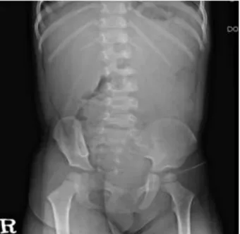
Simple examination of the abdomen showed suspicious hepatosplenomegaly (Fig. 2), but abdominal sonography revealed no remarkable findings. Tests for viral markers revealed surface antigen of the hepatitis B virus negativity, but antibody to hepatitis B surface antigen positivity. Poly- merase chain reaction confirmed the presence of EBV DNA. EBV viral capsid antigen (VCA) IgM antibodies but not IgG antibodies were detec ted in serum. Tests for other viral markers yielded negative results (Hepatitis A virus, Hepatitis C virus, Hepatitis E virus, Herpes simplex virus, Cytomegalovirus). The results of immunofluorescent staining with IgG, IgA, IgM, C3, and fibrinogen were all negative. Ac cordingly, a diagnosis of HSP due to acute EBV hepatitis was rendered.
Treatment with intravenous methylprednisolone (1 mg/
kg/day three divided doses) and hydration was initiated.
Arthralgia, grayish stools, and fatigue disappeared promptly after treatment initiation. The fever subsided on hospital day 2. The swelling on both legs and high GOT/
GPT levels improved slowly during the seven days of ad- mission. At discharge, GOT/GPT levels had decreased to
Fig. 1. Multiple palpable purpuric lesions on lower limbs. Both
feet and ankles were slightly swollen. Fig. 2. Simple abdomen showed hepatosplenomegaly.
Kim HJ, et al. • EBV Hepatitis associated with HSP in a Child 41 www.chikd.org
115/274 IU/L, and the T-bilirubin level was normal. Intra- venous methylprednisolone administered for one day was switched to oral steroids, which were then tapered off over three weeks. The purpura improved, leaving some dark pigmentation on both legs, but disappeared comple tely within one month. Periodic urinalyses were perfor med over the course of one year of follow-up. At two months after discharge, GOT/GPT levels became normal. HSP did not recur and no abnormalities were noted on urinalysis.
Discussion
Hepatic manifestation of EBV infection is commonly transient or self-limited, lasting from a few weeks to several months, but, rarely, can be fatal3). Hepatic manifestations include elevated liver enzymes (50-80%), hepatomegaly (10
%), jaundice (5-7%), chronic hepatitis (rare), or fulminant hepatic failure, which is the main cause of death in severe EBV infection3-5). No direct EBV cytopathic effect on hepa- tocytes has been proven6). EBV infection of T and B lym- phocytes as well as tumor necrosis factor-α (TNF-α), inter- feron-γ, and Fas ligand produced by cytotoxic lympho cytes recruited in the liver have been shown to induce hepatic injury7).
Skin manifestation occurs in 3-15% of patients with primary EBV infection, including petechiae, purpura, morbilliform rash, erythema nodosum, erythema multi- forme, urticaria, and genital ulcer8, 9). Mucocutaneous le- sions are commonly located on the trunk and upper arms;
sometimes they also affect the face and forearms10). These symptoms can result from the responses of EBV-specific cytotoxic T lymphocytes to EBV infection, which lead to systemic inflammation11).
In the 2005, European League Against Rheumatism/
Paediatric Rheumatology European Society (EULAR/PRES) modified the classification criteria for HSP as follows: pal- pable purpura (mandatory criterion) in the presence of at least one of the following four features: 1) diffuse abdominal pain, 2) any biopsy showing predomi nant IgA deposition, 3) arthritis or arthralgia (acute, any joint), and 4) renal in- volvement (any hematuria and/or proteinuria)12). These criteria support the diagnosis of HSP at the patient’s first visit. Acute viral arthritis usually manifests later, rather
than in the early acute stage of infection. While granular deposition of IgA and C3 iden tified by skin biopsy are included in the classic textbook description of HSP, these deposits are not essential diag nostic features13).
Hypersensitivity vasculitis may be difficult to distinguish from HSP only through skin biopsy. In contrast to patients with hypersensitivity vasculitis, patients with HSP usually have as followings: prominent palpable purpura, arthralgia, abdominal pain, gastrointestinal bleeding, hematuria, normal complement levels, evidence of IgA-containing immune complexes on skin or renal biopsy, and/or an ab- sence of recent use of medications that have the potential to cause vasculitis14).
HSP can result from exposure to an antigen from the following organisms: Group A streptococcus, parvovirus B19, Bartonella henselae, Helicobacter pylori, Haemophilus parainfluenza, Coxsackie virus, adenovirus, hepatitis A, B, and C viruses, mycoplasma, Epstein-Barr virus, varicella, campylobacter, and methicillin-resistant Staphylococcus aureus. The exact mechanism of HSP development due to primary EBV infection is not known, but contributing factors may include EBV-infected lymphocytes, as well as host responses to the EBV antigen, that lead to the produc- tion of various cytokines. EBV has been proposed as a po- tential initiating factor for autoim mune diseases like sys- temic lupus erythematosus, rheumatoid arthritis, and pri mary Sjögren’s syndrome, which is characterized by high titers of anti-EBV anti bodies and impaired T-cell immune responses to EBV antigens15). In patients with HSP, the level of regulatory T cells (CD4+CD25+) play a major role in preventing autoimmune and inflammatory diseases by secreting anti-inflammatory cytokines, such as interleukin-10 and transforming growth factor beta-β1, the level of which is lower than in normal controls16). Fur- thermore, TNF-α, a potent pro-inflammatory cytokine, is known to play an important role in the occurrence of HSP and HSP nephritis17).
Kim et al.18) reported twin cases of HSP nephritis associ- ated with acute EBV infection in south Korea, 2004. Their serological findings were anti-EBV VCA IgM nega tive, anti-EBV VCA IgG positive, anti-EBV EA IgG positive, and anti-EBV EBNA IgG negative. The diffe rences with our case are as follows: They did not have any symptom of acute EBV infection. Their HSP provoked at least one month
42 Chil Kidney Dis • 2015;19:39-42 www.chikd.org
after EBV infection by inference from their viral markers.
So, compared with our case who pre sented with HSP ac- companying EBV hepatitis, they may be concerned with delayed hypersensitivity reaction in the pathogenesis of HSP.
In summary, we report the case of a 32 month-old girl with acute EBV hepatitis manifesting as HSP. Thus, pri- mary EBV infection could cause HSP.
Conflict of interest
No potential conflict of interest relevant to this was reported.
Referecnes
1. Cohen JI. Epstein-Barr virus infection. N Engl J Med 2000;343:
481-92.
2. Guissa VR, Aragão PA, Marques HH, Jacob CM, Silva CA. Chronic active Epstein-Barr virus infection mimicking Henoch-Schönlein purpura. Acta Reumatol Port 2010;35:513-7.
3. Shaukat A. Epstein-Barr virus induced hepatitis: An important cause of cholestasis. Hepatol Res 2005;33:24-6.
4. Kimura H. Severe hepatitis caused by Epstein-Barr virus without infection of hepatocytes. Hum Pathol 2001;32:757-62.
5. Feranchak AP, Tyson RW, Narkewicz MR, Karrer FM, Sokol RJ.
Fulminant Epstein-Barr viral hepatitis: orthotopic liver transplan- tation and review of the literature. Liver Transpl Surg 1998;4:469- 76.
6. Michelow P, Wright C, Pantanowitz L. A review of the cytomor- phology of Epstein-Barr virus-associated malignancies. Acta
Cytol 2012;56:1-14.
7. Kimura H, Nagasaka T, Hoshino Y, Hayashi N, Tanaka N, Xu JL, et al. Severe hepatitis caused by Epstein-Barr virus without infection of hepatocytes. Hum Pathol 2001;23:257-62.
8. Cheng CC, Chang LY, Shao PL, Lee PI, Chen JM, Lu CY, et al.
Clinical manifestations and quantitative analysis of virus load in Taiwanese children with Epstein-Barr virus-associated infectious mononucleosis. J Microbiol Immunol Infect 2007;40:216-21.
9. McCarthy JT, Hoagland RJ. Cutaneous manifestations of infectious mononucleosis. JAMA 1964;187:153-4.
10. Mendoza N, Diamantis M, Arora A, Bartlett B, Gewirtzman A, Tremaine AM, et al. Mucocutaneous manifestations of Epstein- Barr virus infection. Am J Clin Dermatol 2008;9:295-305.
11. Catalina MD, Sullivan JL, Bak KR, Luzuriaga K. Differential evolution and stability of epitope-specific CD8+ T cell responses in EBV infection. J Immunol 2001;167:4450-7.
12. Ozen S, Ruperto N, Dillon MJ, Bagga A, Barron K, Davin JC, et al.
EULAR/PReS endorsed consensus criteria for the classification of childhood vasculitides. Ann Rheum Dis 2006;65:936-41.
13. Chan KH, Tang WY, Lo KK. Bullous lesions in Henoch-Schönlein purpura. Pediatr Dermatol 2007;24:325-6.
14. David C. Kimberly M. Hypersensitivity vasculitis in children. In:
UpToDate, Post TW (Ed), UpToDate, Waltham, MA. (Accessed on April 15, 2015)
15. Toussirot E, Roudier J. Epstein-Barr virus in autoimmune diseases.
Best Pract Res Clin Rheumatol 2008;22:883-96.
16. Chen O, Zhu XB, Ren H, Wang YB, Sun R. The imbalance of Th17/
Treg in Chinese children with Henoch-Schönlein purpura. Int Immunopharmaco 2013;16:67-71.
17. Takemura T, Yoshioka K, Murakami K, Akano N, Okada M, Aya N, et al. Cellular localization of inflammatory cytokines in human glomerulonephritis. Virchows Arch 1994;424:459-64.
18. Kim CJ, Woo YJ, Kook H, Choi YY, Ma JS, Hwang TJ. Henoch- Schönlein purpura nephritis associated with Epstein-Barr virus infection in twins. Pediatr Nephrol 2014;19:247-8.