Plateletpheresis: the Process, Devices, and Indicators of Product Quality
Chul-Soo Jang
1†, Sung-In Kim
1†, Hyun-Kyung Kim
1, Chang-Oh Kweon
1, Byung-Won Kim
1, Dong-Chan Kim
1, Yoon Suk Kim
2, Ki-Jong Rhee
2and Jae-Ki Ryu
1*
1
Department of Biomedical Laboratory Science, College of Nursing and Health Science, Gimcheon University, Gimcheon 740-704, Korea
2
Department of Biomedical Laboratory Science, College of Health Science, Yonsei University, Wonju 220-710, Korea
Received June 20, 2014 /Revised July 16, 2014 /Accepted July 25, 2014Platelet products are used to treat hemorrhagic or platelet dysfunction diseases. Plateletpheresis in- volves collecting the platelet components of blood using an apheresis blood-collection system. Various indicators are available for evaluating the qualities of the apheresis platelets. The productivity of pla- telet collection is evaluated through both the collection efficiency and collection rates. Platelet storage quality can be evaluated in vitro using several indicators, including visual appearance, metabolic activ- ities, volume, platelet count, white blood cell count, microparticles, and various platelet activation markers. Platelet activation markers have been used as indicators of storage quality in various studies.
Post-transfusion platelet quality can be evaluated based on the corrected count increment and the per- centage of platelet recovery. Although various studies have investigated the aspects of plateletphe- resis, no article has systemically presented assessments of the platelet products obtained from different plateletpheresis devices. The present study provides a review of plateletpheresis, including the specif- ics of the process, the types of devices employed, the platelet quality, the overall efficacy, and the evaluation indicator qualities. Furthermore, the differences in functionality among the different aphe- resis devices are discussed. Although adverse reactions to the citrate anti-coagulant have been re- ported, apheresis processing may provide a safer option for donors who are at a high risk for pre- syncopal or syncopal reactions related to whole blood collection.
Key words : Plateletpheresis, plateletpheresis devices, platelet quality indicators, platelet activation markers, platelet additive solution
†Authors contributed equally.
*Corresponding author
*Tel : +82-54-420-4041, Fax : +82-54-420-4461
*E-mail : [email protected]
This is an Open-Access article distributed under the terms of the Creative Commons Attribution Non-Commercial License (http://creativecommons.org/licenses/by-nc/3.0) which permits unrestricted non-commercial use, distribution, and reproduction in any medium, provided the original work is properly cited.
Journal of Life Science 2014 Vol. 24. No. 9. 1030~1038 DOI : http://dx.doi.org/10.5352/JLS.2014.24.9.1030
Introduction
Apheresis is the process of collecting blood components such as plasma, platelets, red blood cells, and granulocytes from donor blood. The term “apheresis” is derived from the Latin word “aphaeresis”, which means “withdrawal”.
Apheresis is accomplished using an apheresis instrument termed a cell separator. Whole blood from the donor is sep- arated by the device through centrifugation, based on the specific gravity and/or filtration parameters. The selected component of the blood is retained, while remaining blood components are returned to the donor through automated circulation. The processing time is approximately 1-2 hr [7, 24]. Apheresis instruments are used with various settings,
depending on the specific needs of different cases [8]. Cell
separators employ either continuous flow centrifugation,
which uses 2 venipunctures to collect blood, or intermittent
flow centrifugation, which returns the unwanted blood
components to the donor after it is temporarily collected in
the cell separator. In practice, apheresis presents several
obstacles. For example, apheresis requires a trained cell sep-
arator operator; is difficult to apply in high-capacity, emer-
gency settings; requires large initial expenditures; and can
result in adverse reactions among donors, such as par-
esthesia, tingling, seizure, and muscle cramps. These ad-
verse reactions result from the use of citrate anticoagulants
during apheresis. Nonetheless, the collection and use of
blood components through apheresis has increased over the
past few decades. The transfusion of blood components col-
lected through apheresis has many benefits. Indeed, it pro-
vides larger quantities of components than does whole
blood separation, produces consistent product volumes,
makes efficient use of the same donor, reduces the donor’s
physical stress and blood cell count recovery times, and de-
creases the risk of bacterial contamination. By eliminating
the recipient’s exposure to multiple donors, apheresis addi-
- Review -
tionally reduces the risks of human leukocyte antigen (HLA) alloimmunization and transfusion-transmitted dis- eases [3, 8, 15, 24, 41, 63].
Platelets are anucleate blood cells (150-450×10
3/μl) that form a platelet plug by adhesion and aggregation, thereby contributing to hemostasis [14]. Platelets are transfused for (i) treating hemorrhage due to severe thrombocytopenia, (ii) increasing platelet counts that are less than 20,000 per μl, or (iii) treating platelet dysfunction disease [40, 55].
Plateletpheresis is the process of collecting the platelet com- ponent through apheresis. Various studies have inves- tigated the aspects of apheresis platelet products, including quality evaluation, efficacy, the comparative performance of different devices, and methods designed for different pa- tient symptoms. However, to our knowledge, no article has systematically presented assessments of the quality and effi- cacy of platelets obtained from different plateletpheresis instruments. In the present review, we address the quality and efficacy of apheresis platelets.
Current Status of Methods and Results Donor screening for plateletpheresis
With the exception of the donation interval, the require- ments for apheresis and whole blood donation are the same. Apheresis donors must meet the requirements for whole blood donation and additionally satisfy criteria that are particular to the selected apheresis. Donor screening is required to ensure a safe transfusion for the recipient.
Prospective donors must complete several steps before ac- tual blood donations, including physical examination, a do- nor history questionnaire, and testing for transmissible diseases. The American Association of Blood Banks (AABB) recommends that prospective donors receive physical ex- aminations including an assessment of weight: hemoglobin, hematocrit, ABO and Rh typing: and inspection for marks from intravenous drug use. Tests are performed for the presence of syphilis, human immunodeficiency virus (anti- HIV-1/2 and HIV-1 RNA), hepatitis C virus (anti-HCV and HCV RNA), hepatitis B virus (HBsAg and anti-HBc), hu- man T-cell lymphotropic viruses I and II (anti-HTLV-I/II), Trypanosoma cruzi (anti-Trypanosoma cruzi), and West Nile virus (West Nile virus RNA). The Korean Society of Blood Transfusion recommends the tests for ABO and Rh typing, ABO subtyping, ALT, Total Protein, Antibody Screening Test, HBV (HBs Ag, HBV-NAT), HCV (anti-HCV, HCV-
NAT), HIV (anti-HIV, HIV-NAT), anti-HTLV, anti-Syphilis, and anti-Malaria for the donated blood.
Further, the platelet counts of plateletpheresis donors must be >150×10
3per μl [32]. Individuals can donate 3 days after ingesting aspirin-related medications.
Plateletpheresis instruments
Plateletpheresis instruments include the Fenwal CS3000 and Fenwal CS3000+ (Baxter Healthcare Corp., Round Lake, IL, USA); Fenwal Amicus (Fenwal, Inc., Lake Zurich, IL, USA); Cobe Spectra (Terumo BCT, Lakewood, CO, USA);
Fresenius AS104 and Fresenius COM.TEC (Fresenius AG, Bad Homburg, Germany); Trima (Version 4) and Trima Accel (Terumo BCT, Lakewood, CO, USA); and Haemonet- ics MCS LN9000 and MCS+LN3000 (Haemonetics Corp., Stoughton, MA, USA). These different devices have been compared according to a variety of measures, including pla- telet yield, collection efficiency, and collection rates [3, 8, 9, 10, 58].
Platelet yield
The platelet volume collected by plateletpheresis ranges from 150 to 300 ml and 75% of the collection must meet the standard of 3×10
11platelets/unit. Platelet yields in ex- cess of 6×10
11present several advantages. For example, when large quantities of platelets can be collected from do- nors who have been matched with patients according to HLA type, the collected blood can be divided into two parts and used [5]. Burgstaler et al. [9] reported that plateletphe- resis with a double-needle Amicus provided more double units than did Cobe Spectra double-needle version 5 leukor- eduction systems, Cobe Spectra double-needle version 7 leukoreduction systems, or Fresenius AS 104. The frequency of double units is 65% when Amicus or Trima Accel is used [9, 10]. If pre-donation platelet counts are high, platelet yields are generally high as well.
Reduction of white blood cell count
If a recipient’s white blood cells (WBCs) are exposed to
donor WBCs, allogenic immune reactions may result in
complications. In such cases, the leukoreduction process is
employed to reduce the complications that were induced by
the transfusion. The choice of leukoreduction is motivated
by several major goals: reducing febrile non-hemolytic
transfusion reactions, diminishing HLA sensitization and
platelet refractoriness, reducing the rate of cytomegalovirus
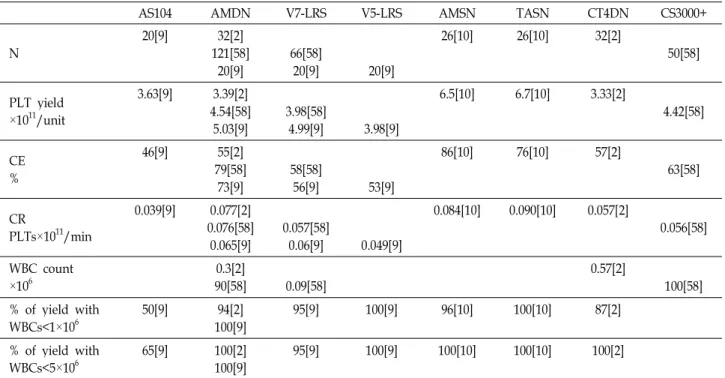
Table 1. Collection efficiency (CE), collection rates (CR), and white blood cell (WBC) content for samples obtained from different plateletpheresis instruments
AS104 AMDN V7-LRS V5-LRS AMSN TASN CT4DN CS3000+
N 20[9] 32[2]
121[58]
20[9] 66[58]
20[9] 20[9]
26[10] 26[10] 32[2]
50[58]
PLT yield
×1011/unit
3.63[9] 3.39[2]
4.54[58]
5.03[9] 3.98[58]
4.99[9] 3.98[9]
6.5[10] 6.7[10] 3.33[2]
4.42[58]
CE
%
46[9] 55[2]
79[58]
73[9] 58[58]
56[9] 53[9]
86[10] 76[10] 57[2]
63[58]
CRPLTs×1011/min
0.039[9] 0.077[2]
0.076[58]
0.065[9] 0.057[58]
0.06[9] 0.049[9]
0.084[10] 0.090[10] 0.057[2]
0.056[58]
WBC count
×106 0.3[2]
90[58] 0.09[58] 0.57[2]
100[58]
% of yield with
WBCs<1×10⁶ 50[9] 94[2]
100[9] 95[9] 100[9] 96[10] 100[10] 87[2]
% of yield with
WBCs<5×10⁶ 65[9] 100[2]
100[9] 95[9] 100[9] 100[10] 100[10] 100[2]
AS104: Fresenius AS104 double-needle, AMDN: Amicus double-needle, V7-LRS: Cobe Spectra double-needle version 7 leukoreduction system, V5-LRS: Cobe Spectra double-needle version 5 leukoreduction system, AMSN: Amicus single-needle, TASN: Trima Accel single-needle, CT4DN: COM.TEC version4, CS3000+: Fenwal CS3000+, N: number of donation, PLT yield: median platelet count of plateletpheresis product, CE: median collection efficiency, CR: median collection rates.
[ ]: reference number
infection, and reducing the rate of transfusion-associated graft versus host disease [33, 48]. Newly developed aphe- resis instruments have built-in leukoreduction systems that eliminate the need for further, post-apheresis reduction of leukocytes counts. Leukoreduced blood products are re- quired to meet AABB, which indicate that WBC counts per unit must be <5×10
6.
Collection efficiency, collection rates, and WBC count Evaluations of platelet collection performance are based on collection efficiency (CE) and collection rate (CR). CE compares the number of platelets in the collection bag with the number of platelets that pass through the device. Is cal- culated according to the following formulas:
CE = platelet yields in collection bag (10
11) ×100/average platelet count × blood volume processed.
Average platelet count = (pre-apheresis platelet count + post-apheresis platelet count)/2
Blood volume processed = total blood volume processed – volume of anticoagulant
CR is defined as the platelet yield per minute of process-
ing time. However, it does not reflect the time that is re- quired to collect platelets. To estimate the time to collect re- quired quantities of platelets, it is necessary to have the CRs for the instruments that are being used. CR is determined by the following formula:
CR = platelet yields in a collection bag (10
11)/min of processing time
Various studies have compared the function and charac- teristics of different apheresis instruments. Table 1 summa- rizes previous reports of CE, CR, and WBC for different apheresis equipment. According to these reports, the sin- gle-needle Amicus and single-needle Trima Accel have the highest CE and CR [3, 8, 9, 10, 58]. Flesch et al. [21] reported that the double-needle Amicus and the single-needle Trima Accel showed similar platelet yields and donation times.
Quality evaluation indicators of apheresis platelet Platelet products that are stored at room temperature (20-24°C) with agitation may remain viable for 5 days [32].
The storage time allowed by the US Food and Drug
Administration (FDA) was increased to 7 days in 1984, but returned to 5 days in 1986 because of events of bacterial transmission by transfused platelets. Slichter et al. [55] re- ported in vivo platelet recovery and survival in a platelet product obtained from MCS+ LN 9,000 and stored for 8 days. The quality of platelets that are stored after donation is influenced by many factors, including differences in blood component processing, gas permeability of the stor- age bag, anticoagulants, agitation, and storage temperature [4, 38, 56]. Platelet quality can be evaluated in vitro using several parameters such as visual appearance, metabolic ac- tivity, volume, platelet count, and WBC count per unit.
After transfusion, platelet quality can be evaluated using corrected count increment (CCI) and percentage platelet re- covery (PPR) [27, 54].
Visual appearance
The visual appearance of platelets, such as evidence of swirling, is used to evaluate platelet functionality [34].
Swirling is caused by the reflection of light when a discoid platelet is exposed to a light source. Generally, platelet con- centrate with a pH of 6.7-7.5 will show swirling when the platelets retain their discoid shape in vitro at the time of transfusion, and are expected to be functional in vivo [6, 34].
Singh et al. [54] found that apheresis-platelet concentrate units from the CS3,000+ instrument showed better swirling and platelet counts than platelet rich plasma-platelet con- centrates; however, Mintz et al. [37] confirmed that spher- ical platelets may revert to the discoid form, and that non-discoid platelets may retain suitable functionality post-transfusion.
Metabolic activity
Metabolic activities of apheresis platelets are mainly measured using pH, pCO
2, pO
2, bicarbonate, glucose, lac- tate, adenosine triphosphate (ATP), and hypotonic shock re- sponse (HSR).
The AABB requires that the pH of the platelet products remains ≥6.2 during storage with agitation at 20-24°C.
During storage, pH decreases as a result of increased lactic acid production due to glycolysis. At pH 6.0, the platelets irreversibly transition from the discoid to the spherical form [39]. Accordingly, pH has been identified as an indicator of stored platelet functionality. However, Tudisco et al. [59]
found that pH had little utility for the quality control of pla- telets produced by MCS + LN 3,000, Amicus, and CS-3,000+
apheresis instruments. Most platelet products obtained
from apheresis collections have a pH ≥6.2 on day 5 of stor- age, and the pH may even exceed 7.0 at 7 days of storage [18, 20, 26, 45, 55].
Assessments of pCO
2, pO
2, and bicarbonate changes in apheresis platelets showed that pCO
2and bicarbonate were both significantly reduced in all platelet concentrates col- lected using MCS+ LN9,000 or Trima instruments.
However, differences in pO
2were observed to increase or decrease, depending on the sample. Results were not sig- nificant in platelets from Trima that had been stored 7-9 days. Furthermore, in apheresis platelets, glucose levels de- crease and lactate significantly increases during storage [18, 20, 26, 45, 55].
ATP is the major energy source for platelet survival.
Naturally, ATP also decreases significantly during storage [18]. ATP was reduced in platelet concentrates collected us- ing Trima [18, 20, 45].
HSR measures the capacity of platelets that have been re- stored to normal morphology after hypotonic shock due to the addition of hypotonic solution. HSR is regarded as a predictor of platelet functionality [20, 46]. The HSR of aphe- resis platelets decreases as storage times increase [20, 26, 45, 55].
Microparticles (MPs)
MPs are membrane vesicles that originate primarily from platelets (PMP) or endothelial cells (EMP). The level of PMP generally increases in plateletpheresis concentrates during storage because PMPs are released by activation [43, 47, 52, 61]. The aim of plateletpheresis is to reduce HLA sensitiza- tion through leukodepletion: however, platelets that are col- lected primarily from plateletpheresis contain MPs, includ- ing EMP, which have high densities of HLA antigen [47, 50]. Therefore, improvements in apheresis technology may be necessary to reduce MP loads.
Platelet activation
Platelets play 2 key roles in hemostasis: platelet adhesion
to exposed subendothelial membrane and platelet ag-
gregation among activated platelets result in platelet plug
formation. Negatively charged platelet surfaces promote the
activation of coagulation factors II and X [8, 39]. Various
studies have investigated platelet activation, which has
been used to evaluate platelet quality during processing
and storage of plateletpheresies products. Activated platelet
markers that have been used in studies of plateletpheresis
include P-selectin (CD62P) [2, 27, 30], CD63 [20, 22, 47], gly-
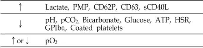
Table 2. Changes in the function and metabolic activity of apheresis platelets after storage
↑ Lactate, PMP, CD62P, CD63, sCD40L
↓ pH, pCO2,Bicarbonate, Glucose, ATP, HSR, GPIbα, Coated platelets
↑or↓ pO2
coprotein (GP)Ibα [25, 30, 36], CD40 ligand (CD40L, CD154) [12, 13, 19, 28, 62], and coated platelets [1, 11, 57].
P-selectin (CD62P) is stored in granules of endothelial cells and platelets. It is expressed on the surface of activated cells, where it functions as a cell adhesion molecule [2, 30].
During storage, increased P-selectin expression on activated platelet surfaces triggers rapid clearance of transfused plate- lets from circulation. Comparisons of platelet concentrates obtained from different apheresis instruments have demon- strated that the double-needle Amicus has higher rates of P-selectin-positive platelets than does the single-needle Trima Accel: this suggests that increased P-selectin ex- pression may result from delayed processing times within the apheresis device [27, 30].
GPIbα is a platelet adhesion molecule that interacts with von Willebrand factor bound at damaged sites of blood ves- sels [25]. GPIbα may function as a platelet activation mark- er, as does P-selectin [30, 36]. GPIbα of activated platelets is redistributed from the platelet surface to the internal membrane during conventional storage: as storage time in- creases, GPIb levels of platelet surface decrease. During storage of the platelet concentrates, platelet products from the Haemonetics PCS-Plus change less than those from pla- telet-rich plasma [36].
CD63 is a 53-kDa lysosomal membrane glycoprotein that is translocated to the plasma membrane after platelet activation. It is considered to be a platelet activation marker [23]. Gutensohn et al. [22] reported that the CD63 levels of platelets collected using Amicus, Cobe Spectra, and Trima devices increased after storage for 5-8 days [20, 47].
CD40L, a member of the tumor necrosis factor family, is a trimeric transmembrane protein that is mainly contained within platelets [12, 28]. CD40L is generally sequestered in- side resting platelets. However, it translocates to the cell surface after activation, and is subsequently cleaved from the platelet surface to generate soluble CD40L (sCD40L).
Therefore, increased sCD40L levels can provide a marker of platelet activation [12, 13, 19, 28, 62]. Platelet products that are collected with MCS+, Trima Accel, and Amicus instru- ments show increased sCD40L levels during storage [19, 28, 62]. Further, the sCD40L levels of platelet products that are collected using the Amicus or Gambro instruments do not differ substantially the sCD40L levels of platelet products that are prepared from whole blood. Coated platelets pro- vide another marker of platelet activation. Coated platelets are activated by collagen and thrombin, representing ap-
proximately 30% of the total platelet population [1, 11]. The surfaces of coated platelets contain procoagulants [1, 11].
The percentage of coated platelets that is obtained from whole blood and apheresis products decreases as post-pro- duction storage time increase. The plateletpheresis method has also been observed to influence platelet activation; in addition to the storage phase itself, the plateletpheresis process conducted with the Trima Blood Collection System decreases the number of coated platelets [11, 57]. Table 2 presents the changes in metabolic activity during apheresis platelet storage [11, 55, 59].
CCI and PPR
When evaluating the in vivo recovery and survival of post-transfusion platelets, CCI and PPR are routinely used to determine the response to platelet transfusions in vivo, as well as to assess platelet transfusion refractoriness. CCI and PPR are calculated according to the following formulas [27, 44].
CCI = (platelet increment/μl) × (body surface area in m
2)/number of platelets transfused (×10
11)
PPR = (platelet count increment/μl) × blood volume (ml)
× 10
3/number of platelets transfused
Successful platelet transfusion is defined as a CCI greater than 7,500 platelets × m
2/μl and a PPR greater than 30%
within 1 hour of transfusion [44]. In a quality evaluation of apheresis platelet products, Pandey et al. [44] found that use of the COM.TEC instrument provided an optimal re- sponse to platelet transfusion, in terms of both CCI and PPR values. However, Julmy et al. [27] reported that the transfusion efficacy of the double-needle Amicus was sig- nificantly lower than that of the single-needle Trima Accel.
Julmy et al. [27] considered this finding to be a consequence
of high platelet concentrations or increased platelet activa-
tion, showing that Amicus is not suitable for high-yield pla-
teletpheresis (≥6.0×10
11). However, CCI and PPR results
are sensitive to the recipient’s medical state and the aphe-
resis process [51]. It has been observed that alloimmunized
patients have difficulty managing their thrombocytopenia
Table 3. Transfusion efficacy after transfusion of apheresis-platelet concentrates
AMDN TASN COM.TEC CS3000+ Cobe Spectra
autologous allogenic CCI 1hour
× 103/μl 7.9[27] 15.6[27]
28[49] 10.1[44] 16.9[53] 15.7[60] 19.8[60]
AMDN: Amicus double-needle, TASN: Trima Accel single-needle Cobe Spectra: Cobe Spectra version 4.0
autologous: cryopreserved in ThromboSol and 2% dimethyl sulfoxide [ ]: reference number
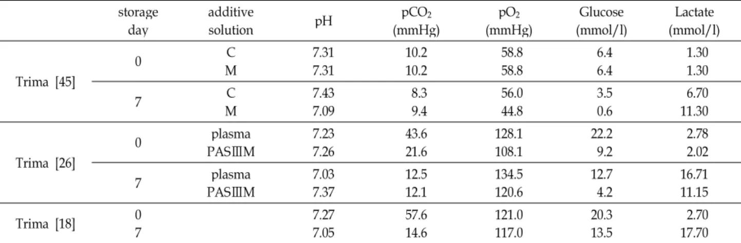
Table 4. Changes in metabolic activity of apheresis platelet according to the use of different additive solutions
storage
day additive
solution pH pCO2
(mmHg) pO2
(mmHg) Glucose
(mmol/l) Lactate (mmol/l)
Trima [45]
0 C
M 7.31
7.31 10.2
10.2 58.8
58.8 6.4
6.4 1.30
1.30
7 C
M 7.43
7.09 8.3
9.4 56.0
44.8 3.5
0.6 6.70
11.30
Trima [26]
0 plasma
PASⅢM 7.23
7.26 43.6
21.6 128.1
108.1 22.2
9.2 2.78
2.02
7 plasma
PASⅢM 7.03
7.37 12.5
12.1 134.5
120.6 12.7
4.2 16.71
11.15
Trima [18] 0
7
7.27
7.05 57.6
14.6 121.0
117.0 20.3
13.5 2.70
17.70 Trima: Trima Accel
M: pathogen reduction technology (PRT) treatment with the Mirasol PRT system C: Mirasol PRT system untreated control
[ ]: reference number
after platelet transfusion, and are refractory to fresh alloge- neic platelets [42, 60]. Additionally, some studies have found decreased CCI values using platelet additive solution (PAS), and further, it has been observed that all patients who were refractory to fresh allogeneic platelet transfusion had successful CCI results to autologous cryopreserved pla- telets [16, 29, 60]. Table 3 presents the transfusion efficacy of apheresis platelets according to different approaches to apheresis, which showed that the successful platelet trans- fusion in all apheresis instruments.
Effect of PAS
When processing the platelet component, the use of a PAS to suspend the platelet concentrates (instead of plas- ma) may reduce the incidence of adverse reactions due to plasma [17, 53]. In a quality comparison of extended plate- let storage (up to 9 days) using the different Trima systems, it was found that platelets collected in PAS were more like- ly to retain acceptable metabolic and cellular characteristics than platelets collected in plasma-stored apheresis units
[26]. However, some studies have reported that platelets suspended in PAS show significantly lower CCI than plate- lets suspended in plasma [16, 29]. Table 4 presents details of the changes in metabolic activity of apheresis platelet ac- cording to the use of different additive solutions when the same device is used [18, 26, 45]. PAS can affect the function- ality of apheresis platelets during storage.
Donor adverse reactions
In the apheresis collection process, the most common do- nor adverse reactions are related to citrate toxicity [31, 35].
Symptoms including tingling, paresthesia, and muscle cramps can result from citrate reactions [63]. Benjamin et al. [5] reported that plateletpheresis using an Amicus in- strument required larger quantities of acid citrate dextrose than did plateletpheresis using a Cobe Spectra instrument.
Benjamin et al. [5] further reported than use of the Amicus
instrument resulted in greater rates of citrate toxicity among
apheresis donors. However, citrate-induced adverse events
that result from apheresis processing can be treated with
calcium supplementation or reductions in the quantity of anticoagulant that is employed. Compared with whole blood collection, blood collection using apheresis may be a safer option for donors who have risk factors for pre- syncopal or syncopal reactions related to whole blood col- lection [3, 63]. Relevant risk factors include younger age, fe- male gender, and small total blood volume.
Conclusion
Plateletpheresis has become more popular because sin- gle-donor platelet products reduce the incidence of adverse reactions and increase the efficiency of blood usage. Various studies have evaluated the quality of platelet products dur- ing apheresis platelet storage. Quality can be assessed using platelet functionality markers such as visual appearance, metabolic activity, MPs, platelet activation, CCI, and PPR.
In particular, various markers of platelet activation have been developed and used for studies related to cell activation. The quality of platelets can also be influenced by the processing method and additive solutions. Additionally, apheresis may provide substantial benefits for donors who have risk factors for presyncopal or syncopal reactions re- lated to whole blood collection.
References
1. Alberio, L., Safa, O., Clemetson, K. J., Esmon, C. T. and Dale, G. L. 2000. Surface expression and functional character- ization of α-granule factor V in human platelets: effects of ionophore A23187, thrombin, collagen, and convulxin.
Blood
95, 1694-1702.2. Altuntas, F., Sari, I., Kocyigit, I., Kaynar, L., Hacioglu, S., Ozturk, A., Oztekin, M., Solmaz, M., Eser, B., Cetin, M. and Unal, A. 2008. Comparison of plateletpheresis on the Fenwal Amicus and Fresenius Com.Tec cell separators.
Transfus Med Hemother
35, 368-373.3. Berger, G., Hartwell, D. W. and Wagner, D. D. 1998. P-se- lectin and platelet clearance.
Blood
92, 4446-4452.4. Baldini, M., Costea, N. and Dameshek, W. 1960. The via- bility of stored human platelets.
Blood
16, 1669-1692.5. Benjamin, R. J., Rojas, P., Christmas, S., Neal, J., Broughton, S., Burgio, C., Barrett, B. and Churchill, W. H. 1999.
Plateletpheresis efficiency: a comparison of spectra LRS and Amicus separators.
Transfusion
39, 895-899.6. Bertolini, F. and Murphy, S. 1996. A multicenter inspection of the swirling phenomenon in platelet concentrates pre- pared in routine practice. Biomedical excellence for safer transfusion (BEST) working party of the international soci- ety of blood transfusion.
Transfusion
36, 128-132.7. Blaney, K. D. and Howard, P. R. 2008.
Basic and applied con- cepts of immunohematology
, pp. 219, 2nd ed. Mosby Elsevier, St. Louis, Missouri, USA8. Burgstaler, E. A. 2006. Blood component collection by apheresis.
J Clin Apher
21, 142-151.9. Burgstaler, E. A., Pineda, A. A. and Bryant, S. C. 1999.
Prospective comparison of plateletapheresis using four apheresis systems on the same donors.
J Clin Apher
14, 163-170.10. Burgstaler, E. A., Winters, J. L. and Pineda, A. A. 2004.
Paired comparison of Gambro Trima Accel versus Baxter Amicus single-needle plateletpheresis.
Transfusion
44, 1612- 1620.11. Charania, R., Smith, J., Vesely, S. K., Dale, G. L. and Holter, J. 2011. Quantitation of coated platelets potential during col- lection, storage, and transfusion of apheresis platelets.
Transfusion
51, 2690-2694.12. Choi, W. S., Jeon, O. H. and Kim, D. S. 2010. CD40 ligand shedding is regulated by interaction between matrix metal- loproteinase-2 and platelet integrin αⅡbβ3.
J Thromb Haemost
8, 1364-1371.13. Danese, S., Katz, J. A., Saibeni, S., Papa, A., Gasbarrini, A., Vecchi, M. and Fiocchi, C. 2003. Activated platelets are the source of elevated level of soluble CD40 ligand in the circu- lation of inflammatory bowel disease patients.
Gut
52, 1435-1441.14. Dale, G. L. 2005. Coated-platelets: an emerging component of the procoagulant response.
J Thromb Haemost
3, 2185-2192.15. Densmore, T. L., Goodnough, L. T., Ali, S., Dynis, M. and Chaplin, H. 1999. Prevalence of HLA sensitization in female apheresis donors.
Transfusion
39, 103-106.16. de Wildt-Eggen, J. and Gulliksson, H. 2003
. In vivo
andin vitro
comparison of platelets stored in either synthetic media or plasma.Vox Sang
84, 256-264.17. de Wildt-Eggen, J., Nauta, S., Schrijver, J. G., van Marwijk Kooy, M., Bins, M. and van Prooijin, H. C. 2000. Reactions and platelet increments after transfusion of platelet concen- trates in plasma or an additive solution: a prospective, randomized study.
Transfusion
40, 398-403.18. Diab, Y. A., Thomas, A. T., Luban, N. L. C., Wong, E. C.
C., Wagner, S. J. and Levy, R. J. 2012. Acquired cytochrome C oxidase impairment in apheresis platelets during storage:
a possible mechanism for depletion of metabolic adenosine triphosphate.
Transfusion
52, 1024-1030.19. Diab, Y., Wong, E., Criss, V. R., Moroff, C. G., Wagner, S.
J. and Luban, N. L. C. 2011. Storage of aliquots of apheresis platelets for neonatal use in syringes with and without agitation.
Transfusion
51, 2642-2646.20. Diedrich, B., Sandgren, P., Jansson, B., Gulliksson, H., Svensson, L. and Shanwell, A. 2008.
In vitro
andIn vivo
ef- fects of potassium and magnesium on storage up to 7 days of apheresis platelet concentrates in platelet additive solution.Vox Sang
94, 96-102.21. Flesch, B. K., Adamzik, I., Steppat, D., Miller, J., Carstensen, L., Schapke, M. and Davey, J. 2010. Paired crossover study of two plateletpheresis systems concerning platelet product
quality and donor comfort.
Transfusion
50, 894-901.22. Gutensohn, K., Geidel, K., Kroeger, N., Eifrig, B. and Crespeigne, N. Kuehnl. P. 2002. Platelet function testing in apheresis products: flow cytometric, resonance thrombo- graphic (RTG) and rotational thromboelastographic (roTEG) analyses.
Transfus Apher Sci
26, 147-155.23. Hamamoto, K., Ohga, S., Nomura, S. and Yasunaga, K. 1994.
Cellular distribution of CD63 antigen in platelets and in three megakaryocytic cell lines.
Histochem J
26, 367-375.24. Hillyer, C. D., Silberstein, L. E., Ness, P. M., Anderson, K.
C. and Roback, J. D. 2006.
Blood banking and transfusion medi- cine: Basic Principles & Practice
, pp. 72, 2nd ed. Churchill Livingstone, Elsevier, Philadelphia, PA, USA25. Hoffmeister, K. M., Felbinger, T. W., Falet, H., Denis, C. V., Bergmeier, W., Mayadas, T. N., von Andrian, U. H., Wagner, D. D., Stossel, T. P. and Hartwig, J. H. 2003. The clearance mechanism of chilled blood platelets.
Cell
112, 87-97.26. Johnson, L., Winterm, K. M., Hartkopf-Theis, T., Reid, S., Kwok, M. and Marks, D. C. 2012. Evaluation of the auto- mated collection and extended storage of apheresis platelets in additive solution.
Transfusion
52, 503-509.27. Julmy, F., Ammann, R. A., Taleghani, B. M., Fontana, S., Hirt, A. and Leibundgut, K. 2008. Effects of high-yield thrombocytapheresis on the quality of platelet products.
Transfusion
48, 442-450.28. Kaufman, J., Spinelli, S. I., Schultz, E., Blumberg, N. and Phipps, R. P. 2007. Release of biologically active CD154 dur- ing collection and storage of platelet concentrates prepared for transfusion.
J Thromb Haemost
5, 788-796.29. Kerkhoffs, J. L., Eikenboom, J. C., Schipperus, M. S., van Wordragen-Vlaswinkei, R. J., Brand, R., Harvey, M. S., de Vries, R. R., Barge, R., van Rhenen, D. J. and Brand, A. 2006.
A multicenter randomized study of the efficacy of trans- fusions with platelets stored in platelet additive solution Ⅱ versus plasma.
Blood
108, 3210-3215.30. Leytin, V., Allen, D. J., Gwozdz, A., Garvey, B. and Freedman, J. 2004. Role of platelet surface glycoprotein Ibα and P-selectin in the clearance of transfused platelet concentrates.
Transfusion
44, 1487-1495.31. Maker, Y. F., Butler, M. O., Cockersole, G. M., Gabra, G.
and Serevitch, J. M. 2002. National audit of citrate toxicity in plateletpheresis donors.
Transfus Med
12, 187-191.32. Makroo, R. N. and Kumar, P. 2006. Platelet transfusion in clinical medicine.
Apollo Medicine
3, 298-300.33. Marschner, S., Fast, L. D., Baldwin, W. M. 3rd., Slichter, S.
J. and Goodrich, R. P. 2010. White blood cell inactivation after treatment with riboflavin and ultraviolet light.
Transfusion
50, 2489-2498.34. Mathai, J., Resmi, K. R., Sulochana, P. V., Sathyabhama, S., Baby Saritha, G. and Krishnan, L. K. 2006. Suitability of measurement of swirling as a marker of platelet shape change in concentrates stored for transfusion.
Platelets
17, 393-396.35. McLeod, B. C., Price, T. H., Owen, H., Ciavarella, D., Sniecinski, I., Randels, M. J. and Smith, J. W. 1998.
Frequency of immediate adverse effects associated with apheresis donation
. Transfusion
38, 938-943.36. Metcalfe, P., Williamson, L. M., Reutelingsperger, C. P., Swann, I. V., Ouwehand, W. H. and Goodall, A. H. 1997.
Activation during preparation of therapeutic platelets af- fects deterioration during storage: a comparative flow cyto- metric study of different production methods.
Br J Haematol
98, 86-95.37. Mintz, P. D., Anderson, G., Avery, N., Clark, P. and Bonner, R. E. 2005. Assessment of the correlation of platelet mor- phology with
in vivo
recovery and survival.Transfusion
45, 72S-80S.38. Murphy, S. and Gardner, F. H. 1969. Effect of storage tem- perature on maintenance of platelet viability-deleterious ef- fect of refrigerated storage.
N Engl J Med
280, 1094-1098.39. Murphy, S. and Gardner, F. H. 1975. Platelet storage at 22°C:
role of gas transport across plastic containers in main- tenance of viability.
Blood
46, 209-218.40. Navarro, J. T., Hernandez, J. A., Ribera, J. M., Sancho, J.
M., Oriol, A., Pujol, M., Milla, F. and Feliu, E. 1998.
Prophylactic platelet transfusion threshold during therapy for adult acute myeloid leukaemia:10000/cumm versus 20000/cumm.
Haematologica
83, 998-1000.41. Ness, P. M. and Campbell-Lee, S. A. 2001. Single donor ver- sus pooled random donor platelet concentrates.
Curr Opin Hematol
8, 392-396.42. Novotný, V. M., Doxiadis, II. and Brand, A. 1999. The reduc- tion of HLA class I expression on platelets: a potential ap- proach in the management of HLA-alloimmunized re- fractory patients.
Transfus Med Rev
13, 95-105.43. Ohto, H. and Nollet, K. E. 2011. Overview on platelet preser- vation: Better controls over storage lesion.
Transfus Apher Sci
44, 321-325.44. Pandey, P., Tiwari, A. K., Sharma, J., Singh, M. B., Dixit, S. and Vimarsh Raina, V. 2012. A prospective quality evalu- ation of single donor platelets (SDP)-An experience of a ter- tiary healthcare center in India
. Transfus Apher Sci
46, 163- 167.45. Picker, S. M., Tauszig, M. E. and Gathof, B. S. 2012. Cell quality of apheresis-derived platelets treated with ribo- flavin-ultraviolet light after resuspension in platelet additive solution.
Transfusion
52, 510-516.46. Plaza, E. M., Lozano, M. L., Guiu, I. S., Egea, J. M., Vicente, V., De Tarán, L. C. and Rivera, J. 2012. Evaluation of platelet function during extended storage in additive solution, pre- pared in a new container that allows manual buffy-coat pla- telet pooling and leucoreduction in the same system.
Blood Transfus
10, 480-489.47. Rank, A., Nieuwland, R., Liebhardt, S., Iberer, M., Grützner, S., Toth, B. and Pihusch, R. 2011. Apheresis platelet concen- trates contain platelet-derived and endothelial cell-derived microparticles.
Vox Sang
100, 179-186.48. Ratko, T. A., Cummings, J. P., Oberman, H. A., Crookston, K. P., DeChristopher, P. J., Eastlund, D. T., Godwin, J. E., Sacher, R. A., Yawn, P. H. and Matuszewski, K. A. 2001.
Evidence-based recommendation for the use of WBC-re-
초록:혈소판성분채집술: 채집과정, 장비, 성분채집혈소판 질의 지표들
김성인
1†․장철수
1†․김현경
1․권창오
1․김병원
1․김동찬
1․김윤석
2․이기종
2․류재기
1* (
1김천대학교 임상병리학과 ,
2연세대학교 임상병리학과 )
혈소판제제는 출혈질환이나 혈소판의 기능장애 치료를 위해 사용되고 있다 . 혈소판성분채집술은 혈액성분채집
기를 사용하여 혈소판 성분을 채집하는 방법으로 , 성분채집혈소판의 질 평가에는 다양한 지표들이 이용되고 있다.
채집된 혈소판의 생산성은 채집효율 (collection efficiency)과 채집률(collection rates)로 평가되고 보관된 혈소판의 질은 in vitro 상에서 시각적 외양, 대사활성도, 양, 혈소판 수, 백혈구 수, 미세입자(microparticles), 그리고 다양한
혈소판 활성 표지자들로 평가된다 . 혈소판 활성 표지자들은 여러 연구분야에서도 이용되고 있다. 수혈된 후의 혈
소판의 질은 교정증가치 (corrected count increment)와 혈소판회복퍼센트(percentage platelet recovery)로 평가된
다 . 본 논문은 혈소판성분채집술의 채집과정, 사용되는 장비, 성분채집혈소판의 질, 전반적인 효율성, 그리고 질
평가 지표들에 대한 리뷰 (review)와 함께 다른 혈액성분채집기 간의 기능의 차이점을 비교하였고, 또한 혈소판성
분채집술은 구연산염 항응고제에 의한 부작용을 일으킬 수는 있지만 전혈 헌혈시에 실신의 전구증상이나 실신을
일으킬 위험이 있는 헌혈자들에게는 더 안전한 방법임을 보여주고 있다 .
duced cellular blood components.
Transfusion
41, 1310-1319.49. Rock, G., Moltzan, C., Alharbi, A., Giulivi, A., Palmer, D.
and Bormanis, J. 2003. Automated collection of blood com- ponents: their storage and transfusion.
Transfuse Med
13, 219-225.50. Sander, T. L., Ou, J. S., Densmore, J. C., Kaul, S., Matus, I., Twigger, S., Halligan, B., Greene, A. S., Pritchard, K. A.
Jr. and Oldham, K. T. 2008. Protein composition of plasmi- nogen activator inhibitor type 1-derived endothelial micro- particles.
Shock
29, 504-511.51. Schlossberg, H. R. and Herman, J. H. 2003. Platelet dosing.
Transfus Apher Sci
28, 221-226.52. Seghatchian, J. and Krailadsiri, P. 2000. Current position on preparation and quality of leucodepleted platelet concen- trates for clinical use.
Transfus Sci
22, 85-88.53. Singh, R. P., Marwaha, N., Malhotra, P. and Dash, S. 2008.
Therapeutic efficacy of different types of platelet concen- trates in thrombocytopenic patients.
Indian J Hematol Blood Transfus
24, 16-22.54. Singh, R. P., Marwaha, N., Malhotra, P. and Dash, S. 2009.
Quality assessment of platelet concentrates prepared by pla- telet rich plasma-platelet concentrate, buffy coat poor-plate- let concentrate (BC-PC) and apheresis-PC methods.
Asian J Transfus Sci
3, 86-94.55. Slichter, S. J., Bolgiano, D., Jones, M. K., Christoffel, T., Corson, J., Rose, L., Foley, J., Popovsky, M., Baril, L. L., Corda, T., Dincecco, D. M. and Snyder, E. L. 2006. Viability and function of 8-day-stored apheresis platelets.
Transfusion
46, 1763-1769.56. Slichter, S. J. and Harker, L. A. 1976. Preparation and storage of platelet concentrates. II Storage variables influencing pla- telet viability and function.
Br J Haematol
34, 403-419.57. Svendsen, M. S., Rojkjaer, R., Kristensen, A. T., Salado- Jimena, J. A., Kjalke, M. and Johansson, P. I. 2007. Impair- ment of the hemostatic potential of platelets during storage as evaluated by flow cytometry, thrombin generation, and thromboelastography under conditions promoting for- mation of coated platelets.
Transfusion
47, 2057-2065.58. Tendulkar, A. and Rajadhyaksha, S. B. 2009. Comparison of plateletpheresis on three continuous flow cell separators.
Asian J Transfus Sci
3, 73-77.59. Tudisco, C., Jett, B. W., Byrne, K., Oblitas, J., Leitman, S.
F. and Stroncek, D. F. 2005. The value of pH as a quality control indicator for apheresis platelets.
Transfusion
45, 773-778.60. Vadhan-Raj, S., Kavanagh, J. J., Freedman, R. S., Folloder, J., Currie, L. M., Bueso-Ramos, C., Verschraegen, C. F., Narvios, A. B., Connor, J., Hoots, W. K., Broemeling, L. D.
and Lichtiger, B. 2002. Safety and efficacy of transfusions of autologous cryopreserved platelets derived from re- combinant human thrombopoietin to support chemo- therapy-associated severe thrombocytopenia: a randomized cross-over study.
Lancet
359, 2145-2152.61. Van Wijk, M. J., Van Bavel, E., Sturk, A. and Nieuwland, R. 2003. Microparticles in cardiovascular diseases.
Cardiovasc Res
59, 277-287.62. Wenzel, F., Günther, W., Baertl, A., Gruber, A., Sorg, R. V., Haas, R. and Giers, G. 2012. Platelet transfusion alters CD40L blood level and release capacity in patients suffering from thrombocytopenia.
Transfusion
52, 1213-1220.63. Yuan, S., Ziman, A., Smeltzer, B., Lu, Q. and Goldfinger, D. 2010. Moderate and severe adverse events associated with apheresis donations: incidences and risk factors.