Identification of Genes Differentially Expressed by Oryctes rhinoceros nudivirus Infection in the Korean Rhinoceros Beetle, Allomyrina dichotoma
Kisang Kwon
1, Bo-Kyung Yoo
2, Hyun-Woo Suh
2, Young Hwa Ko
2, Hong Geun Kim
3, Seokhyun Lee
3, Kwan-Ho Park
3, Ji-Young Choi
3and O-Yu Kwon
2*
1Department of Biomedical Laboratory Science, College of Health & Welfare, Kyungwoon University, Gumi 730-739, Korea
2Department of Anatomy, College of Medicine, Chungnam National University, Daejon 301-747, Korea
3Applied Entomology Division, National Academy of Agricultural Science, RDA, Wanju-gun 560-500, Korea Received July 7, 2015 /Revised July 31, 2015 /Accepted July 31, 2015
The Korean rhinoceros beetle (Allomyrina dichotoma) is popular as a pet and as a food ingredient, and it is commercially distributed in Korea. It is also traditionally regarded as a medicine for liver-related diseases. Recently, the Oryctes rhinoceros nudivirus was introduced from Southeast Asia. This virus is reported as a disease factor for A. dichotoma in mass-rearing facilities, and economic losses due to this viral infection have been increasing in Korea since the 2010s. In this study, we observed serious struc- tural changes in the fat body and the intestine of virus-infected beetles. We report five genes that are up-regulated by the viral infection in the intestine: BTF3H4-like (transcription factor BTF3 homolog 4-like), SPS-like (serine proteinase stubble-like), COPB1 (coatomer protein complex, subunit beta 1), T-CP (T-complex 1 subunit gamma), and HSP70 HSP70 (heat shock protein 70). The results may pro- vide a clue for the early diagnosis and disease-treatment that occurs in mass-rearing facilities. The im- provement of stable productivity will increase the farmers’ income, and quality control of bee- tle-breeding will help industries to utilize this beetle as a promising food ingredient.
Key words :
Allomyrina dichotoma, Korean rhinoceros beetle, Oryctes rhinoceros nudivirus infection
*Corresponding author
*Tel : +82-42-580-8206, Fax : +82-42-586-4800
*E-mail : [email protected]
This is an Open-Access article distributed under the terms of the Creative Commons Attribution Non-Commercial License (http://creativecommons.org/licenses/by-nc/3.0) which permits unrestricted non-commercial use, distribution, and reproduction in any medium, provided the original work is properly cited.
Journal of Life Science 2015 Vol. 25. No. 8. 942~946 DOI : http://dx.doi.org/10.5352/JLS.2015.25.8.942
서 론
2014년 현재 국내 곤충 산업의 시장 규모는 2,000억 원 정도.
곤충 사육 농가는 약 500곳으로 추산된다. 2015년에는 3,000억 원을 넘을 것으로 예상된다. 단백질 공급원, 소자본ㆍ작은 공 간에서 사육가능, 강한 번식력, 상대적으로 쉬운 사육관리, 대 량살상의 윤리문제 회피, 온실가스와 자원비용 절감 등의 장 점을 가지고 있어 곤충산업은 미래 녹색생명산업으로 주목을 받고 있다[9]. 그러나 곤충의 산업화가 진행되면서 건강한 곤 충의 대량사육이 요구되고 있다. Virus에 의한 백강병(white muscardine), 녹강병(green muscardine), 고름병, bacteria에 의한 물렁물렁병등과 함께 fungus, 원충(protozoa), 선충
(nematoda), 응애(mites) 등에 의해서 대량 집단사육 시 질병발생이 발생하여 대규모 피해가 만성적으로 발생하고 있다[6].
그 동안에 농진청에서 진단/판별 kit, 병 유발 유전자탐색, 친 환경 방제법을 발전시켜오고 있다. 그러나 정밀한 진단 및 치 료법이 개발되지 않은 상태이다.
장수풍뎅이(Korean rhinoceros beetle, Allomyrina dichoto-
ma)는 사육되는 대표적인 곤충이다. 한국을 포함한 동남아시
아 등지에 분포하는 장수풍뎅이의 전체 몸길이는 30-85 mm로
매우 단단한 외피에 싸여있다. 몸 색깔은 전체적으로 흑밤색
을 띤다. 수컷은 반짝이는 광택이 나며 머리에 긴 뿔이 나 있는
데, 그 길이가 몸길이의 절반 정도이다. 이들은 주로 낮에는
어두운 썩은 나무속 혹은 땅 속에 숨어 있다가 밤에 나와서
졸참나무(Quercus serrata), 상수리나무(Quercus acutissima)의
수액을 먹는다. 6-8월에 짝짓기를 한 다음에 썩은 부엽토에
30~70개의 알을 낳는다. 유충은 일생 중 두 번 탈피를 한 뒤
월동하였다가 다음해 초여름에 땅 속에서 번데기가 되었다가,
15~20일 뒤에 성충으로 우화한다[8]. 이들은 최근에 관상용으
로 인기가 높으며, 특히 애벌레는 곤충식품으로도 주목을 받
고 있다. 장수풍뎅이의 대량사육 시 발생하는 질병의 원인이
최근에 virus (Oryctes rhinoceros nudivirus)인 것이 보고되었다
[5]. 그러나 아직 정확한 발병기전과 치료방법을 알지 못하여
사육농가에 심각한 경제적 손실을 주고 있다. 본 연구팀은 이
문제를 해결하기 위하여 virus에 감염된 장수풍뎅이의 장
(intestine) 특이적으로 발현하는 유전자를 Differential Display
(DD)-PCR방법으로 동정하였다. 장수풍뎅이의 질병 관련 유
전자에 관한 보고는 본 논문이 최초이다. 이들 유전자에 대한
깊은 연구는 질병예찰과 치료에 도움을 줄 수 있는 실마리를
제공할 것이다. 이는 장수풍뎅이의 안정적인 대량생산을 통해
서 생산성 향상과 소득증대 효과 및 균일화된 대량사육을 통
- Note -
재료 및 방법
곤충사육 및 장(gut) 분리
본 연구에 이용된 장수풍뎅이는 표준 사육기준에 따라 사육 된 3령 larvae를 경기도 시흥시 아이벅스캠프(영농조합법인) 로부터 구입한 후, 약 25℃, 40%의 습도 조건의 발효 톱밥에 보관 하였다. 3령 larvae의 머리 부분을 해부침으로 고정시킨 후 표피를 아래에서 머리 방향으로 한 번에 가른다. Fat body 를 제거한 후 장을 통째로 분리하여 장 내부의 톱밥 등의 내용 물을 제거하고, 차가운 phosphate buffered saline (PBS)에 3회 세척하여 불순물질을 모두 제거 후 4% paraformaldehyde (PFA)로 고정 또는 total RNA를 분리하였다. Virus 감염유무 는 아래의 primer를 사용하여 확인하였다. OrNV-F1; TCCG- GAAATTACACGAGCCAC, OrNV-R1; ATGCCGTACGAG- AGTATAGGTCG.
H&E 염색
4% PFA에 고정한 장 조직을 흐르는 물에 충분히 수세하여 저농도에서 고농도의 에탄올에 순차적으로 담가 탈수시킨 후 자일렌을 이용하여 투명화하였다. 그 다음 파라핀으로 포매하 고 마이크로톰을 이용하여 4 μm 두께로 잘라 슬라이드에 올린 후 파라핀을 제거하였다. Hematoxylin과 eosin을 이용하여 염 색한 후 봉입하여 현미경으로 관찰하였다.
RNA 분리
3 MM paper를 이용하여 수분을 제거한 조직을 1.5 ml tube 에 넣고 500 μl의 RNA isolation buffer를 첨가하여 lysis 시킨 후 100 μl의 chloroform을 넣어 충분히 섞어주었다. 13,000 rpm, 4℃에서 10분 동안 원심분리하여 약 250 μl의 상등액을 취하여 새로운 tube에 옮긴 후 동량의 isopropanol을 첨가하 였다. 실온에서 10분 정도 침전반응 유도한 후 13,000 rpm, 4℃에서 10분 동안 원심분리하고, 75% ethanol을 500 μl로 세 척하여 total RNA을 얻어 RNase-free water로 녹인 후 non- odrop (Thermo Scientific, USA)을 이용하여 정량하였다.
DD-PCR
GeneFishing DEG kit (Seegene, Korea)를 사용하여 PCR- based differential display 방법으로 선별하였다. Total RNA 3 μg과 dT-ACP1 oligomer 를 섞어 80℃에서 3분 동안 열변성 시킨 후, 10x buffer, dNTP, MML-V, RNase inhibitor 등이 포함된 용액과 혼합하여 42℃에서 90분간 반응시켜 cDNA를 합성하였다. 이 cDNA를 주형으로 하여 dT-ACP2와 20가지의 arbitrary ACP를 이용하여 95℃에서 1분, 50℃에서 3분, 72℃
에서 1분 동안 반응하여 double-strand cDNA 를 합성하였다.
오려서 Gel elution kit (ELPIS Biotech, Korea)를 이용하여 DNA를 회수한 후 pGEM-T easy vector (Promega, USA)를 이용하여 cloning 하였다. Plasmid miniprep kit (ELPIS Biotech, Korea)를 이용하여 DNA를 추출한 후 솔젠트회사에 의뢰하여 염기서열을 결정하였다(Solgent, Korea).
RT-PCR
발현차이를 보인 유전자에 해당하는 primer를 이용하여 re- verse transcription-PCR을 수행하였다. Total RNA 3 μg과 oli- go-dT를 섞어 80℃에서 3분 동안 열 변성 시킨 후, 10x buffer, dNTP, MML-V, RNase inhibitor 등이 포함된 용액과 혼합하 여 42℃에서 90분간 반응시켜 cDNA를 합성하였다. cDNA를 증폭시키기 위해서 해당 primer를 이용하여 95℃에서 30초, 56℃에서 30초, 72℃에서 30초로 28회 반응하여 전기영동으로 확인하였다. 사용된 primer는 다음과 같다. BTF3H4-like; F (5'-TACCGGATTTGGTGGAGAAC)/R(5'-CACATTCCCGA CTGAAACAA), SPS; F(5'-GATGCATGCACGGGTGAC)/R (5'-GCAACCAAGACCCCATGTTA), COPB1; F(5'-GGCCA- CGTACGAGTTAGAGC)/R(5'-CATGCCTTAAATTGGCCT TT), T-cp1; F(5'-CGGGGAACTAGTCGAACAAA)/R(5'-CC- ATTTCCGAGGGTTTAGGT), Hsp70; F(5'-AAATCGGAAG- AGGTGCAAGA)/R(5'-CAAGACGCCTGGTTGGTTAT), α- Tublin; F(5'-TGGTGTCCAACAGGTTTCAA)/R(5'-AAATC- TTCACGGGCTTCTGA).
결과 및 고찰
Oryctes rhinoceros nudivirus에 감염된 장수풍뎅이의 3령
larvae는 정상적인 것에 비하여 체장의 변화는 없다. 그러나
육안으로도 감염된 것은 몸 전체가 투명하지 않으며 진한 노
란 색깔을 띤다. 가장 특징적인 것은 탈장을 확인할 수 있다
(Fig. 1A). 본 실험에서는 virus sequence를 이용하여 RT-PCR
방법으로 감염유무를 확인하였다(Fig. 1B). 곤충의 fat body는
포유동물의 liver와 같이 지질대사와 해독작용에 관여하는 기
관이다. 결국 virus감염에 의해서 fat body가 방어시스템을 작
동하였기에 정상적인 fat body보다가 크게 팽창한 것으로 생
각된다. 해부하여 각각의 fat body를 HE염색하여 현미경하에
서 조직을 관찰하였다. Virus에 감염된 것의 fat body가 아주
팽창되어 있고 부분적으로 터져있는 것을 확인할 수 있었다
(Fig. 1C). 이와 같은 결과는 정상적인 신진대사가 일어나지
않아서 탈장이 유도되는 것을 생각된다. 이때에 장(intestine)
의 구조적인 변화를 관찰하기 위하여 각각의 장을 HE염색하
여 조직학적인 구조변화를 현미경하에서 관찰하였다. 정상적
인 장구조에 비하여 virus감염된 장구조는 기저층에 있는 근
A B
C D
E
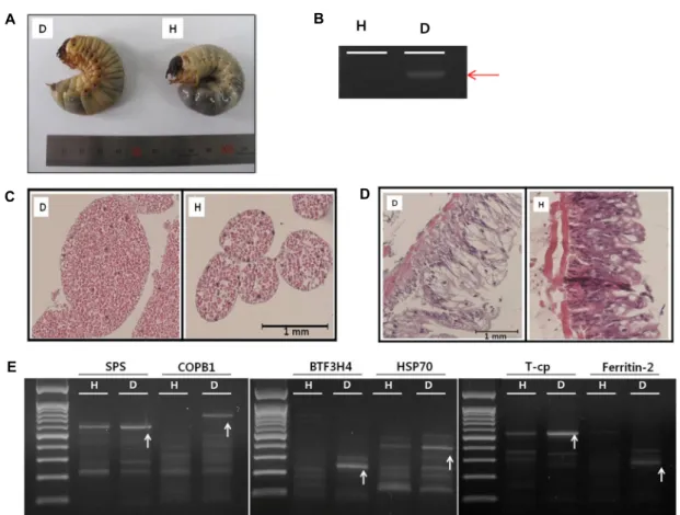
Fig. 1. (A) Visual comparison of both a virus-infected larva (left) and a healthy larva (right). The virus-infected larva appears more yellowish than the healthy one, and its abdomen is swollen with an intestinal hernia. (B) Virus infection confirmed by RT-PCR.
(C) Histological changes of the fat body from the virus-infected larva (left) and that from the healthy larva (right). Fat body infected by virus is swollen and partially exploded. (D) Histological changes of intestine from the virus-infected larva (left) and that from the healthy one (right). Intestine from the virus-infected larva shows weak lamina muscular compared to that from a healthy larva. Virus-infected epithelium glands almost lost its goblet cells. (E) The result of DD-PCR, arrows indicate differentially expressed DNA fragments.
육층(lamina musclaris)이 아주 얇아져 있고 점막(tunica mu- sclaris)층이 빈약해져 있다. 그 결과 점액(mucin)을 분비하는 술잔세포(goblet cell)의 숫자가 현저히 감소되어 거의 관찰되 지 않는다(Fig. 1D). 점액은 산성단백질로 미세융모(microvil- lus)의 당질층(glycocalyx)에 보호막을 형성하여 상피(epithe- lium)표면을 윤활하게 하고 상피를 보호하는 역할을 담당하고 있다. 이처럼 virus감염에 의해서 장의 정상적인 기능이 작동 할 수 없는 상태가 질병유발의 주요 원인인 것으로 생각된다.
장수풍뎅이가 Oryctes rhinoceros nudivirus에 감염되었을 때 에 장에서 일어나는 분자기전을 이해하기 위하여 DD-PCR방 법을 이용하여, 특이적으로 발현하는 유전자를 탐색하였다.
GeneFishing DEG kit (Seegene, Korea)는 포유동물의 특이유 전자탐색에 사용되는 것이지만, 곤충을 상대로 했을 때에도 잘 적용되는 것을 확인하였다. Virus에 감염된 장과 비 감염된 장으로부터 total RNA를 얻어서 사용하였다. 그 결과 다수의 1차 결과를 얻었지만 reverse PCR결과 많은 false가 확인되었 지만 특이적인 DNA단편이 확인되었다(Fig. 1E). 그러나 Fig.
2의 결과에서 보는 것과 같이 5개의 유전자는 virus감염에 의 해서 특이적으로 발현이 상승하는 것이 확인되었다. BTF3H4- like (transcription factor BTF3 homolog 4-like)의 발현은 vi- rus감염에 의해서 2이상 상승하였다. 그러나 이 단백질의 생체 내 기능은 아직 알려진 것이 없다. 단지 일반적인 유전자전사 인자로서 RNA polymerase II complex와 안정적인 관계를 유 지한다는 것 정도가 알려져 있다[2]. SPS-like (serine protei- nase stubble-like)의 발현은 virus감염에 의해서 3배 상승하였 다. 이 단백질의 주된 생체내 기능은 serine-type endopepti- dase활성을 가지고 있는 것으로 hormone 의존적으로 epi- thelial morphogenesis에 관여한다[3]. COPB1 (coatomer pro- tein complex, subunit beta 1)의 발현은 virus감염에 의해서 4배 증가하였다. Golgi-to-ER transport 시스템에 중요한 단백 질로서 분비vesicles형성에 관여한다[4]. T-CP (T-complex 1 subunit gamma)의 발현은 virus감염에 의해서 4배 상승한다.
이 단백질은 molecular chaperone으로서 ATP hydrolysis에
의해서 단백질의 folding을 돕는다, 특히 actin and tubulin
D E
Fig. 2. Five genes up-regulated by virus infection in the intestine. H;
healthy intestine, D; Virus-infect- ed intestine.
folding에 중요하다[1]. HSP70 (heat shock protein 70)의 발현 은 virus감염에 의해서 6배 상승하였다. 어디에서나 발현하는 heat shock protein으로서 protein folding, 외부 스트레스로부 터 세포보호에 중요한 역할을 한다[7].
본 연구 결과는 장수풍뎅이(Korean Rhinoceros Beetle, Allomyrina dichotoma)가 Oryctes rhinoceros nudivirus에 감염되 면 fat body와 장에서 조직적인 구조변화가 일어나는 것을 관 찰되었다. 이와 함께 장에서 특이적으로 발현이 상승하는 유 전자로 BTF3H4-like, SPS-like, COPB1, T-CP, HSP70를 보고한 다. 이 결과는 장수풍뎅이의 대량사육 시 발생하는 질병의 조 기진단과 치료에 새로운 기회를 제공할 것이다. 그리고 안정 적인 생산성 향상을 통해서 소득증대 효과 및 균일화된 대량 사육을 통해서 식품원료로 인정받을 수 있을 것이다.
감사의 글
본 성과물(논문)은 농촌진흥청 연구사업(세부과제번호: PJ 01086401)의 지원에 의해 이루어진 것임.
References
1. Bhaskar, Kumari, N. and Goyal N. 2012. Cloning, character- ization and sub-cellular localization of gamma subunit of T-complex protein-1 (chaperonin) from Leishmania donovani.
Biochem. Biophys. Res. Commun. 429, 70-74.
2. http://www.uniprot.org/uniprot/Q96K17 3. http://www.ncbi.nlm.nih.gov/gene/100158786 4. http://www.uniprot.org/uniprot/P53618
5. Lee, S., Park, K. H., Nam, S. H., Kwak, K. W. and Choi, J. Y. 2015. First report of Oryctes rhinoceros nudivirus (Coleoptera: Scarabaeidae) causing severe disease in Allo- myrina dichotoma in Korea. J. Insect Sci. 15: DOI: 10.1093/ji- sesa/iev002.
6. Manley, R., Boots, M. and Wilfert, L. 2015. Emerging viral disease risk to pollinating insects: ecological, evolutionary and anthropogenic factors. J. Appl. Ecol. 52, 331-340.
7. Mayer, M. P. 2013. Hsp70 chaperone dynamics and molec- ular mechanism. Trends Biochem. Sci. 38, 507-514.
8. Suh, H. J., Kim, S. R., Lee, K. S., Park, S. and Kang, S. C.
2010. Antioxidant activity of various solvent extracts from Allomyrina dichotoma (Arthropoda: Insecta) larvae. J.
Photochem. Photobiol. B. 99, 67-73.
9. van Huis, A. 2013. Potential of insects as food and feed in assuring food security. Annu. Rev. Entomol. 58, 563-583.
초록:장수풍뎅이(Korean Rhinoceros Beettle,
Allomyrina dichotoma)에
Oryctes rhinoceros nudivirus감염 특이적으로 발현하는 유전자 동정
권기상
1․유보경
2․서현우
2․고영화
2․김홍근
3․이석현
3․박관호
3․최지영
3․권오유
2(1경운대학교 임상병리학과, 2충남대학교 의학전문대학원 해부학교실, 3농진청 국립농업과학원 곤충산업과)