구순구개열 Vol. 5, No. 2 2002
Anatomic Study on the Lacrimal Duct using Computerized Tomograph
Pill-Hoon Choung, Ui-Lyong Lee, Jong-Rak Hong
Department of Oral and Maxillofacial Surgery, Maxillofacial Deformity Clinic, Craniofacial Tissue Engineering Lab. of BK21 Human Life Science and
Biointeface Engineering Research Center, College of Dentistry, Seoul National University, Seoul, Korea
구순
구개 열환자의 악교정 성형수술로
구강내Le Fort II
골절단술이나비중격 성형술이
많이이용되는에, 이 때
비골의외측 골절단술을
요하며,이에 대한 술후 합병증으로
비루관의폐쇄나
비골의분쇄골절등이
발생할수
있어, 악안면기형환자를다루는 구강악안면외과의사에게 비루관의 응용해
부학적 연구는중요하다. 본 연구의
목적은교합
면을기준으로 촬영된컴퓨터 단층
촬영에서 비루관의 위치와 크기를 조사하는데 있다.2000
년7월부터 2003
년2
월까지 서울대학교병원 구강악안면방사선과에서
컴퓨터 단층촬영을시행한환자 62
명을대상으로 비상악봉합선에서비루관까지의 최단거리와
비루관의 최대반경과 최소반경을 측정하였다. 우측 비상악
봉합선에서 비루관까지의 거리는 5.68mm이고, 좌측은 5.67mm
였다.좌우
및 성별간의차이는 없었다.
이의해부학적
지견은악기형수술시의 비 루관
폐쇄라는합병증의 예방책으로
기여하리라생각된다.This study was supported by a grant of the Korea Health 21 R&D project, Ministry of Healdi 8l Welfere, Republic of Korea(00-PJl- PG1-CH11-0004)
Introduction
Transient period of nasolacrimal obstruction occurs relatively often during the early postoperative phases rhinoplasty utilizing lateral osteotomies, LeFort II osteotomy and maxillary sinus surgery. As an example, Flowers and Anderson1 reported that 21 of 27 rhinoplasty patients, who were examined on the second postoperative day, had epiphoranon one or both sides. This was usually secondary to edema, creating a functional blockage to the passage of tears.
Only rarely does actual surgical disruption of some
portion of the system lead to a permanent obstruction.
The most common affection of the lacrimal pathways is obstruction, either congenital or acquired. Aquired lacrimal drainage system obstruction is caused mainly by trauma, inflammation and primary or secondary tumours of the lacrimal apparatus.2
The sequelae of lacrimal drainage system obstruction are troublesome for patients: (1) pathologic epiphora impairs the performance of tasks which require correct vision; (2) spectacles cause discomfort; (3) contact lenses cannot be worn and (4) recurrent infections of the eyelids, conjunctiva, lacrimal sac and naso-lacrimal duct are dangerous not
5:109~112, 2002
109
Pill-Hoon Choung, Ui-Lyong Lee, Jong-Rak Hon
only in respect of vision but even for the patient’s life.3
The aim of this paper is to present the average length from nasomaxillary suture to lacrimal duct and size of lacrimal duct which were measured on computed tomogram.4
Materials and Methods
A retrospective study of axial cut maxillofacial computed tomographic scans conducted. One level of the bony nasolacrimal duct system ; most inferior level of bony orbit, on the right and left sides were measured. The maxillofacial computed tomographic scans completed at the Department of Oral and Maxillofacial Surgery, College of Dentistry, Seoul National University between July 4, 2000, and Februry 25,2003 were reviewed.
The patients had facial deformities; for example, mandibular prognathism or maxillary canting and so forth. So these maxillofacial computed tomogram were for conducting accurate pre-operative plans.
There was no evidence of pathologic changes of nasolacrimal drainage system on any CT scans.
The MaroView software was used to measure the shortest distance from nasomaxillary groove to nasolacrimal duct(Fig 1), the longest diameter and the shortest diameter of nasolacrimal duct(Fig 2). This section is at the level of the most inferior portion of orbit
Statistical analysis was completed using the 2-tail t test, analysis of variance, and linear regression.
Results
The mean ages for the 24 men and 37 wemen were 23.7 and 22.3 years, respectively. The overall average age of 62 patients was 23.0 years.
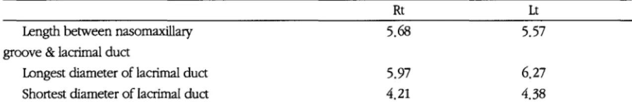
The overall average of right and left length between nasomaxillary groove and nasolacrimal duct were 5.68 and 5.57 respectively.(Thble 1)
Qualitatively, a difference in sizes of nasolacrimal drainage systems at the most inferior level of orbit in regard to the longest and shortest diameter of nasolacrimal duct was observed on scans of male and female nasolacrimal duct There was no significant sex difference in length between nasomaxillary groove and nasolacrimal duct. Although slight differences are noted between right and left length
Fig 1.
Axial computed tomographic scan demonstrates
Fig2. Axial computed tomographic scan demonstrates the shortest distance from nasolacrimal duct to the longest diameter and the shortest diameter of nasomaxillary suture, (shown as a line) THs section is at nasolacrimal duct(shown as a line) This section is at the the level of the most inferior portion orbit. level of the most inferior portion oibit.
110
구순구개열 Vol. 5, No. 2 2002
Table 1.
Overall average length from nasomaxillary groove to lacrimal duct, the shortest and the longest diameter of lacrimal duct( 62 patients)
Rt Lt
Length between nasomaxillary 5.68 5.57
groove & lacrimal duct
Longest diameter of lacrimal duct 5.97 6.27
Shortest diameter of lacrimal duct 4.21 4.38
lacrimal duct( 24 male)
Table 2.
Average length from nasomaxillary groove to lacrimal duct, the shortest and the longest diameter of
Rt Lt
Length between nasomaxillary 5.85 5.66
groove & lacrimal duct
Longest diameter of lacrimal duct 6.47 6.95
Shortest diameter of lacrimal duct 4.51 4.63
lacrimal duct (36 female)
Table 3.
Average length from nasomaxillary groove to lacrimal duct, the shortest and the longest diameter of
Rt Lt
Length between nasomaxillary 5.66 5.64
groove & lacrimal duct
Longest diameter of lacrimal duct 5.57 5.78
Shortest diameter of lacrimal duct 3.95 4.17
-onp
raE-
= 0 -
pue©aoojb
-(l'
=x'U OJS rau
ceeAMeq
=
* 6 u e _
Fig. 1.
The length from nasomaxillary groove to lacrimal duct. No positive or negative slope in the line equation was observed
between nasomaxillary groove and lacrimal duct, the longest and shortest diameter of nasolacrimal duct, no significant differences were observed.
There was no significant relation between age and the length between nasomaxillary groove and nasolacrimal duct(figure 3)
111
Pill-Hoon Choung, Ui-Lyong Lee, Jong-Rak Hon
Discussion
The nasolacrimal duct, about 18mm long, descends from the lacrimal sac to open anteri이y in the inferior meatus at an expanded orifice; a mucosal lacrimal fold forms an imperfect valve just above this opening.
The duct runs down an osseous canal formed by the maxilla, lacrimal bone and inferior nasal concha; it is narrowest in the middle and directed down, back and a little laterally. The mucosa of the lacrimal sac and the nasolacrimal duct has a bilaminar columnar epithelium, ciliated in places.
A surrounding plexus of veins, forming erectile tissue, may, when engorged, obstruct the duct.1
Lacrimal fluid enters the conjunctival sac at its superolateral angle and, by capillary and blinking, is carried across the eye to the lacus lacrimalis, mainly between the lower palpebral margin and the eyeball.
From the lacus it enters the lacrimal canaliculi.
Contraction of the orbicularis oculi press the puncta lacrimalia more firmly into the lacus and capillary attraction draws the secretion into the lacrimal sac.
Sudden dilatation of the sac, produced by the lacrimal part of the orbicularis oculi during blinking, probably aids this. Normally the tarsal secretion prevents tear fluid from overflowing and also covers the capillary film of fluid on the cornea and sclera, perhaps delaying evaporation.
The lacrimal bone at the lateral nasal wall was found to be just anterior to the mid-third of the uncinate process. The average length and width was 7.4mm and 2.5mm, respectively. The position of the lacrimal passage covered by the lacrimal bone corresponded to the postero-medial aspect of the upper lacrimal duct and the lower lacrimal sac.
Anatomy of the lacrimal apparatus and its relationship to the surrounding bony vault is discussed and well illustrated in several standard texts. The surgeon is concerned with the lacrimal bones, nasal bones, and frontal process of the maxilla during the creation of the osteotomy.2
Our study illustrated the idea that osteotomy can be done safely, regardless of the type of osteotome. This damage to the nasolacrimal duct from osteotomizing on the lateral bone, which leads to safe positioning along the nasomaxillary groove. In 1968, Flowers and Anderson reported a study of osteotomies performed in 37 fresh autopsy specimens. Dacryocystograms were made and 23% showed evidence of injury to the lacrimal apparatus. Anatomic study of the nasolacrimal system has shown that two areas are particularly vulnerable to inadvertent surgical trauma.
In endonasal procedures, the structure most likely to be damaged is the distal orifice of the lacrimal duct, sometimes called Hasner’s valve.
Reference
1. Flower RS, Anderson R ; Injury to the lacrimal apparatus during ihinoplasty. Past Reconstr Sing 42 : 577-581,1968
2. Lavine DM, Lehman JA, Jackson T : Is the lacrimal apparatus injured following cosmetic ihinoplasty. Arch Otolaiyngol 105:
刀9-720,1979
3. Thomas JR, Griner N : The relationship of lateral osteotomies in rhinoplasty to the lacrimal drainage system. Otolaryngol Head Neck Sing 94 : 362-367, 1986
4. Sarah A : An anatomical basis for primary acquired nasolacrimal duct obstruction. Aih Otolaiyngol 115 :
刀-74
저자연락저
서울시
종로구연건동
28번지 서울대학교 치과대학 구강악안면외과
이외룡우편번호)110-744 전화: 02-760-2638 E-mail: choungph@snu.ac.kr112