METASTATIC GESTATIONAL TROPHOBLASTIC NEOPLASM PRESENTING AS SPONTANEOUS RENAL AND CEREBRAL HEMORRHAGE WITH LOW TITER OF HCG: A CASE REPORT OF AN UNUSUAL CASE
Hee Hyoun Moon, MD, Seok Mo Kim, MD, Jong Ho Moon, MD, Woo Dae Kang, MD, Ho Sun Choi, MD
Department of Obstetrics and Gynecology, Chonnam National University Medical School, Gwangju, Korea
Gestational trophoblastic neoplasm includes tumor spectrum of four entities: hydatidiform mole (complete and partial), invasive mole, choriocarcinoma and placental site trophoblastic tumor. The hydatidiform mole is usually benign, but it is regarded as a pre-malignant disease. The other three conditions are malignant and are termed gestational trophoblastic tumor. Although most molar pregnancies behave in a benign fashion, metastatic tumors develop after complete molar pregnancy in 4% of patients.
However, even when the disease is spread to many distal organs, it is highly curable with chemotherapy in most cases. We recently encountered an unusual case of metastatic gestational trophoblastic neoplasm following complete mole, presenting as spontaneous renal and cerebral hemorrhage with a fatal course.
Keywords: Metastatic gestational trophoblastic disease; Complete mole; Invasive mole; Low titer of hCG, Renal hemorrhage;
Cerebral hemorrhage
Received: 2012.3.26. Revised: 2012.6.18. Accepted: 2012.6.21.
Corresponding author: Seok Mo Kim, MD
Department of Obstetrics and Gynecology, Chonnam National University Medical School, 42 Jebong-ro, Dong-gu, Gwangju 501-757, Korea
Tel: +82-62-220-6370 Fax: +82-62-227-1637 E-mail: [email protected]
This is an Open Access article distributed under the terms of the Creative Commons Attribution Non-Commercial License (http://creativecommons.org/licenses/
by-nc/3.0/) which permits unrestricted non-commercial use, distribution, and reproduction in any medium, provided the original work is properly cited.
Copyright © 2012. Korean Society of Obstetrics and Gynecology
Gestational trophoblastic disease (GTD) is a spectrum of cellular proliferations arising from the placental villous trophoblast encom- passing 4 main clinicopathologic forms: hydatidiform mole (com- plete and partial), invasive mole, choriocarcinoma, and placental site trophoblastic tumor. The term “gestational trophoblastic neo- plasia (GTN)” has been applied collectively to the latter 3 condi- tions, which can progress, invade, metastasize, and lead to death if left untreated [1].
Historically, GTD and GTN were associated with high mortality and morbidity of up to 15% and almost 100%, respectively, due to catastrophic and uneventful hemorrhages. However, after intro- duction of a proper chemotherapeutic agent, gestational tropho- blastic neoplasm became one of the most curable diseases even in the condition of widespread metastatic lesions.
Another characteristic finding of GTD is a high level of human chorionic gonadotropin produced by the molar tissue, which can be used in the initial diagnosis of GTD and for surveillance of recur- rence after treatment of GTN. It is known that approximately over 50% of patients of complete mole shows high preevacuation hu- man chorionic gonadotropin (hCG) levels over 100,000 mIU/mL.
To our knowledge, spontaneous renal and cerebral hemorrhage as a complication of multiple metastases of GTN followed by com- plete mole with low serum hCG level (below 2,300 mIU/mL) has not yet been reported in English literature.
We report a case of a peri-menopausal woman with an aggressive metastatic gestational trophoblastic neoplasm followed by com- plete mole, presented as spontaneous renal and cerebral hemor- http://dx.doi.org/10.5468/KJOG.2012.55.10.766
pISSN 2233-5188 · eISSN 2233-5196
rhage with a fatal outcome, despite the low serum hCG level and the short term period from the initial diagnosis of complete mole.
Case Report
A 52-year-old Korean woman, gravid 8, para 3, was referred to Chonnam National University Hwasun Hospital for vaginal bleed- ing with an abnormal ultrasonographic finding from a local medi- cal clinic on December 15, 2010.
She presented with mild vaginal bleeding followed by vaginal spotting for several days with a positive result in urine pregnancy test. Her last delivery was 11 years ago, and because she was at the perimenopausal age and had been having irregular menstrual cycle for the last several months, she couldn’t exactly remember her last menstrual period. She had used no contraception, and de- nied any recent sexual activity, except for one event four months ago. Clinically she was in a stable condition. The uterus was not enlarged and an ultrasonography showed thickened endometrium of 11 mm, and did not show the typical pattern of molar pregnan- cy which was observed at the local clinic. However, pelvic exami- nation showed a dilated cervix with moderate amount of necrotic, foul smelling, grape-cluster like tissue, with a high possibility of it being a molar tissue. The evacuation was done without any com- plications by suction curettage and the obtained tissue revealed complete mole on histological examination (Fig. 1). Because she was in a clinically stable condition, after checking the chest X-ray and several serologic markers, she made a reservation of the next visit one week after and went home. Pre-evacuation serum hCG was measured using a commercial radioimmunoassy kit (KP14CT β-hCG IRMA Radim, Rome, Italy), and was found to be 2,269 mlU/mL. Other laboratory findings were within normal ranges.
After a week, she presented to the emergency department with a history of acute onset of severe right flank pain that started 3 hours ago. At that time of the visit, hemoglobin was 8.2 g/dL, and decreased to 5.1 g/dL within one hour. Serum hCG was 1,711 mlU/mL. Chest and Abdominopelvic computed tomography (CT) was checked promptly and revealed multiple metastatic lesions in the lung, liver and right kidney with hemorrhage, due to a suspicious rupture of a metastasis (Fig. 2). Brain magnetic reso- nance imaging was also checked and showed a suspicious early metastatic lesion in the leptomeninges but there were no definite lesions. After several pints of transfusion and embolization of the right posterior segmental renal artery with gelfoam and microcoils, she returned to stable conditions with hemoglobin level of 9.9 g/
dL and showed no more evidence of active bleeding. We decided to start chemotherapy as soon as possible, if she continued under stable condition. However, at 72 hours after embolization, hemo- globin level again decreased, from 9.9 g/dL to 6.6 g/dL, with a slight increase in size of renal hematoma on abdomen CT. After a few hours, she suddenly presented global aphasia with right-sided hemiplegia.
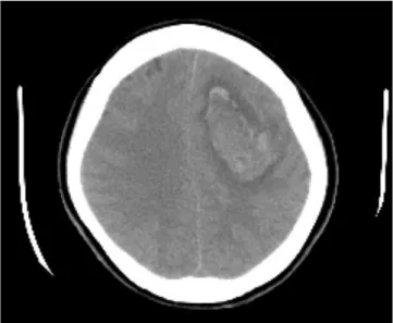
Brain CT revealed acute intracranial hemorrhage in the left frontal lobe with intraventricular hemorrhage and midline shifting (7 mm) (Fig. 3). Craniotomy, tumor and hematoma removal was performed (Fig. 4A) and metastatic invasive mole was confirmed by histology (Fig. 4B). Postoperative hCG was 990 mlU/mL. The postoperative period was uneventful for the first 72 hours and she gradually recovered her sensorium and motor abilities. However, on the fifth postoperative day, the patient developed marked pyrexia (39.6
oC) which was thought to be due to aspiration pneumonia. Although she was treated with adequate broad spectum intravenous antibi- otics, she lapsed into septic shock and could not be recovered.
Discussion
It is known that there are wide regional variations in the incidence
of hydatidiform mole through multiple epidemiologic studies, and
the incidence is found to be higher in oriental and developing
countries than Western and developed countries. The incidence
of molar pregnancy has decreased in South Korea from 4.4 cases
per 1,000 births in the 1960s to 1.6 cases per 1,000 births in the
Fig. 1. Microscopic examination of the evacuated molar tissue. Enlarged
villous with central cavitation and surrounding trophoplastic hyperplasia
are shown. Complete mole was diagnosed (H&E, ×100).
Fig. 2. Abdominopelvic computed tomography showing renal hemor- rhage with multiple metastases.
1990s [2], although the figure is still higher than that of Western countries.
Most women died from this malignant disease just about 60 years ago, but now, overall cure rates have exceeded over 90% even if the disease accompanies multiple metastatic lesions.
This advancement is the result of the inherent sensitivity of tro- phoblastic neoplasm to chemotherapy, the effective use of the bio marker hCG for initial diagnosis of the disease and monitoring of the effectiveness of therapy, the identification of prognostic factors that predict treatment responses and enhance individualization of therapy, and use of combined modality treatment with chemother-
apy, radiation, and surgery in the group of patients of the highest
risk [3]. Recently, angiographic embolization has also been proven
to be a safe and highly effective alternative procedure for massive
bleeding [4-10]. This technique offers several advantages including
avoidance of major surgery and general anesthesia and preserva-
tion of fertility [6,7]. Moodley and Moodley [8], in their series of 4
patients, reported that embolization was successful in managing
bleeding secondary to GTN. Keepanasseril et al. [9] reported 8
cases of patients with GTN, who presented massive bleeding and
underwent angiographic embolization with the success rate of
85.7%. The largest series was reported by Lim et al. [10] involv-
ing 14 patients who underwent embolization for hemorrhage secondary to GTN. In their series, 79% of the patients achieved successful control of bleeding with embolization alone. Despite these advancements in GTD, there are still many patients who die from these diseases presenting metastasis in the liver and brain.
Metastatic GTN occurs in about 4% of patient after evacuation of complete mole, but it is seen more often when GTN is developed after nonmolar pregnancy [11]. Metastatic GTN is highly vascular and prone to have severe spontaneous bleeding. The most com- mon sites of metastases are the lungs (80%), vagina (30%), pelvis
(20%), liver (10%), brain (10%) and less frequently, kidney [11,12].
Because trophoblastic tumors are often perfused by fragile vessels, they are frequently hemorrhagic and the symptoms of metastases may result from spontaneous bleeding from metastatic foci. All pa- tients with stage IV disease (hepatic, cerebral metastasis) should be treated with primary intensive combination chemotherapy and the selective use of radiation therapy and surgery [11]. Metastases of GTD respond well to chemotherapy, and there are also few reports on successful intervention trials to control severe hemor- rhages in metastatic GTN [13-15]. However, the prognosis of liver or brain metastasis is still poor [4,13-15]. Lal et al. [13] reported a case of spontaneous renal hemorrhage in a patient with metastat- ic choriocarcinoma presenting with gross hematuria that was suc- cessfully managed with angioembolization. The patient was start- ed on chemotherapy, but developed gastrointestinal bleeding due to jejunal metastasis and subsequently succumbed to sepsis. In our case, the patient was also presented with spontaneous renal hemorrhage, which was controlled by angiographic embolization.
However, she could not receive chemotherapy due to subsequent cerebral hemorrhage that emerged very shortly, and succumbed to sepsis. Lurain et al. [14] reported 48 fatal GTN cases, including 9 patients with liver metastases. Lok et al. [15] reported the util- ity of embolization of the hepatic artery to control the bleeding from liver metastases in GTN in 2 cases. Due to the characteristic of GTN, clinical manifestations can be varied like in the present patient who presented symptoms other than the typical symptoms of GTN, which can cause confusion in managing patients.
Another pitfall is the inaccuracy of the measurement of hCG. It is
Fig. 4. (A) Macroscopic finding of metastatic lesion in the brain and (B) Microscopic examination of the removed tumoral bleeding from the brain (H&E,
×200). Metastatic invasive mole was diagnosed.
A B
Fig. 3. Brain computed tomography showing acute intracranial hemor-
rhage in the left frontal lobe.
well known that human chorionic gonadotropin is a disease spe- cific tumor marker produced by hydatidiform moles and gestation- al trophoblastic neoplasms. Due to its easy quantitative measure- ment and correlation with the burden of disease, hCG is used as a specific and valuable tumor marker. In general, hydatidiform moles are commonly associated with a markedly elevated hCG level above those of normal pregnancy. Approximately 50% of patients with complete mole have pre evacuation hCG levels >100,000 mIU/mL [16,17]. However, in the present case, despite the severity and rapid progression of the disease, the measured level of hCG was only less than 2,300 mlU/mL. Several forms of hCG exist, including at least 6 major variants that can be detected in serum, hyperglycosylated, nicked, absent C-terminal of the ß subunit, free ß subunit, nicked ß subunit, and free α subunit [3]. Also, the hCG molecules in GTD are more heterogenous and pond to degrade than those in normal pregnancy, therefore, ordinary laboratory tests can give rise to false negative or misleadingly low serum hCG levels, so an assay that can detect all main forms of hCG and its multiple fragments should be considered to be used in follow up patients with GTD [18-20]. There is also another cause for massive underestimation of serum hCG, called “High dose hook effect”, in which there is a paradoxical return to a low response of the assay at high levels of the substance to be measured [21]. For example, when using “sandwich assays”, in cases where excessive amounts of hCG are present, there will not be enough antibodies in the solution to bind these molecules and hence much of it will be rinsed away without being measured. This false low level of hCG can mislead clinicians to misdiagnose and perform inappropriate management for patients with GTN. There have been reports of such treatment on false grounds in patients with GTN [18,21,22].
O’Reilly and Rustion [21] reported about a patient with multiple metastatic choriocarcinoma, whose management was adversely influenced by the falsely low serum hCG level measured by a com- mercial kit. The patient who had an intermittent abnormal vaginal bleeding for about a year presented with multiple pulmonary met- astatic lesions of unknown origin. Hysterectomy was performed and revealed choriocarcinoma. Her initial serum hCG was only 202 mlU/mL, thus repeated serum hCG was checked due to the inconsistency with the diagnosis of choriocarcinoma and the level was in fact 149,072 mlU/mL. This “high dose hook effect” can be avoided by performing the assay at several dilutions of the sample serum [23]. In our case, we could not make the dilutions due to the rapidly devastating condition of the patient. There still remains a doubt if her “real” serum hCG was that low.
In the present case, we can see how diverse and aggressive mani-
festations could be seen in GTN, so gynecologists should be aware of all possible metastatic sites of GTN and be alert and capable of diagnosing this fatal condition. Moreover, if there is a symptomatic or radiological evidence of metastasis even though the low level of checked hCG, other hCG molecules such as hyperglycosylated hCG should be checked.
References
1. Seckl MJ, Sebire NJ, Berkowitz RS. Gestational trophoblastic disease. Lancet 2010;376:717-29.
2. Martin BH, Kim JH. Changes in gestational trophoblastic tu- mors over four decades. A Korean experience. J Reprod Med 1998;43:60-8.
3. Lurain JR. Gestational trophoblastic disease I: epidemiology, pathology, clinical presentation and diagnosis of gestational trophoblastic disease, and management of hydatidiform mole.
Am J Obstet Gynecol 2010;203:531-9.
4. Crawford RA, Newlands E, Rustin GJ, Holden L, A’Hern R, Bag- shawe KD. Gestational trophoblastic disease with liver me- tastases: the Charing Cross experience. Br J Obstet Gynaecol 1997;104:105-9.
5. Carlini L, Villa A, Busci L, Trezzi G, Agazzi R, Frigerio L. Selec- tive uterine artery embolization: a new therapeutic approach in a patient with low-risk gestational trophoblastic disease.
Am J Obstet Gynecol 2006;195:314-5.
6. Pearl ML, Braga CA. Percutaneous transcatheter embolization for control of life-threatening pelvic hemorrhage from gesta- tional trophoblastic disease. Obstet Gynecol 1992;80:571-4.
7. Takemura M, Yamasaki M, Tanaka F, Shimizu H, Okamoto E, Hisamatu K, et al. Transcatheter arterial embolization in the management of gynecological neoplasms. Gynecol Oncol 1989;34:38-42.
8. Moodley M, Moodley J. Transcatheter angiographic emboliza- tion for the control of massive pelvic hemorrhage due to ges- tational trophoblastic disease: a case series and review of the literature. Int J Gynecol Cancer 2003;13:94-7.
9. Keepanasseril A, Suri V, Prasad GR, Gupta V, Bagga R, Aggar- wal N, et al. Management of massive hemorrhage in patients with gestational trophoblastic neoplasia by angiographic em- bolization: a safer alternative. J Reprod Med 2011;56:235-40.
10. Lim AK, Agarwal R, Seckl MJ, Newlands ES, Barrett NK, Mitch-
ell AW. Embolization of bleeding residual uterine vascular mal-
formations in patients with treated gestational trophoblastic
tumors. Radiology 2002;222:640-4.
11. Berkowitz RS, Goldstein DP. Gestational trophoblastic disease.
In: Berek JS, Novak E, editors. Berek & Novak’s gynecology.
14th ed. Philadelphia (PA): Lippincott Williams & Wilkins;
2007. p.1581-604.
12. Li MC. Trophoblastic disease: natural history, diagnosis, and treatment. Ann Intern Med 1971;74:102-12.
13. Lal A, Singhal M, Kumar S, Bag S, Singh SK, Khandelwal N.
Bilateral renal and jejunal metastasis of choriocarcinoma pre- senting as spontaneous renal hemorrhage. Cancer Imaging 2009;9:56-8.
14. Lurain JR, Brewer JI, Mazur MT, Torok EE. Fatal gestational trophoblastic disease: an analysis of treatment failures. Am J Obstet Gynecol 1982;144:391-5.
15. Lok CA, Reekers JA, Westermann AM, Van der Velden J. Embo- lization for hemorrhage of liver metastases from choriocarci- noma. Gynecol Oncol 2005;98:506-9.
16. Menczer J, Modan M, Serr DM. Prospective follow-up of pa- tients with hydatidiform mole. Obstet Gynecol 1980;55:346- 9.
17. Genest DR, Laborde O, Berkowitz RS, Goldstein DP, Bernstein MR, Lage J. A clinicopathologic study of 153 cases of com- plete hydatidiform mole (1980-1990): histologic grade lacks
prognostic significance. Obstet Gynecol 1991;78:402-9.
18. Flam F, Hambraeus-Jonzon K, Hansson LO, Kjaeldgaard A. Hy- datidiform mole with non-metastatic pulmonary complications and a false low level of hCG. Eur J Obstet Gynecol Reprod Biol 1998;77:235-7.
19. Cole LA, Butler SA, Khanlian SA, Giddings A, Muller CY, Seckl MJ, et al. Gestational trophoblastic diseases: 2. Hyperglycosyl- ated hCG as a reliable marker of active neoplasia. Gynecol Oncol 2006;102:151-9.
20. Cole LA, Khanlian SA, Muller CY, Giddings A, Kohorn E, Berkowitz R. Gestational trophoblastic diseases: 3. Hu- man chorionic gonadotropin-free beta-subunit, a reliable marker of placental site trophoblastic tumors. Gynecol Oncol 2006;102:160-4.
21. O’Reilly SM, Rustin GJ. Mismanagement of choriocarcinoma due to a false low HCG measurement. Int J Gynecol Cancer 1993;3:186-8.
22. Levavi H, Neri A, Bar J, Regev D, Nordenberg J, Ovadia J. “Hook effect” in complete hydatidiform molar pregnancy: a falsely low level of beta-HCG. Obstet Gynecol 1993;82:720-1.
23. Hoffman KL. Optimization of sandwich immunometric assay. J Clin Immunoassay 1985;8:237-44.
저역가의 융모성 성선자극호르몬을 보이는 전이성 임신성 영양막세포질환에서 발생한 자연콩팥출혈 및 뇌 출혈 1예
전남대학교 의과대학 산부인과교실 문희현, 김석모, 문종호, 강우대, 최호선
임신성 영양막세포질환은 포상기태(완전포상기태와 부분포상기태), 침윤성기태, 융모막암종, 태반부착부위 융모상피성 종양과 같은 네 가지의 질환을 포함하는 질환의 범주이다. 포상기태는 대개는 양성이지만, 전암성 병변으로 여겨지기도 하며, 그 외의 세 가지 질환은 악 성으로, 임신성 영양모아세포성 종양이라고 일컫는다. 대부분의 포상기태 임신이 양성 질환의 추이를 보이나, 완전 포상기태의 4% 정도 에서는 전이가 확인되기도 한다. 하지만 타 장기까지의 광범위한 전이를 보이는 경우에도, 대부분의 경우 화학요법에 아주 반응이 좋은 것으로 알려져 있다. 최근 저자들은 낮은 융모성 성선자극호르몬 수치에도 불구하고, 완전 포상기태에 이어 발생한 전이성 임신성 영양모 아세포성 종양에서 자연콩팥출혈 및 뇌출혈로 사망한 1예를 경험하였기에 문헌고찰과 함께 이를 보고하는 바이다.
중심단어: 전이성 임신성 영양막세포질환, 완전 포상기태, 침윤성 기태, 융모성 성선자극호르몬, 콩팥출혈, 뇌출혈