Articles published in Obstet Gynecol Sci are open-access, distributed under the terms of the Creative Commons Attribution Non-Commercial License (http://creativecommons.
org/licenses/by-nc/3.0/) which permits unrestricted non-commercial use, distribution, and reproduction in any medium, provided the original work is properly cited.
Copyright © 2014 Korean Society of Obstetrics and Gynecology http://dx.doi.org/10.5468/ogs.2014.57.5.373
pISSN 2287-8572 · eISSN 2287-8580
Received: 2013.11.27. Revised: 2014.4.23. Accepted: 2014.5.19.
Corresponding author: Byung Chul Jee
Department of Obstetrics and Gynecology, Seoul National University Bundang Hospital, 82 Gumi-ro 173beon-gil, Bundang- gu, Seongnam 463-707, Korea
Tel: +82-31-787-7254 Fax: +82-31-787-4054 E-mail: [email protected]
Introduction
Recent progress in in vitro fertilization (IVF) techniques involves the culture of human embryos to the blastocyst stage. Blasto- cyst transfer has been known to improve uterine and embry- onic synchronicity and enable self-selection of viable embryos, resulting in higher implantation rates [1,2]. To obtain more blastocysts with a high implantation potential, optimal in vitro culture condition might be essential.
The growth and development of the preimplantation embryo is regulated by various cytokines and endogenous growth-promoting substances which are secreted from lu-
Dose-dependent embryotrophic effect of recombinant granulocyte-macrophage colony-stimulating factor and brain-derived neurotrophic factor in culture medium for mouse preimplantation embryo
Jee Hyun Kim
1,2, Hyun Ju Lee
3, Eun Jeong Yu
3, Byung Chul Jee
1,2, Chang Suk Suh
1,2, Seok Hyun Kim
2,3Department of Obstetrics and Gynecology, 1Seoul National University Bundang Hospital, Seongnam; 2Seoul National University College of Medicine, Seoul; 3Seoul National University Hospital, Seoul, Korea
Objective
To evaluate the dose effect of recombinant mouse granulocyte-macrophage colony-stimulating factor (rmGM-CSF) or brain-derived neurotrophic factor (BDNF) in culture medium on the development of in vitro fertilized mouse embryos.
Methods
Mature oocytes were retrieved from superovulated female BDF1 mice and inseminated by sperm from male BDF1 mice. On day 1, two-cell stage embryos were divided and cultured until day 5 in the embryo maintenance medium supplemented with 0, 1, 2, 5, or 10 ng/mL of rmGM-CSF or supplemented with 0, 5, 10, or 20 ng/mL of BDNF. Blastocyst formation rate and their cell numbers were assessed.
Results
The blastocyst formation rate and the total cell count in blastocyst was similar in all the rmGM-CSF treatment groups when compared with the control. However, the blastocyst formation rate and the total cell count was significantly higher in the group supplemented with 10 ng/mL of BDNF compared with the control (63.9%, 45.8±11.5 vs. 52.3%, 38.0±6.8; P<0.05, respectively).
Conclusion
Supplementation of 10 ng/mL of BDNF enhanced the developmental potential of mouse preimplantation embryos, but supplementation of rmGM-CSF did not.
Keywords: Brain-derived neurotrophic factor; Culture medium; Embryotrophic effects; Granulocyte-macrophage colony-stimulating factor
minal epithelial cells of oviducts in human and other species [3]. Corresponding receptors for these growth factors have been known to be expressed by preimplantation embryos, supporting the close interaction between growth factors and embryos. Many studies have demonstrated the addition of growth factors to embryo culture medium can improve the development of the preimplantation embryo and the efficacy of IVF [4-6].
Granulocyte-macrophage colony-stimulating factor (GM- CSF) is a multifunctional cytokine originally identified as a regulator of the proliferation, differentiation, and activation of myeloid hemopoietic cells [7]. In hemopoietic precursors, GM- CSF acts as a survival factor through suppressing apoptosis [8- 10]. In reproduction, GM-CSF, synthesized by estrogen-primed epithelial cells in human uterus and oviducts [11,12] has been known to improve the embryo development. Previous experi- ments showed that in vitro culture under GM-CSF increases the blastulation rate and blastomeres in mouse and human embryos [13,14].
Brain-derived neurotrophic factor (BDNF) is a member of the neurotrophin family of proteins, which activate the high- affinity tyrosine kinase receptor (TrkB) and the low-affinity pan-neurotrophin receptor p75 [15]. It is widely distributed throughout the brain in diverse cell types and mediates the neuronal survival and differentiation [16]. Recently, it has been shown that BDNF also plays a role in the development of vari- ous non-neuronal tissues including reproductive systems [17].
Previous studies suggested that BDNF regulates folliculogen- esis [18], promotes oocyte maturation and in vitro develop- ment of zygotes to preimplantation embryos [19,20] and the development of preimplantation embryos to the blastocysts with the increased proliferation and decreased apoptosis [21].
Several reports denoted a different effect according to the concentration of recombinant GM-CSF (rGM-CSF) for the em- bryo development in the different species [22,23]. In the study of porcine embryos, highest proportion of blastulation was observed in the presence of 10 ng/mL of rGM-CSF among different concentrations of 2, 10, and 100 ng/mL [23]. In naturally fertilized murine embryos, blastulation was impeded at the concentration of GM-CSF in the culture media as high as 5 ng/mL [22]. Regarding BDNF, there has been no study to evaluate the dose effect of BDNF as far as we know.
Herein, we attempted to evaluate the effect of adding dif- ferent concentrations of recombinant mouse (rm) GM-CSF and BDNF to blastocyst culture in the in vitro fertilized murine embryos. To determine the developmental competence, the
blastulation rate and the total number of blastomeres were assessed.
Materials and methods
1. Animal and in vitro fertilization protocol
Five- to six-week-old female BDF1 mice (Orient Co., Seoul, Korea) were used in this study. Animal care and use were in accordance with the guidelines established by the Institutional Animal Care and Use Committee of Seoul National University of Bundang Hospital. Superovulation and oocyte retrieval from mice was performed as previously described [24]. Briefly, BDF1
female mice were injected with 5 IU of Pregnant Mare’s Serum Gonadotropin (Sigma-Aldrich, St Louis, MO, USA), followed by intraperitoneal 5 IU of human Chorionic Gonadotropin (Sigma-Aldrich) 48 hours later. After 13 to 14 hours, the mice were sacrificed by cervical dislocation. Oocytes were collected from the ampullae and incubated in 1 mL of modified mouse tubal fluid (mMTF) containing 0.4% bovine serum albumin (BSA, Sigma-Aldrich).
The epididymal sperm were retrieved from the cauda epidid- ymis of 8- to 10-week-old BDF1 mice, and the sperm suspen- sions were preincubated for 1.5 hours in capacitation medium (mMTF supplemented with 0.8% BSA). The oocytes were then inseminated by sperm at a final dilution of 2×10⁶/mL and incubated at 37℃ in humidified 5% CO2 in air. Inseminated oocytes were washed by pipetting 6 hours later and cultured in mMTF medium supplemented with 0.8% BSA overnight.
On day 1, fertilization was assessed by the formation of 2-cell stage.
2. Culture medium
Embryos reached 2-cell stage were transferred to embryo maintenance medium (Global medium supplemented with 10% human serum albumin; Life Global, Guilford, CT, USA) containing 0, 1, 2, 5, or, 10 ng/mL of rmGM-CSF (Sigma- Aldrich) or containing 0, 5, 10, or, 20 ng/mL of BDNF (Sigma- Aldrich). On day 5 after insemination, development to blasto- cyst was assessed. In all cultures, groups of up to 10 embryos were placed in 50-μL microdrops of medium under mineral oil (Sigma-Aldrich) in 35×10-mm Petri dishes (Falcon; Becton Dickinson, Franklin Lakes, NJ, USA) and were placed at 37℃
in humidified 5% CO2 in air. All produced blastocysts were mounted on the slides and stained by 4,6-diamidino-2-phenyl- indole (DAPI; Vector-mounting medium for fluorescence with
DAPI/H-1200) for 15 minutes for nuclear counting.
3. Statistical analysis
It was calculated that at least 150 in vitro fertilized 2-cell em- bryos in each group achieves 80% power to reject the null hypothesis using an error of 0.05 and the chi-square test, as- suming a baseline blastocyst rate of 50% and a difference of 5% between the two groups. Data were analyzed with SPSS version 17.0 (SPSS Inc., Chicago, IL, USA). The percentage of blastocyst in each treatment group was compared with that in the untreated (control) group by using the chi-square test.
Numeric data were compared by the Student’s t-test after per- forming the normality test. The result was considered signifi- cant when the P-value was less than 0.05.
Results
Sixty mice were used for rmGM-CSF experiment; 1,120 ma- ture oocytes were obtained and overall IVF rate was 74.5%.
The blastocyst formation rate and the total cell count in blas- tocyst was similar in all rmGM-CSF treatment groups when compared with control (Table 1). The blastocyst formation rate was highest in the group supplemented by 2 ng/mL of rmGM- CSF (57.2%), however, the differences did not reach a statisti- cal significance when compared with control. The blastocyst formation rate was lowest in the 5-ng/mL-rmGM-CSF treat- ment group (43.4%) and this was significantly different when compared with 2-ng/mL-rmGM-CSF treatment group (P=0.02).
For BDNF experiment, fifty mice were used; 1,330 mature oocytes were obtained and overall IVF rate was 54.8%. In the
Table 1. Developmental outcomes after culture of mouse preimplantation embryos supplemented with rmGM-CSF rmGM-CSF (ng/mL)
0 1 2 5 10
No. 2-cell 162 162 159 159 160
No. blastocyst 80 86 91 69 79
Early (%) 60.3 48.0 54.3 44.4 61.5
Expanded (%) 36.5 49.3 44.3 51.9 33.8
Hatching (%) 3.2 2.7 1.4 3.7 4.6
% per 2-cell (pooled) 49.4 53.1 57.2 43.4a) 49.4
% per 2-cellb) 46.6±17.8 53.9±22.2 55.6±23.2 45.3±18.7 49.9±25.9
No. blastomeresb) 35.0±15.6 27.0±7.4 30.8±12.6 32.8±11.0 31.2±14.5
Fourteen replicates were performed; Pooled percentages were not significant when compared with the control.
rmGM-CSF, recombinant mouse granulocyte-macrophage colony-stimulating factor.
a)P=0.02 when compared with 2-ng/mL-rmGM-CSF treatment group; b)Presented as mean±standard deviation and no differences were found when compared with each other.
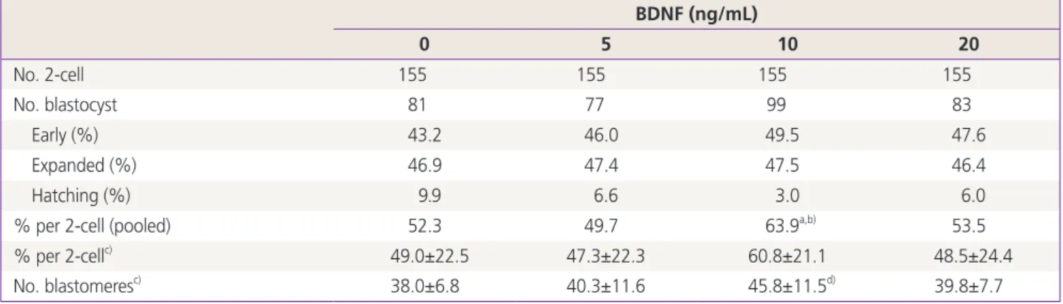
Table 2. Developmental outcomes after culture of mouse preimplantation embryos supplemented with BDNF BDNF (ng/mL)
0 5 10 20
No. 2-cell 155 155 155 155
No. blastocyst 81 77 99 83
Early (%) 43.2 46.0 49.5 47.6
Expanded (%) 46.9 47.4 47.5 46.4
Hatching (%) 9.9 6.6 3.0 6.0
% per 2-cell (pooled) 52.3 49.7 63.9a,b) 53.5
% per 2-cellc) 49.0±22.5 47.3±22.3 60.8±21.1 48.5±24.4
No. blastomeresc) 38.0±6.8 40.3±11.6 45.8±11.5d) 39.8±7.7
Twelve replicates were performed.
BDNF, brain-derived neurotrophic factor.
a)P=0.049 when compared with the control; b)P=0.017 when compared with 5-ng/mL-BDNF treatment group; c)Presented as mean±standard deviation and no differences were found when compared with each other except; d)P=0.015 when compared with the control.
group supplemented by 10 ng/mL of BDNF, the blastocyst for- mation rate (63.9%) was significantly higher when compared with control (52.3%) and 5-ng/mL-BDNF treatment group (49.7%) (Table 2). Total cell count in 10-ng/mL-BDNF treat- ment group (45.8±11.5) was significantly higher when com- pared with the control.
Discussion
Our results suggest that BDNF may play embryotrophic role in a specific concentration, 10 ng/mL, but rmGM-CSF may not.
Our study showed that adding GM-CSF to the culture media did not have any significant positive impact on the blastula- tion rate or cell number, which is in accordance with previous report [22,25,26]. Behr et al. [25] tested the effect of a range of concentrations of GM-CSF (0.0625–2 ng/mL) on mouse embryo development and found that concentrations from 0.25 to 2 ng/mL did not promote blastocyst development and the best effect was reported in the 0.125 ng/mL group. Elaimi et al. [22] reported no difference in the blastulation potential was noted with the addition of 1 and 2 ng/mL of GM-CSF compared with the controls but, GM-CSF exerted a negative impact on the blastulation rate at 5 and 10 ng/mL concentra- tions. On the other hand, some studies reported significant ef- fects of adding 2 ng/mL of GM-CSF to embryo culture [14,27].
These different effects might come from the use of a different type of culture medium. Karagenc et al. [28] examined the effect of adding GM-CSF (0, 2, 4, 8, and 16 ng/mL) on the development of mouse embryos from different strains and under different culture conditions. They found no marked ef- fect of supplementing the media with GM-CSF using different concentrations. However, in the absence of protein source, the stimulatory effect of GM-CSF was observed on the total number of blastomeres.
Regarding BDNF in our study, supplementation of 10 ng/mL of BDNF showed the highest blastocyst formation rate and the total cell number when compared with the control. However, when cultured with 20 ng/mL of BDNF, the embryotrophic ef- fect was eliminated. This peculiar tendency was also observed by other researcher [21]; hatched blastocyst formation rate was the highest under 10 ng/mL rather than 30 ng/mL. This observation could be partly explained by the negative regula- tion of the receptors when the growth factors are present at high concentrations.
The mechanisms underlying the embryotrophic actions of
GM-CSF was suggested by Sjoblom et al. [29]. They suggested that GM-CSF acts mediated via an interaction with the GM- CSF-receptor alpha subunit (GM-Rα) that may occur indepen- dently of GM-CSF-receptor beta common subunit (ßc). In that study, by using reverse transcription-polymerase chain reaction and immunocytochemistry, expression of mRNA and protein for GM-Rα was identified in embryos from the first-cleavage through blastocyst stages of development, but the GM-CSF- receptor ßc could not be detected at any stage.
When neutralizing antibodies reactive with GM-Rα were added to embryo culture medium, the development-promot- ing effect of GM-CSF was ablated [29]. In contrast, GM-CSF activity in embryos was not inhibited either by antibodies to ßc or by E21R, a synthetic GM-CSF analogue that acts to an- tagonize ßc-mediated GM-CSF signaling. Unexpectedly, E21R was found to mimic native GM-CSF in promoting blastulation.
These indicate that GM-CSF regulates cell viability through the GM-Rα that is independent of ßc. GM-CSF receptor has been known to act through ßc, which forms a high-affinity complex when associated with ligand-coupled GM-Rα. Thus, in embryos, GM-Rα may act with ligation stimulating glucose uptake through a kinase-independent pathway.
Kawamura et al. [21] demonstrated the embryotrophic ac- tions of BDNF. According to their study, BDNF reached at its highest levels in the blastocyst stages and the corresponding receptor, TrkB were detectable throughout the early embry- onic stages with an increase after the early blastocyst stage.
Both BDNF and TrkB are expressed in trophectoderm cells and ligand-binding studies indicated the specific binding of BDNF to trophectoderm cells. Treatment with BDNF promoted the development of 2-cell stage embryos into blastocysts showing increased proliferation and decreased apoptosis. The effects of BDNF were blocked by the TrkB ectodomain or a Trk receptor inhibitor.
Among the several growth factors, recombinant human GM-CSF has been applying to the human IVF clinics. A recent research using commercially available culture medium contain- ing recombinant human GM-CSF (2 ng/mL) was accomplished and showed the beneficial effect on reducing miscarriage rate in patients with a previous pregnancy loss [30]. For the wider application of GM-CSF-containing culture medium to hu- man IVF, safety concerns should be resolved. A previous mu- rine study denoted that GM-CSF does not affect mosaicism/
aneuploidy, but increases the percentage of aneuploid cells within the mosaic embryos [22]. The latter finding might be attributed to anti-apoptotic effect of GM-CSF, by which more
cells with aneuploid cells in the mosaic embryos would sur- vive. Although a recent human study indicates no increment of cytogenetically abnormal embryos [31], adding GM-CSF to the culture media for clinical purpose requires further studies either on human or animal models to evaluate its long-term effects.
The present study demonstrated that GM-CSF exerted no impact on the blastulation rate at any concentration but, BDNF showed the embryotrophic effects of at a specific con- centration. For the clinical application of BDNF, safety profiles such as chromosomal constitution and long-term fetal effects should be studied in advance. Further studies are needed re- garding the effective concentration in human embryo culture and whether combination of these growth factors could im- prove the embryo development more than one.
Conflict of interest
No potential conflict of interest relevant to this article was reported.
Acknowledgments
This work was supported by grant no. 800-20130224 from the Seoul National University College of Medicine Research Fund.
References
1. Gardner DK, Lane M, Calderon I, Leeton J. Environment of the preimplantation human embryo in vivo: metabo- lite analysis of oviduct and uterine fluids and metabolism of cumulus cells. Fertil Steril 1996;65:349-53.
2. Gardner DK, Vella P, Lane M, Wagley L, Schlenker T, Schoolcraft WB. Culture and transfer of human blasto- cysts increases implantation rates and reduces the need for multiple embryo transfers. Fertil Steril 1998;69:84-8.
3. Richter KS. The importance of growth factors for pre- implantation embryo development and in-vitro culture.
Curr Opin Obstet Gynecol 2008;20:292-304.
4. Aflalo ED, Sod-Moriah UA, Potashnik G, Har-Vardi I. EGF increases expression and activity of PAs in preimplanta-
tion rat embryos and their implantation rate. Reprod Biol Endocrinol 2007;5:4.
5. Papayannis M, Eyheremendy V, Sanjurjo C, Blaquier J, Raffo FG. Effect of granulocyte-macrophage colony stimulating factor on growth, resistance to freezing and thawing and re-expansion of murine blastocysts. Reprod Biomed Online 2007;14:96-101.
6. Zhou P, Liu DJ, Cang M, Ma YZ, Yang DS, Li HJ, et al. TG- Falpha and EGFR in ovine preimplantation embryos and ef- fects on development. Anim Reprod Sci 2008;104:370-81.
7. Ruef C, Coleman DL. Granulocyte-macrophage colony- stimulating factor: pleiotropic cytokine with potential clinical usefulness. Rev Infect Dis 1990;12:41-62.
8. Caceres-Cortes J, Rajotte D, Dumouchel J, Haddad P, Ho- ang T. Product of the steel locus suppresses apoptosis in hemopoietic cells: comparison with pathways activated by granulocyte macrophage colony-stimulating factor. J Biol Chem 1994;269:12084-91.
9. Rajotte D, Haddad P, Haman A, Cragoe EJ Jr, Hoang T.
Role of protein kinase C and the Na+/H+ antiporter in suppression of apoptosis by granulocyte macrophage colony-stimulating factor and interleukin-3. J Biol Chem 1992;267:9980-7.
10. Williams GT, Smith CA, Spooncer E, Dexter TM, Tay- lor DR. Haemopoietic colony stimulating factors pro- mote cell survival by suppressing apoptosis. Nature 1990;343:76-9.
11. Giacomini G, Tabibzadeh SS, Satyaswaroop PG, Bonsi L, Vitale L, Bagnara GP, et al. Epithelial cells are the major source of biologically active granulocyte macrophage colony-stimulating factor in human endometrium. Hum Reprod 1995;10:3259-63.
12. Zhao Y, Chegini N. Human fallopian tube expresses gran- ulocyte-macrophage colony stimulating factor (GM-CSF) and GM-CSF alpha and beta receptors and contain im- munoreactive GM-CSF protein. J Clin Endocrinol Metab 1994;79:662-5.
13. Robertson SA, Sjoblom C, Jasper MJ, Norman RJ, Sea- mark RF. Granulocyte-macrophage colony-stimulating factor promotes glucose transport and blastomere vi- ability in murine preimplantation embryos. Biol Reprod 2001;64:1206-15.
14. Sjoblom C, Wikland M, Robertson SA. Granulocyte-macro- phage colony-stimulating factor promotes human blasto- cyst development in vitro. Hum Reprod 1999;14:3069-76.
15. Barbacid M. The Trk family of neurotrophin receptors. J Neurobiol 1994;25:1386-403.
16. Kafitz KW, Rose CR, Thoenen H, Konnerth A. Neuro- trophin-evoked rapid excitation through TrkB receptors.
Nature 1999;401:918-21.
17. Seifer DB, Feng B, Shelden RM, Chen S, Dreyfus CF. Brain- derived neurotrophic factor: a novel human ovarian fol- licular protein. J Clin Endocrinol Metab 2002;87:655-9.
18. Dissen GA, Hirshfield AN, Malamed S, Ojeda SR. Expres- sion of neurotrophins and their receptors in the mam- malian ovary is developmentally regulated: changes at the time of folliculogenesis. Endocrinology 1995;136:4681-92.
19. Anderson RA, Bayne RA, Gardner J, De Sousa PA. Brain- derived neurotrophic factor is a regulator of human oocyte maturation and early embryo development. Fertil Steril 2010;93:1394-406.
20. Kawamura K, Kawamura N, Mulders SM, Sollewijn Gelpke MD, Hsueh AJ. Ovarian brain-derived neuro- trophic factor (BDNF) promotes the development of oo- cytes into preimplantation embryos. Proc Natl Acad Sci U S A 2005;102:9206-11.
21. Kawamura K, Kawamura N, Fukuda J, Kumagai J, Hsueh AJ, Tanaka T. Regulation of preimplantation embryo de- velopment by brain-derived neurotrophic factor. Dev Biol 2007;311:147-58.
22. Elaimi A, Gardner K, Kistnareddy K, Harper J. The effect of GM-CSF on development and aneuploidy in murine blastocysts. Hum Reprod 2012;27:1590-5.
23. Kwak SS, Jeung SH, Biswas D, Jeon YB, Hyun SH. Effects of porcine granulocyte-macrophage colony-stimulating factor on porcine in vitro-fertilized embryos. Therio- genology 2012;77:1186-97.
24. Jee BC, Jo JW, Lee JR, Suh CS, Kim SH, Moon SY. Effect of trichostatin A on fertilization and embryo develop- ment during extended culture of mouse oocyte. Zygote 2012;20:27-32.
25. Behr B, Mooney S, Wen Y, Polan ML, Wang H. Prelimi- nary experience with low concentration of granulocyte- macrophage colony-stimulating factor: a potential regu- lator in preimplantation mouse embryo development and apoptosis. J Assist Reprod Genet 2005;22:25-32.
26. Desai N, Kattal N, AbdelHafez FF, Szeptycki-Lawson J, Goldfarb J. Granulocyte-macrophage colony stimulat- ing factor (GM-CSF) and co-culture can affect post-thaw development and apoptosis in cryopreserved embryos. J Assist Reprod Genet 2007;24:215-22.
27. Sjoblom C, Roberts CT, Wikland M, Robertson SA. Gran- ulocyte-macrophage colony-stimulating factor alleviates adverse consequences of embryo culture on fetal growth trajectory and placental morphogenesis. Endocrinology 2005;146:2142-53.
28. Karagenc L, Lane M, Gardner DK. Granulocyte-mac- rophage colony-stimulating factor stimulates mouse blastocyst inner cell mass development only when me- dia lack human serum albumin. Reprod Biomed Online 2005;10:511-8.
29. Sjoblom C, Wikland M, Robertson SA. Granulocyte- macrophage colony-stimulating factor (GM-CSF) acts independently of the beta common subunit of the GM- CSF receptor to prevent inner cell mass apoptosis in hu- man embryos. Biol Reprod 2002;67:1817-23.
30. Ziebe S, Loft A, Povlsen BB, Erb K, Agerholm I, Aasted M, et al. A randomized clinical trial to evaluate the effect of granulocyte-macrophage colony-stimulating factor (GM- CSF) in embryo culture medium for in vitro fertilization.
Fertil Steril 2013;99:1600-9.
31. Agerholm I, Loft A, Hald F, Lemmen JG, Munding B, Sorensen PD, et al. Culture of human oocytes with gran- ulocyte-macrophage colony-stimulating factor has no effect on embryonic chromosomal constitution. Reprod Biomed Online 2010;20:477-84.