Attenuation of Peripheral Regulatory T-Cell Suppression of Skin-Homing CD8 + T Cells in Atopic Dermatitis
Bao-Xiang Zhang,
1Jun-Cheng Lyu,
2Hai-Bo Liu,
3Dian-Qin Feng,
1Dian-Cai Zhang,
1Xing-Jie Bi,
4Zhi-Wu Duan,
1and Gang Ding
51Department of Dermatology, Yidu Central Hospital, Weifang Medical University, Weifang;
2Department of Health Statistics, Public Health College of Weifang Medical University, Weifang;
3Department of Clinical Laboratory, Yidu Central Hospital, Weifang Medical University, Weifang;
4Department of Dermatology, Weifang Skin Disease Hospital, Weifang;
5Department of Stomatology, Yidu Central Hospital, Weifang Medical University, Weifang, P.R. China.
Received: March 6, 2014 Revised: April 23, 2014 Accepted: April 23, 2014
Corresponding author: Dr. Gang Ding, Department of Stomatology, Yidu Central Hospital, Weifang Medical University,
No.4138 South Linglongshan Road, Qingzhou, Shandong Province 262500, P.R. China.
Tel: 86-536-3275691, Fax: 86-536-3279421 E-mail: [email protected]
∙ The authors have no financial conflicts of interest.
© Copyright:
Yonsei University College of Medicine 2015 This is an Open Access article distributed under the terms of the Creative Commons Attribution Non- Commercial License (http://creativecommons.org/
licenses/by-nc/3.0) which permits unrestricted non- commercial use, distribution, and reproduction in any medium, provided the original work is properly cited.
Purpose: Cutaneous lymphocyte-associated antigen (CLA)-expressing CD8+T cells have been known to play an important role in the pathogenesis of atopic der- matitis (AD). However, the mechanisms underlying the loss of self-tolerance re- main unclear. Regulatory T cells (Tregs) play a key role in the development of ho- meostasis in the immune system. We, therefore, hypothesized that a reduced ability of Tregs to inhibit autologous CD8+CLA+T cells might be underlying mechanism in AD. Materials and Methods: CD8+CLA+T cells and Tregs were obtained from the peripheral blood of AD patients and control volunteers. The fre- quencies of CD8+CLA+T cells were evaluated. The proliferative responses of CD8+CLA+T cells were assessed by flow cytometry, and the levels of transforming growth factor-β1 (TGF-β1) and interleukin-10 (IL-10) in culture supernatants were detected by enzyme-linked immunosorbent assay. Results: Our results revealed higher frequency and increased expression of perforin and granzyme-B in periph- eral CD8+CLA+T cells in AD, and lower inhibitory ability of Tregs on proliferation of CD8+CLA+T cells in AD. Meanwhile, the levels of TGF-β1 produced by Tregs were significantly lower in AD, and anti-TGF-β1 abolished such suppression.
Conclusion: The attenuated inhibitory ability of Tregs on hyper-activated autolo- gous CD8+CLA+T cells, mediated by TGF-β1, plays an important role in the pathogenesis of AD.
Key Words: Cutaneous lymphocyte-associated antigen, CD8+T cell, regulatory, T cells, atopic dermatitis
INTRODUCTION
Atopic dermatitis (AD) is a T-cell-mediated highly pruritic chronic inflammatory skin disease.1 Recent findings have suggested that CD8+T cells are required for the development of AD lesion.2,3 Cutaneous lymphocyte-associated antigen (CLA) is an important skin-homing receptor displayed by memory/effector T cells which recog-
CLA-FITC, anti-CD4-FITC, anti-CD25-PECY7, FOXP3- APC, purified anti-CD3 NA/LE and anti-CD28 NA/LE (anti-CD3/CD28), and FACSCalibur Flow Cytometer, all of which were obtained from BD Biosciences (Franklin Lakes, NJ, USA). CD8+T Cell Isolation Kit, anti-CLA MicroBead Kit, CD4+CD25+Regulatory T Cell Isolation Kit, 5-(and 6)-carboxyfluorescein diacetate succinimidyl ester (CFSE), MS/LD Columns, and MiniMACS separator were purchased from Miltenyi Biotec (Cologne, Germany). Biotin-conjugat- ed anti-transforming growth factor-β1 (anti-TGF-β1) or bio- tin-conjugated chicken IgG for a negative control were from Calbiochem (Darmstadt, Germany). RPMI 1640 medium, 100 U/mL penicillin G and 100 μg/mL streptomycin were purchased from Invitrogen (Carlsbad, CA, USA).
Detection of peripheral CD8+CLA+T cell frequencies and associated marker expressions
Peripheral blood samples were drawn in ethylene diamine tet- raacetic acid (EDTA)-treated tubes, and the samples were processed within 24 h after collection for flow cytometry.
Briefly, for the surface staining, appropriate blood was in- cubated for 15 min in the dark with the following antibod- ies: anti-CD3, anti-CD8, and anti-CLA. A lysin solution was then added to the tubes. After 10-min incubation, the cells were centrifuged, and the supernatants were discarded.
The remainder was then washed in phosphate buffered saline (PBS), the cells were centrifuged again, and the supernatant was discarded. Next, the cells were re-suspended in PBS and were analysed immediately. For intracellular labeling of perforin or granzyme-B, appropriate blood was incubat- ed for 15 min in the dark with the following antibodies: an- ti-CD3, anti-CD8, and anti-CLA. Then, Reagent A was added to the tubes and incubated for 5 min according to the manufacturer’s protocol of IntraSure Kit. Next, lysing solu- tion was added and incubated for 10 min. The mixture was centrifuged and supernatants were then discarded. Reagent B and anti-perforin or anti-granzyme-B were added to the tubes and incubated for 15 min. PBS was added to wash the cells, the cells were centrifuged again and the superna- tant discarded. Lastly, the cells were re-suspended in PBS and were analysed immediately. Isotype-matched controls were used for non-specific staining and analysis gates. Data were collected and analyzed.
Cell purification
Peripheral blood mononuclear cells (PBMCs) were isolated on Ficoll-Hypaque gradient. CD8+CLA+T cells were isolat- nize skin-related allergens.4 In peripheral blood, CD8+subsets
of CLA+CD45RO+T cells in AD are in an activated state, and the CD8+CLA+ memory/effector T cells are capable of responding to superantigenic stimulation.5 Furthermore, in- creased frequencies of CLA-positive CD8+T cells produc- ing type 2 cytokines have been detected in the blood of pa- tients with AD.6 However, the mechanisms involved in the induction and activation of CD8+CLA+T cells in AD are not clear.
Regulatory T cells (Tregs) play a crucial role in the main- tenance of immunologic-self-tolerance via dominant control of self-reactive T cells.7 It has been suggested that Tregs are able to suppress the proliferation of effector T cells in vitro and to inhibit the development and progression of systemic T cell-mediated autoimmune disorders in vivo; Foxp3-in- fected T cells can be successfully used to treat autoimmune and inflammatory disorders in mice.8-10 In AD, a dysregula- tion of disease-causing effector T cells is observed in skin biopsies, in association with an impaired Tregs infiltration.11 In contrary, however, the presence of Tregs in infiammatory lesions in AD has also been observed,12-14 suggesting the role of Tregs in the AD pathogenesis. Therefore, we hypothe- sized that an imbalance between activated CD8+CLA+T cells and Tregs exists in peripheral blood of patients with AD. However, the basic mechanisms of suppression medi- ated by Tregs in AD have not been reported. The purpose of this study is to analyze the inhibition ability of peripheral Tregs on autologous CD8+CLA+T cells in AD, and investi- gate the mechanisms involved.
MATERIALS AND METHODS
Participants
In this study, thirty patients (median age 25.6 years; 16 fe- male, 14 male) with history of AD of Yidu Central Hospital of Weifang and 25 healthy controls, age- and gender-matched, were enrolled in the study. AD was diagnosed according to the criteria proposed by Hanifin and Rajka.15 All volunteers had no other autoimmune-associated disorders and no med- ications had been used for at least 4 weeks prior to the study.
All participants signed informed consent and the study was approved by the Ethics Committee.
Reagents and equipment
The following monoclonal antibodies were used for flow cy- tometry experiments: anti-CD3-APC, anti-CD8-PerCP, anti-
namely, cells cultured in the absence (5×10⁴ cells/well) and presence (5×10⁴ cells/well) of unlabeled autologous Tregs.
After incubation for 4 days, the cells were harvested and washed in PBS containing 0.5 mM EDTA. CFSE labeled cells were resuspended and analyzed on the Flow Cytome- ter. According to the fluorescence intensity of CFSE, the proliferating (CFSEDIM) and non-proliferating (CFSEBRIGHT) populations of T cells were gated.
Detection of the levels of TGF-β1 and IL-10
The levels of TGF-β1 and interleukin-10 (IL-10) in culture supernatants were measured by enzyme-linked immunosor- bent assay (ELISA) kits (Endogen, Rockford, IL, USA) ac- cording to the manufacturer instructions.
Statistical analysis
Statistical differences between groups were analyzed by Student’s t-test, and data were expressed as mean±SD. A value of p<0.05 was considered significant. Statistical anal- ysis was performed using software SPSS 13.0 (SPSS Inc., Chicago, IL, USA).
RESULTS
Increased peripheral CD8+CLA+ cytotoxic T lymphocytes in patients with AD
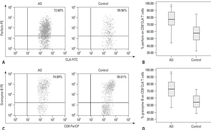
PBMCs obtained from 30 AD patients and 25 normal con- trols were analyzed for the frequencies of CD8+CLA+T cells and the expression of perforin and granzyme-B by flow cy- tometry. There was significantly higher number of peripher- al CD8+CLA+T cells in the AD group than the control group (p=0.000) (Fig. 2). Furthermore, the expression levels of perforin on CD8+CLA+T cells were significantly higher in the AD group than the control group (p=0.003). Similarly, the expression levels of granzyme-B on CD8+CLA+T cells were also significantly higher in the AD group than the con- trol group (p=0.001) (Fig. 3).
Analysis of proliferation of peripheral CD8+CLA+T cells in AD patients and healthy volunteers
CD8+CLA+T cells from either AD patients (n=8) or healthy volunteers (n=8) were labelled with CFSE and stimulated with anti-CD3/CD28 in the absence of Tregs for 4 days.
Flow cytometric analysis of CFSE intensity revealed no significant difference in the percentages of proliferating CD8+CLA+T cells between the two groups (Fig. 4).
ed by a two-step procedure; CD8+T cells were purified by negtive selection (using CD8+T Cell Isolation Kit), and then CLA+T cells were isolated by positive selection (using anti- CLA MicroBead Kit) on the MidiMACS magnetic separa- tion system, according to the manufacturer’s instructions, with final purities >93%. Tregs were isolated by a two-step procedure using CD4+CD25+ Regulatory T Cell Isolation Kit, according to the manufacturer’s instructions, and the purities were confirmed >95% (Fig. 1).
CFSE labeling
For the proliferation assay, freshly isolated CD8+CLA+T cells or Tregs were labeled with CFSE immediately. Brief- ly, cells were resuspended in pre-warmed (37°C) PBS/0.1%
albumin from bovine serum (BSA) at a final concentration of 1×10⁶ cells/mL, and CFSE solution was added for a final working concentration of 2.5 μM. Cells were incubated at 37°C for 10 min. Then, staining was quenched by adding 5 volumes of ice-cold culture media (RPMI 1640 supplement- ed with 10% fetal bovine serum (FBS), 100 U/mL penicillin G and 100 μg/mL streptomycin) and the cells were incubat- ed on ice for 5 min. Cells were then washed 3 times and re- suspended in the culture media.
T cells culture and proliferation assay
CFSE labeled cells were plated in 96-well U-bottom plates, precoated overnight with 5 μg/mL anti-CD3 NA/LE and 5 μg/mL anti-CD28 NA/LE (anti-CD3/CD28), at 4°C. CFSE labeled CD8+CLA+T cells were divided into two groups;
Fig. 1. The purities of isolated Tregs (CD4+CD25+T cells). PBMCs were iso- lated on Ficoll-Hypaque gradient. According to the manufacturer’s instruc- tions of CD4+CD25+ Regulatory T Cell Isolation Kit, CD4+T cells were purified by negtive selection, and then CD25+T cells were isolated by positive selec- tion. The purities were confirmed >95%. A representative profile of purified Tregs was showed. PBMC, peripheral blood mononuclear cell; FITC, fluo- rescein isothiocyanate; PE, phycoerythrin.
CD4 FITC 100
101 102 103 104
CD25 PE
100 101 102 103 104
99.04%
centages of proliferating CD8+CLA+T cells were analyzed and found to be significantly higher in the AD group than the control group (Fig. 5). Furthermore, Tregs showed an attenuated suppressive function on the proliferation of CD8+ CLA+T cells in the AD group.
The suppressive function of Tregs is mediated by TGF-β1 We determined the ability of CD8+CLA+T cells and Tregs to produce cytokines under the above established condition 4 Effects of reduced suppressive function of Tregs on the
proliferation of CD8+CLA+T cells in AD
Our preliminary analysis showed no significant difference in the frequencies of Tregs between the two groups (data not shown): Tregs were confirmed not to proliferate after stimulation with anti-CD3/CD28. Therefore, CFSE labeled CD8+CLA+T cells from either 8 AD patients or 8 normal control subjects were stimulated as described above and co- cultured with autologous Tregs. Four days later, the per-
Fig. 2. Frequencies of peripheral CD8+CLA+T cells. The frequencies of CD8+CLA+T cells in peripheral blood were measured by flow cytometry, and the repre- sentative results (A) from AD patients and healthy volunteers were shown by dot plot graphs. (B) The data showed the frequencies of CD8+CLA+T cells were significantly higher in the AD group (n=30) compared to the control group (n=25, *p<0.05). CLA, cutaneous lymphocyte-associated antigen; AD, atopic der- matitis; PE, phycoerythrin.
Fig. 3. Expression levels of cytotoxic molecules (perforin and granzyme-B) on peripheral CD8+CLA+T cells. The percentages of cytotoxic molecules (perforin and granzyme-B) on CD8+CLA+T cells in peripheral blood were measured by flow cytometry, and the representative results (A and C) from AD patients and healthy volunteers were shown by dot plot graphs. (B and D) The data showed the expression levels of both perforin and granzyme-B on CD8+CLA+T cells were significantly higher in the AD group (n=30) compared to the control group (n=25, *p<0.05). CLA, cutaneous lymphocyte-associated antigen; AD, atopic dermatitis; PE, phycoerythrin; FITC, fluorescein isothiocyanate.
CD8 PerCP
CD8 PerCP CLA FITC 100
100 100
100
100 100 101
101 101
101
101 101 102
102 102
102
102 102 103
103 103
103
103 103 104
104 104
104
104 104
CLA PEGranzyme-B PEPerforin PE
100
100 100
100
100 100 101
101 101
101
101 101 102
102 102
102
102 102 103
103 103
103
103 103 104
104 104
104
104 104 AD
AD AD
Control
Control Control 4.39%
74.89%
72.60%
2.01%
56.01%
59.56%
0.00 1.00 2.00 3.00 4.00 5.00 6.00 7.00
% CLA on CD8+ T cells
AD Control
*
30.00 40.00 50.00 60.00 70.00 80.00 90.00 100.00
% granzyme-B on CD8+ CLA+ T cells
AD Control
* 30.00
40.00 50.00 60.00 70.00 80.00 90.00 100.00
% perforin on CD8+ CLA+ T cells
AD Control
*
A
C A
B
D B
in the cocultures of Tregs and CD8+CLA+T cells, Tregs could produce high levels of TGF-β1 and IL-10 in both groups.
Nevertheless, the levels of TGF-β1 were significantly lower days later. Results showed that CD8+CLA+T cells stimulated
with anti-CD3/CD28 alone secreted undetectable amounts of TGF-β1 and only minimal amounts of IL-10. However,
Fig. 4. Proliferation of peripheral CD8+CLA+T cells. CFSE-labeled peripheral CD8+CLA+T cells stimulated with anti-CD3/CD28 in the absence of Tregs for 4 days. According to the CFSE intensity by flow cytometry, the percentages of proliferating CD8+CLA+T cells were detected. (A) Representative CFSE profiles from an AD patient and a healthy volunteer were shown. (B) The data showed the percentages of proliferating CD8+CLA+T cells were of no significant differ- ence between the AD group (n=8) and the control group (n=8). CFSE, carboxyfluorescein diacetate succinimidyl ester; CLA, cutaneous lymphocyte-associ- ated antigen; AD, atopic dermatitis.
CFSE
0 0
20 10
20 60
40 30
80
40 50
100 60
CD8+ CLA+ T cells counts
100 101 102 103 104 100 101 102 103 104 70.00%
M1
50.00 60.00 70.00 80.00
% CD8+ CLA+ T cells proliferation
AD Control
A B
67.44%
M1
Fig. 5. Tregs suppress proliferation of autologous CD8+CLA+T cells. CFSE-labeled peripheral CD8+CLA+T cells stimulated with anti-CD3/CD28 in the presence of autologous Tregs for 4 days. According to the CFSE intensity by flow cytometry, the percentages of proliferating CD8+CLA+T cells were detected. (A) Representative CFSE profiles from an AD patient and a healthy volunteer were shown. (B) The data showed the percentages of proliferating CD8+CLA+T cells were significantly higher in the AD group (n=8) compared to the control group (n=8, *p<0.05). (C) The suppressive function of Tregs on the proliferation of autologous CD8+CLA+T cells was significantly lower in the AD group (n=8) compared to the control group (n=8, *p<0.05). CFSE, carboxyfluorescein diace- tate succinimidyl ester; Tregs, regulatory T cells; CLA, cutaneous lymphocyte-associated antigen; AD, atopic dermatitis.
CFSE
0 0
10 30
30
20 60
50 120
40 90
60 150
CD8+ CLA+ T cells (+Tregs) counts
100 101 102 103 104 100 101 102 103 104
AD Control
50.95% 36.98%
M1 M1
A
30.00 10
0 40.00
20 30 50.00
40 50
60.00 60
70
% CD8+CLA+T cells (+Tregs) proliferation % Tregs suppression on CD8+CLA+T cells
AD Control AD Control
*
*
B C
Fig. 6. Levels of TGF-β1 and IL-10 in culture supernatants of Tregs and/or CD8+CLA+T cells. 5×10⁴ Tregs co-cultured with 5×10⁴ autologous CD8+CLA+T cells stimulated with anti-CD3/CD28 in 96-well plates. Four days later, the amount of TGF-β1 (A) and IL-10 (B) in culture supernatant was measured by ELISA. The levels of TGF-β1 produced by Tregs were significantly lower in the AD group (n=8) compared to the control group (n=8, *p<0.05). However, the levels of IL-10 produced by Tregs showed no difference between the two groups. And 5×10⁴ CD8+CLA+T cells stimulated with anti-CD3/CD28 in 96-well plates for 4 days, se- creted undetectable amounts of TGF-β1 and minimal amounts of IL-10 in the two groups. CLA, cutaneous lymphocyte-associated antigen; AD, atopic derma- titis; Tregs, regulatory T cells; ELISA, enzyme-linked immunosorbent assay; TGF-β1, transforming growth factor-β1; IL-10, interleukin-10.
CD8+CLA+T cells Tregs
0 30 60 90 120 150 180
Levels of TGF-β1 (pg/mL)
AD Control AD Control
A B
*
n.d. n.d.
CD8+CLA+T cells Tregs
0 500 1000 1500 2000 2500 3000
Levels of IL-10 (pg/mL)
AD Control AD Control
AD Control
Akdis, et al.5 demonstrated that CLA+CD8+T cells isolated from the skin or peripheral blood of AD patients responded to superantigenic stimulation to the same extent as CD4+T cells: they spontaneously proliferated ex vivo, secreting high levels of IL-5 and IL-13, and therefore, were capable of preventing spontaneous eosinophil apoptosis and enhanc- ing IgE. In the present study, we showed that patients with AD have a higher frequency of CD8+CLA+T cells and in- creased expression of perforin and granzyme-B. Similarly, Yawalkar, et al.21 found that perforin and granzyme-B are cytotoxic molecules produced by activated cytotoxic CD8+T cells, which are strongly expressed in AD lesional skin.
Moreover, CD8+granzyme-B+T cells infiltrating the epider- mis are associated with the development of positive atopy patch test.3 These results together with the present study are consistent with the hypothesis that these hyper-activated cytotoxic T cells are responsible for the apoptosis of kerati- nocytes and for the epidermal spongiosis, which are both pathological hallmarks of AD.22,23
The functional hallmark of Tregs is their remarkable ca- pacity to suppress T effector/memory (Teff/mem) cell activa- tion including T-cell proliferation.24 Recent reviews have up- dated the idea that Tregs can inhibit activated T cells to maintain peripheral tolerance.25,26 The suppressive capacity of Tregs is diminished in patients with autoimmune diseases such as psoriasis27 and bullous pemphigoid,28 and Tregs showed a dysfunction to CD4+CD25-T cells27 or CD8+T cells in theses diseases.29 Considering the fact that CD8+CLA+T cells are hyper-activated in AD, to assay the suppressive function of Tregs on CD8+CLA+T cells appears to be highly significant. Our study showed that proliferative responses of peripheral CD8+CLA+T cells in the absence of autologous Tregs stimulated with anti-CD3/CD28 had no significant dif- ference between AD and control groups. However, after co- in the AD group than the control group (p=0.025), whereas
the levels of IL-10 were not significantly different between the two groups (Fig. 6), demonstrating that the levels of TGF-β1 produced by Tregs in the cocultures were decreased in AD.
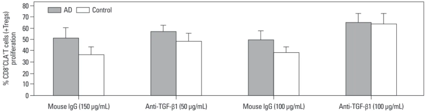
We further investigated whether such production mediates immunosuppression: whether Tregs mediate immunosup- pression in the presence or absence of anti-TGF-β1. Fig. 7 shows that, in cultures containing 50 μg/mL of anti-TGF-β1, the Tregs-mediated suppression decreased and the CD8+ CLA+T cells showed increased proliferation. More impor- tantly, in the cultures containing 100 μg/mL of anti-TGF-β1, Tregs-mediated suppression was greatly weakened, while the percentages of proliferating CD8+CLA+T cells were fur- ther increased, with no significant difference between the two groups. These results suggest that the suppression of CD8+CLA+T cells proliferation by Tregs is mediated, at least in part, by TGF-β1 in AD.
DISCUSSION
It has been proven that CD8+CLA+T cells play an important role in immune-related diseases, such as psoriasis and alope- cia areata.16,17 Recently, Hennino, et al.2,3 showed that CD8+T cells are recruited early to allergen exposure sites in atopy patch test reactions in human atopic dermatitis and aller- gen-primed CD8+T cells initiate atopic dermatitis lesions.
Furthermore, CLA+T cells isolated from peripheral blood of atopic individuals were shown to contain and spontane- ously release cytokines IL-4 and IL-13,18,19 and staphylo- coccal enterotoxin B treatment of atopic PBMC in vitro re- sulted in up-regulation of CLA expression on T cells, thereby facilitating the homing of T cells in the AD skin.20
Fig. 7. Suppression of CD8+CLA+T cells proliferation by Tregs is mediated by TGF-β1. 5×10⁴ Tregs co-cultured with 5×10⁴ autologous CD8+CLA+T cells from AD patients (n=8, gray bars) or healthy volunteers (n=8, white bars) stimulated with anti-CD3/CD28 in 96-well plates in the presence of anti-TGF-β1 or control IgG. Four days later, the percentages of proliferating CD8+CLA+T cells were detected. The CD8+CLA+T cells exhibited increased proliferation in the presence of 50 μg/mL of anti-TGF-β1, and their proliferation were further increased and of no significant difference between the two groups in the presence of 100 μg/
mL of anti-TGF-β1. Tregs, regulatory T cells; CLA, cutaneous lymphocyte-associated antigen; AD, atopic dermatitis; TGF-β1, transforming growth factor-β1.
0 10 20 30 40 50 60 70 80
% CD8+ CLA+ T cells (+Tregs) proliferation
Mouse lgG (150 µg/mL) Anti-TGF-β1 (50 µg/mL) Mouse lgG (100 µg/mL) Anti-TGF-β1 (100 µg/mL) AD Control
allergen exposure sites in atopy patch test reactions in human atopic dermatitis. J Allergy Clin Immunol 2011;127:1064-7.
3. Hennino A, Vocanson M, Toussaint Y, Rodet K, Benetière J, Schmitt AM, et al. Skin-infiltrating CD8+ T cells initiate atopic dermatitis lesions. J Immunol 2007;178:5571-7.
4. Sallusto F, Lenig D, Förster R, Lipp M, Lanzavecchia A. Two subsets of memory T lymphocytes with distinct homing potentials and effector functions. Nature 1999;401:708-12.
5. Akdis M, Simon HU, Weigl L, Kreyden O, Blaser K, Akdis CA.
Skin homing (cutaneous lymphocyte-associated antigen-positive) CD8+ T cells respond to superantigen and contribute to eosino- philia and IgE production in atopic dermatitis. J Immunol 1999;
163:466-75.
6. Teraki Y, Hotta T, Shiohara T. Increased circulating skin-homing cutaneous lymphocyte-associated antigen (CLA)+ type 2 cyto- kine-producing cells, and decreased CLA+ type 1 cytokine-pro- ducing cells in atopic dermatitis. Br J Dermatol 2000;143:373-8.
7. Sakaguchi S. Naturally arising CD4+ regulatory t cells for immu- nologic self-tolerance and negative control of immune responses.
Annu Rev Immunol 2004;22:531-62.
8. Loser K, Hansen W, Apelt J, Balkow S, Buer J, Beissert S. In vi- tro-generated regulatory T cells induced by Foxp3-retrovirus in- fection control murine contact allergy and systemic autoimmunity.
Gene Ther 2005;12:1294-304.
9. Fontenot JD, Gavin MA, Rudensky AY. Foxp3 programs the de- velopment and function of CD4+CD25+ regulatory T cells. Nat Immunol 2003;4:330-6.
10. Hori S, Nomura T, Sakaguchi S. Control of regulatory T cell de- velopment by the transcription factor Foxp3. Science 2003;299:
1057-61.
11. Verhagen J, Akdis M, Traidl-Hoffmann C, Schmid-Grendelmeier P, Hijnen D, Knol EF, et al. Absence of T-regulatory cell expres- sion and function in atopic dermatitis skin. J Allergy Clin Immu- nol 2006;117:176-83.
12. Caproni M, Antiga E, Torchia D, Volpi W, Barletta E, Gitti G, et al. FoxP3-expressing T regulatory cells in atopic dermatitis le- sions. Allergy Asthma Proc 2007;28:525-8.
13. Schnopp C, Rad R, Weidinger A, Weidinger S, Ring J, Eberlein B, et al. Fox-P3-positive regulatory T cells are present in the skin of generalized atopic eczema patients and are not particularly affect- ed by medium-dose UVA1 therapy. Photodermatol Photoimmunol Photomed 2007;23:81-5.
14. Szegedi A, Baráth S, Nagy G, Szodoray P, Gál M, Sipka S, et al.
Regulatory T cells in atopic dermatitis: epidermal dendritic cell clusters may contribute to their local expansion. Br J Dermatol 2009;160:984-93.
15. Hanifin JM, Rajka G. Diagnostic features of atopic dermatitis.
Acta Derm Venereol (Stockh 92) 1980;(Suppl 92):44-7.
16. Sigmundsdóttir H, Gudjónsson JE, Jónsdóttir I, Lúdvíksson BR, Valdimarsson H. The frequency of CLA+ CD8+ T cells in the blood of psoriasis patients correlates closely with the severity of their disease. Clin Exp Immunol 2001;126:365-9.
17. Yano S, Nakamura K, Okochi H, Tamaki K. Analysis of the ex- pression of cutaneous lymphocyte-associated antigen on the pe- ripheral blood and cutaneous lymphocytes of alopecia areata pa- tients. Acta Derm Venereol 2002;82:82-5.
18. Santamaria Babi LF, Picker LJ, Perez Soler MT, Drzimalla K, Flohr P, Blaser K, et al. Circulating allergen-reactive T cells from patients with atopic dermatitis and allergic contact dermatitis ex- press the skin-selective homing receptor, the cutaneous lympho-
cultured with autologous Tregs, proliferative responses of CD8+CLA+T cells were significantly higher in the AD group, demonstrating that the suppressive effect of Tregs on CD8+ CLA+T cells was decreased in AD patients. On the other hand, however, there is a possibility that the CD8+CLA+T cells are resistant to the suppression in AD patients, and crisscross experiments are required to rule it out (e.g., to as- say the proliferation of CD8+CLA+T cells of a patient with AD in the presence of Tregs of healthy donor). However, it is difficult to perform such experiments and beyond the scope of the present study.
In many model systems, the cell contact‒dependent im- munosuppression by Tregs is mediated by inhibitory cyto- kines including TGF-β and IL-10.30 Two major modes of action exist for these cytokines to promote Tregs suppres- sive functions. First, these cytokines can directly inhibit ac- tivation and/or survival of Teff cells themselves, thereby dampening autoreactive Teff cell activation in the setting of autoimmunity. Secondly, these cytokines can also generate peripherally derived Tregs and contribute to the peripheral homeostasis and survival of these cells.31 Nakamura, et al.32 found that Tregs exert immunosuppression by cell‒cell in- teraction and via presentation of TGF-β to TGF-βR on tar- get cells. Similarly, we found that the TGF-β1 levels in cul- ture supernatants in the presence of anti-CD3/CD28 were decreased in AD, suggesting that TGF-β1 may play a role in the pathogenesis of AD. Furthermore, our present data showed that Tregs-mediated suppression was weakened in the presence of anti-TGF-β1 dose-dependently, and the dif- ference in the proliferation of CD8+CLA+T cells co-cul- tured with Tregs between the two groups was abolished in the presence of 100 μg/mL of anti-TGF-β1. These results suggest that the suppressive function of Tregs in AD is me- diated, at least in part, by TGF-β1.
In summary, our results reveal hyper-activated CD8+ CLA+T cells and lower inhibitory ability of Tregs in AD, which are mediated by TGF-β1, and support the hypothesis that attenuated suppressive function of Tregs on autologous CD8+CLA+T cells plays an important role in the pathogene- sis of AD.
REFERENCES
1. Bieber T. Atopic dermatitis. N Engl J Med 2008;358:1483-94.
2. Hennino A, Jean-Decoster C, Giordano-Labadie F, Debeer S, Van- bervliet B, Rozières A, et al. CD8+ T cells are recruited early to
mune diseases. Clin Rev Allergy Immunol 2008;34:338-44.
26. Costantino CM, Baecher-Allan CM, Hafler DA. Human regulato- ry T cells and autoimmunity. Eur J Immunol 2008;38:921-4.
27. Sugiyama H, Gyulai R, Toichi E, Garaczi E, Shimada S, Stevens SR, et al. Dysfunctional blood and target tissue CD4+CD25high regulatory T cells in psoriasis: mechanism underlying unrestrained pathogenic effector T cell proliferation. J Immunol 2005;174:164- 73.
28. Antiga E, Quaglino P, Volpi W, Pierini I, Del Bianco E, Bianchi B, et al. Regulatory T cells in skin lesions and blood of patients with bullous pemphigoid. J Eur Acad Dermatol Venereol 2013. [Epub ahead of print]
29. Lili Y, Yi W, Ji Y, Yue S, Weimin S, Ming L. Global activation of CD8+ cytotoxic T lymphocytes correlates with an impairment in regulatory T cells in patients with generalized vitiligo. PLoS One 2012;7:e37513.
30. Umetsu DT, Akbari O, Dekruyff RH. Regulatory T cells control the development of allergic disease and asthma. J Allergy Clin Im- munol 2003;112:480-7.
31. Goodman WA, Cooper KD, McCormick TS. Regulation genera- tion: the suppressive functions of human regulatory T cells. Crit Rev Immunol 2012;32:65-79.
32. Nakamura K, Kitani A, Strober W. Cell contact-dependent immu- nosuppression by CD4(+)CD25(+) regulatory T cells is mediated by cell surface-bound transforming growth factor beta. J Exp Med 2001;194:629-44.
cyte-associated antigen. J Exp Med 1995;181:1935-40.
19. Akdis M, Akdis CA, Weigl L, Disch R, Blaser K. Skin-homing, CLA+ memory T cells are activated in atopic dermatitis and regu- late IgE by an IL-13-dominated cytokine pattern: IgG4 counter- regulation by CLA- memory T cells. J Immunol 1997;159:4611-9.
20. Leung DY, Gately M, Trumble A, Ferguson-Darnell B, Schlievert PM, Picker LJ. Bacterial superantigens induce T cell expression of the skin-selective homing receptor, the cutaneous lymphocyte-as- sociated antigen, via stimulation of interleukin 12 production. J Exp Med 1995;181:747-53.
21. Yawalkar N, Schmid S, Braathen LR, Pichler WJ. Perforin and granzyme B may contribute to skin inflammation in atopic derma- titis and psoriasis. Br J Dermatol 2001;144:1133-9.
22. Pastore S, Mascia F, Girolomoni G. The contribution of keratino- cytes to the pathogenesis of atopic dermatitis. Eur J Dermatol 2006;16:125-31.
23. Akdis M, Trautmann A, Klunker S, Daigle I, Kucuksezer UC, De- glmann W, et al. T helper (Th) 2 predominance in atopic diseases is due to preferential apoptosis of circulating memory/effector Th1 cells. FASEB J 2003;17:1026-35.
24. Hilchey SP, Bernstein SH. Use of CFSE to monitor ex vivo regu- latory T-cell suppression of CD4+ and CD8+ T-cell proliferation within unseparated mononuclear cells from malignant and non- malignant human lymph node biopsies. Immunol Invest 2007;36:
629-48.
25. Toubi E. The role of CD4+CD25+ T regulatory cells in autoim-