INTRODUCTION
Laparoscopic surgery is widely used for the treatment of a va- riety of benign disorders, such as gallbladder stones and in- guinal hernia, and the advantages of this minimally invasive technique include less pain, quicker recovery, and shorter hos- pitalization time.1,2 In recent years, it has been proposed that laparoscopic procedures may also be a safe alternative to open surgery for the treatment of malignant diseases.3-5 However, concerns have been raised in light of numerous case reports documenting port-site recurrences after diagnostic laparosco- py or laparoscopic resection in patients with malignancies,6-10 and these concerns have limited the use of laparoscopic tech- niques for the resection of intra-abdominal malignancies. There-
Port-Site Metastases and Chimney Effect
of B-Ultrasound-Guided and Laparoscopically-Assisted Hyperthermic Intraperitoneal Perfusion Chemotherapy
Ming-Chen Ba
1, Hui Long
2, Xiang-Liang Zhang
1, Yuan-Feng Gong
1, Zhao-Fei Yan
1, Shuai Wang
1, Yun-Qiang Tang
1, and Shu-Zhong Cui
11Intracelom Hyperthermic Perfusion Therapy Center, Cancer Center of Guangzhou Medical University, Guangzhou;
2Department of Pharmacy, Guangzhou Dermatology Institute, Guangzhou, P.R. China.
Purpose: CO2 leakage along the trocar (chimney effect) has been proposed to be an important factor underlying port-site metas- tasis after laparoscopic surgery. This study aimed to test this hypothesis by comparing the incidence of port-site metastasis be- tween B-ultrasound-guided and laparoscopically-assisted hyperthermic intraperitoneal perfusion chemotherapy (HIPPC).
Materials and Methods: Sixty-two patients with malignant ascites induced by gastrointestinal or ovarian cancer were divided into two groups to receive either B-ultrasound-guided or laparoscopically-assisted HIPPC. Clinical efficacy was assessed from the objective remission rate (ORR), the Karnofsky Performance Status (KPS) score, and overall survival. The incidence of port-site metastasis was compared between the two groups.
Results: Patients in the B-ultrasound (n=32) and laparoscopy (n=30) groups were comparable in terms of age, sex, primary dis- ease type, volume of ascites, and free cancer cell (FCC)-positive ascites. After HIPPC, there were no significant differences between the B-ultrasound and laparoscopy groups in the KPS score change, ORR, and median survival time. The incidence of port-site metastasis after HIPPC was not significantly different between the B-ultrasound (3 of 32, 9.36%) and laparoscopy (3 of 30, 10%) groups, but significantly different among pancreatic, gastric, ovarian, and colorectal cancer (33.33, 15.79, 10.00, and 0.00%, p<0.001).
Conclusion: The chimney effect may not be the key reason for port-site metastasis after laparoscopy. Other factors may play a role, including the local microenvironment at the trocar site and the delivery of viable FCCs (from the tumor or malignant ascites) to the trauma site during laparoscopic surgery.
Key Words: Chimney effect, laparoscopy, B-ultrasound, HIPPC, port-site metastasis
pISSN: 0513-5796 · eISSN: 1976-2437
Received: July 14, 2015 Revised: October 10, 2015 Accepted: October 26, 2015
Co-corresponding authors: Dr. Ming-Chen Ba, Intracelom Hyperthermic Perfusion Therapy Center, Cancer Center of Guangzhou Medical University, No. 78 Hengzhigang Road, Guangzhou 510095, P.R. China.
Tel: 86-020-83489984, Fax: 86-20-83509106, E-mail: bamingchen@126.com and Dr. Hui Long, Department of Pharmacy, Guangzhou Dermatology Institute, No. 56 Hengfu Road, Guangzhou 510095, P.R. China.
Tel: 86-020-83509285, Fax: 86-20-83489984, E-mail: 798587414@qq.com
•The authors have no financial conflicts of interest.
© Copyright: Yonsei University College of Medicine 2017
This is an Open Access article distributed under the terms of the Creative Com- mons Attribution Non-Commercial License (http://creativecommons.org/licenses/
by-nc/4.0) which permits unrestricted non-commercial use, distribution, and repro- duction in any medium, provided the original work is properly cited.
Yonsei Med J 2017 May;58(3):497-504 https://doi.org/10.3349/ymj.2017.58.3.497
fore, further studies of the factors contributing to port-site me- tastasis are needed to overcome this serious problem.11-14
The development of port-site metastases after laparoscopic surgery is likely multifactorial.15-18 One factor may be the leak- age of CO2 along a trocar, the so-called “chimney effect.” Initial experimental data suggested that cell aerosolization due to pneumoperitoneum and port-site contamination via the chim- ney effect may play a role.19-22 Several studies have shown that pneumoperitoneum increases the risk of port-site metastasis, compared to gasless laparoscopy.23-25 Leakage around a trocar during laparoscopic procedures could result in a high local flow of free-floating tumor cells.26-29 Of course, leakage along trocar openings can be overcome simply by placing a purse-string suture around the trocar.13 However, the fact that a significantly higher incidence of port-site metastasis is observed at leakage sites than at intact purse-string sites suggests that leakage is not a major contributor to port-site metastases.13 Another fac- tor that may increase the risk of tumor growth at the trocar site is local tissue trauma.16,30-32 The traumatized trocar wound ap- pears to be a good medium for the implantation and growth of tumor cells, due to the abundance of growth factors in the traumatized tissue.12,13,16,30-32 However, the presence of free via- ble tumor cells in the peritoneal cavity and transportation of these cells to the trocar wounds is required, and these hypoth- eses remain controversial. Although laparoscopic surgery causes less tissue trauma than open surgery, tumor recurrence in abdominal wounds has often been described after laparos- copy, whereas open surgery is associated with a lower inci- dence of tumor metastases at incision sites.6,7 This is difficult to explain based on the local tissue trauma theory alone. Clear- ly, the occurrence of tumor implantation and growth at port sites is a complex process involving many factors.
Hyperthermic intraperitoneal perfusion chemotherapy (HIPPC) has been shown to have good clinical efficacy for the treatment of peritoneal carcinomatosis.33-35 Laparoscopically- assisted placement of perfusion tubes for HIPPC has also achieved satisfactory results in the treatment of malignant as- cites not amenable to cytoreductive surgery.33-37 On the basis of our successful experience using laparoscopically-assisted HIPPC,36 we have also achieved satisfactory outcomes using B-ultrasound-guided HIPPC for the treatment of malignant ascites secondary to gastric cancer (GC), colorectal cancer (CRC), ovarian cancer (OC), and pancreatic cancer (PC).37,38 However, our study demonstrated that B-ultrasound-guided HIPPC could not avoid the problem of port-site metastasis, de- spite the fact that a CO2-pneumoperitoneum is not required for this technique. This implies that the chimney effect may be not a key factor underlying port-site metastasis.37
In one of our previous studies, we compared treatment out- comes (surgery duration, cost, efficacy, and quality of life) be- tween B-ultrasound-guided and laparoscopically-assisted HIPPC.37 In the present study, we used the same cohort of pa- tients to make a preliminary comparison of the incidence of
port-site metastasis between B-ultrasound-guided HIPPC and laparoscopically-assisted HIPPC, in order to reveal whether CO2 leakage (the chimney effect) is a major factor causing port-site metastasis after laparoscopic surgery.
MATERIALS AND METHODS
Patients
The participants were from a prospective study of patients with peritoneal carcinomatosis exhibiting malignant ascites, treat- ed in the Cancer Hospital of Guangzhou Medical University, China between March 2007 and August 2014.37 All patients had confirmed primary tumors. Written informed consent was obtained from all patients. The study was approved by the Medical Ethics Committee of the Cancer Hospital of Guang- zhou Medical University (No. GZCH 200825).
The inclusion criteria for enrollment in the study were as fol- lows: 1) age ≥18 years; 2) peritoneal carcinomatosis originat- ing from GC, OC, PC, or CRC and diagnosed via computed to- mography (CT), magnetic resonance imaging, serum tumor biomarker examination, ascites cytology, and/or laparotomy;
and 3) malignant ascites confirmed via B-ultrasound and/or CT. Patients satisfying any of the following criteria were ex- cluded: 1) minimal or no malignant ascites; 2) limited encap- sulation of intraperitoneal effusions; 3) extensive abdominal adhesions due to multiple previous operations; and 4) com- plete intestinal obstruction.
Study design and grouping
The included patients were divided into two groups using a computer-generated random number table: a B-ultrasound group (patients treated with B-ultrasound-guided HIPPC) and a laparoscopy group (patients treated with conventional lapa- roscopically-assisted HIPPC).
B-ultrasound guided placement of catheters for chemotherapy
B-ultrasound guided placement of catheters for chemotherapy was performed as described previously.36,37 Pethidine hydro- chloride (75 mg) and promethazine hydrochloride (25 mg) were administered via intramuscular injection prior to place- ment of the HIPPC catheters, with the patient in a supine posi- tion. Intravenous propofol was administered continuously at 3–8 mL/h, according to patient status.
All four abdominal quadrants were examined using B-ultra- sound to select an optimal single puncture point in the region with the most ascites yet without adherence between the ab- dominal wall and tissues of the peritoneal cavity and without any signs of tumor or previous abdominal incision. After local administration of 0.5% lidocaine (as an anesthetic agent), a 1.2-cm incision was made at the puncture point using a scalpel, and a Hasson trocar (1.2 cm in diameter) was inserted into
the peritoneal cavity.
Outflow catheters with multiple side holes (inner diameter 0.8 cm, outer diameter 1.0 cm, length 100 cm) were placed in the left and right lower quadrants of the intraperitoneal cavity.
Infusion catheters (internal length 40–80 cm) were positioned in the left and right upper quadrants of the intraperitoneal cavity. The perfusion catheters were fixed to the abdominal wall using cutaneous sutures (Fig. 1A). The ascitic fluid was extracted as completely as possible.
Laparoscopic examination and chemotherapeutic catheter placement
Laparoscopic examination and chemotherapeutic catheter placement were as described previously.36,37 Briefly, patients were placed in a supine position, and following conventional endotracheal anesthesia, a 1.2-cm incision was made using a scalpel at the intersection of the left midclavicular line and the transverse line positioned approximately two finger-widths be- low the umbilicus. The ascitic fluid was extracted as completely as possible. An artificial pneumoperitoneum was established via a closed procedure with a pressure of 15 mm Hg (1 mm Hg=0.133 kPa), and a trocar (1.2 cm diameter) was inserted into the abdominal cavity via the working port. Subsequently, a laparoscope (10 mm and 30°) was inserted via the trocar to examine the abdominal viscera and tumors. The site, size, and clinical stage of the tumor were examined laparoscopically.
Three new ports were prepared under laparoscopic guid- ance. On the right side, the second and third ports (both 1.2 cm long) were prepared at the intersections of the midclavicular line and the transverse lines positioned approximately two fin- ger-widths above and below the umbilicus, respectively. On the left side, the fourth port (1.2 cm long) was prepared at the intersection of the midclavicular line and the transverse line positioned approximately two finger-widths below the umbi- licus. Subsequently, trocars (1.2 cm diameter) were inserted into the abdominal cavity via the incisions, under laparoscop- ic guidance.
Perfusion catheters were placed in the left and right superior abdominal cavities via the third and fourth working ports, re- spectively. One drainage catheter was placed in the Douglas cavity, the lowest region in the pelvic cavity, via the second work- ing port. The laparoscope was then placed in the inferior ab- domen, and the trocar inserted completely. The laparoscope was then removed, and the perfusion catheter was placed in the Douglas cavity using the trocar for guidance (Fig. 1B).
HIPPC procedures
HIPPC was performed as described previously, using our self- developed Bao Rui-Tiqiangreguanzhu (BR-TRG)-II-type high- precision hyperthermic intraperitoneal perfusion treatment system.36-38 Our system has a precision of ±0.15°C for tempera- ture control and ±5% for flow control and has been approved by the State Food Drug Administration of China (approval no.
2009-3260924). Notably, the device was coupled to an auto- matic cooling apparatus.
HIPPC therapy was conducted over three sessions. The first session was completed in the operating room under the origi- nal anesthesia, and post-placement of perfusion catheters, guided via B-ultrasound or laparoscopic assistance, was con- ducted on the same day. The second and third sessions were performed in the intensive care unit on consecutive days fol- lowing the first session. A 0.9% saline solution (equivalent to the cavity volume, ≈3000 to 6500 mL) was added to the custom- made infusion bag and delivered via the infusion tubes over 90 min, at a flow rate of 450–600 mL/min. The in-flow tempera- ture was 43°C, resulting in interior abdominal temperatures of 41.5–42.5°C. The hyperthermic intraperitoneal chemothera- peutic agents added to the perfusion fluid were: 1) cisplatin (50 mg/m2 of body surface) and doxorubicin (50 mg/m2 of body surface) for ascites originating from OC or 2) mitomycin-C (12.5 mg/m2 of body surface) for ascites originating from GC, PC, or CRC. The specified dosages were applied during all three HIPPC sessions.
Upon completion of the third session, the peritoneal perfu- sion liquid and ascites were drained, and the two infusion cath- eters and single outflow catheter were removed, with only a single outflow catheter retained for drainage on days 1–3. At 2 weeks after the final HIPPC, all patients underwent 4–6 cycles of systemic chemotherapy, according to the confirmed primary tumor type.
Follow-up and outcome measures
The following patient demographic and clinical characteristics were recorded: age, sex, origin of ascites (i.e., primary tumor type), volume of ascites, and the presence or absence of free cancer cells (FCCs) in the ascitic fluid. Treatment efficacy, post- operative complications, and incidence of port-site metasta- ses were compared between the two groups by blinded observ- ers. Patients were followed up for 21 months or until death. B- ultrasound or CT examinations were performed during follow- A
Fig. 1. Placement sites for the infusion and outflow catheters, for B-ultra- sound-guided HIPPC (A) and laparoscopically-assisted HIPPC (B). a mark the two infusion catheters, b mark the two outflow catheters, and the white clips mark the loop circuit for HIPPC preparation. HIPPC, hyperther- mic intraperitoneal perfusion chemotherapy.
B
up at a frequency of at least once per month, and the amount of ascites and tumor progression were assessed at each exam- ination. Clinical efficacy was divided into the following three grades, based on the modified World Health Organization cri- teria for efficacy assessment in malignant tumors:37 1) com- plete remission (CR), in which the ascites showed CR after a 4-week treatment; 2) partial remission (PR), in which the asci- tes was reduced by 50% after a 4-week treatment; and 3) no consequence (NC), in which the ascites was not obviously re- duced or had increased after treatment. The objective remis- sion rate (ORR) was determined as: ORR=CR+PR. Both the amount of ascites and survival time were assessed from pa- tient follow-up examinations. The Karnofsky Performance Sta- tus (KPS) score was used as an indicator of functional impair- ment and prognosis. The detection and characterization of port-site metastases were achieved based on abdominal CT and histopathological and immunohistochemical examina- tion of the resected tumor. If CT imaging detected nodules in previous trocar sites in the right or left quadrants of the abdom- inal wall or if the pathology of these masses revealed meta- static carcinoma with a degree of differentiation that was con- sistent with the primary tumor, immunohistochemistry was performed to confirm that these metastases were in fact from the primary tumor. These assessments were performed by blinded observers.
Statistical analysis
Data are presented as numbers and percentages, or as means±
standard deviations. Comparisons of median survival between the B-ultrasound and laparoscopy groups, as well as between patients with different types of malignancy, were made using the Kaplan-Meier method and log-rank tests. Comparisons of quantitative data, including KPS scores, between the B-ultra- sound and laparoscopy groups were conducted using Stu- dent’s t-test. The chi-square test was used for comparisons of qualitative data, including the incidences of port-site metasta- ses in the various groups. p values of less than 0.05 were con- sidered to be statistically significant.
RESULTS
Demographic and clinical characteristics of the patients
The characteristics of the study patients have been reported
previously.37 A total of 62 patients with peritoneal carcinoma- tosis exhibiting malignant ascites (23 males and 39 females), with a median age of 51 years (ranging from 37 to 76 years), were enrolled. All patients had confirmed primary tumors, in- cluding GC (8 cases) or peritoneal carcinomatosis post-GC resection (11 cases), OC (10 cases) or peritoneal carcinomato- sis from OC (10 cases), CRC (11 cases) or peritoneal carcino- matosis from CRC (9 cases), and PC (3 cases). All patients ex- hibited approximately 3000 to 6500 mL of ascites, measured after peritoneal drainage.
As reported previously,37 patients in both the B-ultrasound and laparoscopy groups did not differ significantly in terms of their demographic characteristics, primary disease types, vol- umes of ascites, and rates of FCC-positive ascites. The intra- operative course was uneventful in all patients in both treat- ment groups. No postoperative deaths or complications related to the HIPPC procedure were reported in either group.
Clinical efficacy Objective remission rate
As previously reported,37 clinical CR, PR, and NC were obtained in 84.38% (27 of 32), 9.38% (3 of 32), and 6.25% of patients (2 of 32), respectively, in the B-ultrasound group. The correspond- ing values in the laparoscopy group were 86.67% (26 of 30), 6.67% (2 of 30), and 6.67% (2 of 30), respectively. The ORR was 93.75% in the B-ultrasound group and 93.34% in the laparos- copy group (p>0.05), indicating that the clinical efficacy of the treatment was similar in the two groups (Table 1).
KPS score
As previously reported,37 the mean KPS score in the B-ultra- sound group increased significantly from 54.06 before treat- ment to 77.19 after HIPPC (p<0.001). Similarly, the mean KPS score in the laparoscopy group was significantly elevated from 54.09 before treatment to 76.73 after HIPPC (p<0.001). The change in KPS score was not significantly different between the two groups (23.13% vs. 22.64%, p>0.05), indicating that the two HIPPC procedures resulted in a similar improvement in patient quality of life.
Survival time
All patients completed follow-up (up to 21 months or death).
In the B-ultrasound group, 29 of 32 patients died, and survival times ranged from 2 to 30 months with a median survival of 9 Table 1. Comparison of Clinical Efficacy in Patients with Malignant Ascites Treated with B-Ultrasound-Guided or Laparoscopically-Assisted HIPPC
Group Clinical efficacy, ORR* (%) Median survival time (months) Metastasis to puncture hole (%)
B-ultrasound-guided HIPPC (n=32) 93.75 9 9.36
Laparoscopically-assisted HIPPC (n=30) 93.34 8 10.00
p value >0.05 >0.05 >0.05
HIPPC, hyperthermic intraperitoneal perfusion chemotherapy; ORR, objective remission rate; CR, complete remission; PR, partial remission.
*ORR=CR+PR.
months; the remaining three patients were still alive at the 21-month follow-up examination. In the laparoscopy group, 27 of 30 patients died, and survival times ranged from 2 to 20 months with a median survival of 8 months; the remaining three patients were still alive at 21 months. There were no sig- nificant differences in median survival time (9 months vs. 8 months) between the two treatment groups (p>0.05). Notably, patients with differing cancer types (PC, GC, OC, and CRC) had significantly different survival times (11 months vs. 6 months vs. 17 months vs. 9 months, p=0.01) (Table 2).
Port-site metastasis
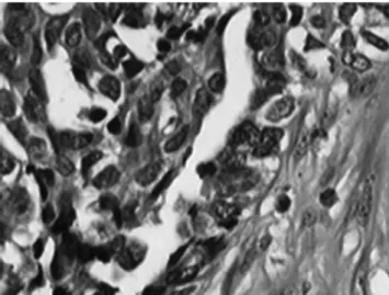
The overall incidence of port-site metastasis was 9.68% (6 of 62). The six patients with port-site metastasis exhibited subcu- taneous nodules corresponding to the port site 1–3 months af- ter surgery. CT showed a new subcutaneous mass around the abdominal port site (Fig. 2). The patients exhibiting port-site metastasis included one patient with GC whose ascites re- curred 3 months after B-ultrasound-guided HIPPC (Fig. 3). In three patients, histological analysis of a transcutaneous biopsy of the port-site mass confirmed port-site metastasis and es- tablished that the pathological type and degree of tumor dif- ferentiation was the same as that of the primary tumor (Fig. 4).
Tumor biomarker expression (carcinoembryonic antigen) of the port-site mass was shown via immunohistochemistry to be the same as that of the primary tumor (Fig. 5).
Port-site metastasis was found in three patients in each of the B-ultrasound and laparoscopy groups, and the incidence of port-site metastasis was not significantly different between the two groups (9.36% vs. 10.00%, p>0.05) (Table 2). In the laparoscopy group, the survival times of the three patients with port-site metastasis ranged from 2 to 7 months, with a median survival of 3 months. In the B-ultrasound group, the survival times of the three patients with port-site metastasis ranged from 3 to 6 months, with a median survival of 3 months. There was no significant difference between the two groups in terms of the median survival time (3 months vs. 3 months) of pa- tients with port-site metastasis (p>0.05).
Notably, the rates of FCC-positive ascites differed signifi- cantly between the various primary cancer types (PC, 100.00%;
GC, 78.95%; OC, 70.00%; CRC, 20.00%; p<0.05) (Table 2). The incidence of port-site metastasis also differed significantly be- tween the various cancer types and correlated with the rate of
FCC-positive ascites (PC, 33.33%; GC, 15.79%; OC, 10.00%;
CRC, 0.00%; p<0.001) (Table 2). As might be expected, patient prognosis for the various cancer types correlated with the in- cidence of port-site metastasis and the rate of FCC-positive ascites, with the exception of OC (Table 2).
DISCUSSION
The use of HIPPC in the treatment of malignant ascites induced by peritoneal carcinomatosis has been reported to improve Table 2. Comparison of the Incidence of Port-Site Metastasis between Different Tumor Types
Original tumor (n) FCC-positive ascites, % (n) Incidence of port-site metastasis, % (n) Median survival time (months)
PC (3) 100.00 (3/3) 33.33 (1/3) 11
GC (19) 78.95 (15/19) 15.79 (3/19) 6
OC (20) 70.00 (14/20) 10.00 (2/20) 17
CRC (20) 20.00 (4/20) 0.00 (0/20) 9
Total 58.10 (36/62) 9.68 (6/62) 8.6
p value <0.05 <0.001 <0.05
CRC, colorectal cancer; FCC, free cancer cell; GC, gastric cancer; OC, ovarian cancer; PC, pancreatic cancer.
Fig. 2. Abdominal CT scan (axial) showing a port-site metastasis (arrow) on the right abdominal wall.
Fig. 3. Abdominal CT scan (axial) in a patient with gastric cancer exhibiting port-site metastasis (arrow) and recurrence of ascites 3 months after B- ultrasound-guided hyperthermic intraperitoneal perfusion chemotherapy.
patient quality of life and prolong survival.33-35 The main find- ings of our previous study were that the clinical efficacies of B- ultrasound-guided HIPPC and laparoscopically-assisted HIP- PC, assessed from the ORR, KPS score, and overall survival, were comparable. In the present study, the incidence of port- site metastasis was found to be similar in the B-ultrasound and laparoscopy groups, although it varied depending on the primary tumor type. Taken together, these data indicate that B-ultrasound-guided HIPPC is a useful alternative to laparo- scopically-assisted HIPPC.
The incidence of port-site metastasis after diagnostic lapa- roscopy or laparoscopic resection is difficult to establish.8,9,11 Several review articles have described unacceptably high levels of more than 10%, while others have estimated the incidence to range from 3.3% to 5.3%.30,32 In the present study, the inci-
dence of port-site metastasis following B-ultrasound-guided and laparoscopically-assisted HIPPC was 9.68%, much higher than that reported previously in the literature. The high rate of FCC-positive ascites (58.10%) observed in the present study may be relevant to the high incidence of port-site metastasis.
Viable FCCs in the peritoneal cavity and their transportation to the trocar wounds are key to port-site metastasis. The high level of viable tumor cells, possibly including tumor stem cells, in the ascitic fluid of the patients in our study may have contributed to the high incidence of port-site metastasis. Sev- eral investigations have shown that a deficient supply of blood and oxygen in ascites provides favorable conditions for the growth and survival of tumor stem cells.39,40 Indeed, the pres- ence of malignant ascites during and after surgery has been reported to increase the risk of port-site metastasis.12 Another reason may be that tumor cells in the ascitic fluid flow easily into the port site during the B-ultrasound-guided or laparo- scopically-assisted placement of catheters for HIPPC. The de- livery of FCCs to the trauma site may promote their implanta- tion at the port site. Thus, one of the key factors underlying port-site metastasis may be the implantation of viable tumor cells (present in the ascitic fluid) in the trocar wounds during surgery. Allardyce, et al.14 found more tumor cells at operating ports than at assisting ports, implying that wound implanta- tion was caused by contamination of the instruments. Meta- static growth at port sites used for specimen extraction sug- gests that contamination due to direct contact with the resected specimen may be responsible.17
There is evidence that a direct influence of CO2 on tumor growth is a key factor underlying port-site metastasis after lap- aroscopic surgery. Jacobi, et al.20 showed that insufflation of CO2 promoted tumor growth in a rat model when compared to helium or controls. Using a rat model of laparoscopy, Tseng, et al.13 also found enhanced tumor growth in a CO2 insufflation group compared to a gasless group. However, Hubens, et al.21 failed to demonstrate any effect of a CO2 pneumoperitoneum on tumor cell implantation or growth. The observation that a purse-string suture around the trocar can increase the incidence of port-site metastasis suggests that leakage contributes less to tumor growth than tissue trauma induced by the purse-string suture.13 A purse-string suture may itself induce tissue trauma that enhances tumor adherence and growth.13 In our study, the incidence of port-site metastasis was not significantly different between the B-ultrasound and laparoscopy groups. Given that a CO2 pneumoperitoneum is required for the latter yet not for the former, this suggests that the chimney effect may not be a major factor underlying the occurrence of port-site metastasis.
Several researchers have proposed that the microenviron- ment of the port site could be an important factor in tumor growth and that local factors at the port-site may play an im- portant role in the mechanism of port-site metastasis.20,32 A mi- croenvironment conducive to cancer cell growth likely facili- tates port-site metastasis, and tissue trauma and local peritoneal Fig. 5. Immunohistochemical assessment of a metastatic nodule in the ab-
dominal wall (hematoxylin and eosin staining, ×100). The carcinoembryonic antigen expression of the metastatic nodule was the same as that of the primary tumor.
Fig. 4. Histopathological examination of a resected tumor (hematoxylin and eosin staining, ×400). The metastatic nodule resected from the abdominal wall was a well-differentiated adenocarcinoma; the pathological type and degree of differentiation were the same as those of the primary tumor.
factors may be as important as cell aerosolization. Local peri- toneal immunity has been recognized as having an important influence on cell anchorage and proliferation. The study of Tseng, et al.13 investigated the effects of tissue trauma and sug- gested that local peritoneal factors induced by the trocar could promote port-site metastasis. Paracentesis is an efficient tech- nique for attenuating the symptoms induced by malignant as- cites; although paracentesis tubes are much smaller than HIP- PC catheters, our clinical experience is that metastasis to sites around the puncture holes is much more common after para- centesis than after laparoscopy-assisted HIPPC (data not shown). In the current study, B-ultrasound-guided HIPPC and laparoscopy-assisted HIPPC exhibited a comparable incidence of port-site metastasis; both techniques used 1.2-cm incisions and a 1.2-cm diameter trocar. Thus, we believe that the micro- environment around the trocar sites may promote the implan- tation of tumor cells at the port sites.
Cancers differ in their malignant potential and ability to metastasize. Therefore, the incidence of port-site metastasis would also be expected to vary between different types of tu- mors. In this study, the incidence of post-HIPPC port-site me- tastasis differed between the various primary tumor types and correlated with the rate of FCC-positive ascites that was ob- served for the various cancer types. These observations indi- cate that the incidence of port-site metastasis for different tu- mor types parallels the incidence of FCC-positive ascites, suggesting that these tumors vary in their ability to metasta- size to port sites.
Our study is not without its limitations. The current study enrolled a relatively small patient cohort that may not be rep- resentative of the general patient population; additional in- vestigations in larger cohorts are thus required. Furthermore, although the presence of FCCs in ascitic fluid appeared to correlate with port-site metastasis, this association was not spe- cifically investigated (for example, with regression analysis).
Additionally, the roles of other factors, such as the microenvi- ronment at the trocar site, were not assessed in any detail.
In conclusion, CO2 leakage around the trocar (chimney ef- fect) caused by an artificial pneumoperitoneum during lapa- roscopic surgery may not be a key mechanism underlying port-site metastasis. The delivery of FCCs from the tumor or malignant ascites to the site of trauma, as well as the local mi- croenvironment at the trocar site, may promote the implanta- tion of tumor cells at port sites.
ACKNOWLEDGEMENTS
This study was supported by the Breakthroughs in Key Areas of Guangdong and Hong Kong Projects (No. 2006Z1-E6041), and by the Guangdong Provincial Science and Technological Programs (No. 2009A030301013).
REFERENCES
1. Holloway RW, Ahmad S, DeNardis SA, Peterson LB, Sultana N, Bigsby GE 4th, et al. Robotic-assisted laparoscopic hysterectomy and lymphadenectomy for endometrial cancer: analysis of surgi- cal performance. Gynecol Oncol 2009;115:447-52.
2. Young-Fadok T, Talac R, Nelson H. Laparoscopic colectomy for cancer; the need for trials. Semin Colon Rectal Surg 1999;19:94- 101.
3. Kim JW, Kim WS, Cheong JH, Hyung WJ, Choi SH, Noh SH. Safety and efficacy of fast-track surgery in laparoscopic distal gastrectomy for gastric cancer: a randomized clinical trial. World J Surg 2012;
36:2879-87.
4. Fagotti A, Vizzielli G, Costantini B, Lecca A, Gallotta V, Gagliardi ML, et al. Learning curve and pitfalls of a laparoscopic score to de- scribe peritoneal carcinosis in advanced ovarian cancer. Acta Ob- stet Gynecol Scand 2011;90:1126-31.
5. Lanowska M, Mangler M, Spek A, Grittner U, Hasenbein K, Chi- antera V, et al. Radical vaginal trachelectomy (RVT) combined with laparoscopic lymphadenectomy: prospective study of 225 pa- tients with early-stage cervical cancer. Int J Gynecol Cancer 2011;
21:1458-64.
6. Curet MJ, Putrakul K, Pitcher DE, Josloff RK, Zucker KA. Laparo- scopically assisted colon resection for colon carcinoma: periop- erative results and long-term outcome. Surg Endosc 2000;14:
1062-6.
7. Zmora O, Weiss EG. Trocar site recurrence in laparoscopic sur- gery for colorectal cancer. Myth or real concern? Surg Oncol Clin N Am 2001;10:625-38.
8. Berretta R, Rolla M, Patrelli TS, Gramellini D, Fadda GM, Nardelli GB. Incidence of port-site metastasis after laparoscopic manage- ment of borderline ovarian tumors: a series of 22 patients. Eur J Gynaecol Oncol 2009;30:300-2.
9. Zivanovic O, Sonoda Y, Diaz JP, Levine DA, Brown CL, Chi DS, et al.
The rate of port-site metastases after 2251 laparoscopic procedures in women with underlying malignant disease. Gynecol Oncol 2008;111:431-7.
10. Eng MK, Katz MH, Bernstein AJ, Shikanov S, Shalhav AL, Zorn KC.
Laparoscopic port-site metastasis in urologic surgery. J Endourol 2008;22:1581-5.
11. Kruitwagen RF, Swinkels BM, Keyser KG, Doesburg WH, Schijf CP. Incidence and effect on survival of abdominal wall metastases at trocar or puncture sites following laparoscopy or paracentesis in women with ovarian cancer. Gynecol Oncol 1996;60:233-7.
12. Agostini A, Robin F, Aggerbeck M, Jaïs JP, Blanc B, Lécuru F. Influ- ence of peritoneal factors on port-site metastases in a xenograft ovarian cancer model. BJOG 2001;108:809-12.
13. Tseng LN, Berends FJ, Wittich P, Bouvy ND, Marquet RL, Kazem- ier G, et al. Port-site metastases. Impact of local tissue trauma and gas leakage. Surg Endosc 1998;12:1377-80.
14. Allardyce RA, Morreau P, Bagshaw PF. Operative factors affecting tumor cell distribution following laparoscopic colectomy in a por- cine model. Dis Colon Rectum 1997;40:939-45.
15. Ziprin P, Ridgway PF, Peck DH, Darzi AW. The theories and realities of port-site metastases: a critical appraisal. J Am Coll Surg 2002;
195:395-408.
16. Wang PH, Yuan CC, Lin G, Ng HT, Chao HT. Risk factors contrib- uting to early occurrence of port site metastases of laparoscopic surgery for malignancy. Gynecol Oncol 1999;72:38-44.
17. Whelan RL, Lee SW. Review of investigations regarding the etiolo- gy of port site tumor recurrence. J Laparoendosc Adv Surg Tech A 1999;9:1-16.
18. Iwanaka T, Arya G, Ziegler MM. Mechanism and prevention of port-site tumor recurrence after laparoscopy in a murine model. J Pediatr Surg 1998;33:457-61.
19. Hewett PJ, Thomas WM, King G, Eaton M. Intraperitoneal cell movement during abdominal carbon dioxide insufflation and lap- aroscopy. An in vivo model. Dis Colon Rectum 1996;39(10 Suppl):
S62-6.
20. Jacobi CA, Sabat R, Böhm B, Zieren HU, Volk HD, Mûller JM.
Pneumoperitoneum with carbon dioxide stimulates growth of ma- lignant colonic cells. Surgery 1997;121:72-8.
21. Hubens G, Pauwels M, Hubens A, Vermeulen P, Van Marck E, Eyskens E. The influence of a pneumoperitoneum on the peritone- al implantation of free intraperitoneal colon cancer cells. Surg En- dosc 1996;10:809-12.
22. Canis M, Botchorishvili R, Wattiez A, Mage G, Pouly JL, Bruhat MA. Tumor growth and dissemination after laparotomy and CO2 pneumoperitoneum: a rat ovarian cancer model. Obstet Gynecol 1998;92:104-8.
23. Mathew G, Watson DI, Rofe AM, Ellis T, Jamieson GG. Adverse impact of pneumoperitoneum on intraperitoneal implantation and growth of tumour cell suspension in an experimental model.
Aust N Z J Surg 1997;67:289-92.
24. Gutt CN, Riemer V, Kim ZG, Jacobi CA, Paolucci V, Lorenz M. Im- pact of laparoscopic colonic resection on tumour growth and spread in an experimental model. Br J Surg 1999;86:1180-4.
25. Allendorf JD, Bessler M, Kayton ML, Oesterling SD, Treat MR, Nowygrod R, et al. Increased tumor establishment and growth af- ter laparotomy vs laparoscopy in a murine model. Arch Surg 1995;
130:649-53.
26. Yamaguchi K, Hirabayashi Y, Shiromizu A, Shiraishi N, Adachi Y, Kitano S. Enhancement of port site metastasis by hyaluronic acid under CO2 pneumoperitoneum in a murine model. Surg Endosc 2001;15:504-7.
27. Watson DI, Mathew G, Ellis T, Baigrie CF, Rofe AM, Jamieson GG.
Gasless laparoscopy may reduce the risk of port-site metastases following laparascopic tumor surgery. Arch Surg 1997;132:166-8.
28. Bouvy ND, Giuffrida MC, Tseng LN, Steyerberg EW, Marquet RL, Jeekel H, et al. Effects of carbon dioxide pneumoperitoneum, air pneumoperitoneum, and gasless laparoscopy on body weight and tumor growth. Arch Surg 1998;133:652-6.
29. Bouvy ND, Marquet RL, Jeekel H, Bonjer HJ. Impact of gas(less) laparoscopy and laparotomy on peritoneal tumor growth and ab- dominal wall metastases. Ann Surg 1996;224:694-700.
30. Murthy SM, Goldschmidt RA, Rao LN, Ammirati M, Buchmann T, Scanlon EF. The influence of surgical trauma on experimental me- tastasis. Cancer 1989;64:2035-44.
31. Little D, Regan M, Keane RM, Bouchier-Hayes D. Perioperative immune modulation. Surgery 1993;114:87-91.
32. Sporn MB, Roberts AB. Peptide growth factors and inflammation, tissue repair, and cancer. J Clin Invest 1986;78:329-32.
33. Facchiano E, Scaringi S, Kianmanesh R, Sabate JM, Castel B, Fla- mant Y, et al. Laparoscopic hyperthermic intraperitoneal chemo- therapy (HIPEC) for the treatment of malignant ascites secondary to unresectable peritoneal carcinomatosis from advanced gastric cancer. Eur J Surg Oncol 2008;34:154-8.
34. Garofalo A, Valle M, Garcia J, Sugarbaker PH. Laparoscopic intra- peritoneal hyperthermic chemotherapy for palliation of debilitat- ing malignant ascites. Eur J Surg Oncol 2006;32:682-5.
35. Valle M, Van der Speeten K, Garofalo A. Laparoscopic hyperther- mic intraperitoneal peroperative chemotherapy (HIPEC) in the management of refractory malignant ascites: a multi-institutional retrospective analysis in 52 patients. J Surg Oncol 2009;100:331-4.
36. Ba MC, Cui SZ, Lin SQ, Tang YQ, Wu YB, Wang B, et al. Chemo- therapy with laparoscope-assisted continuous circulatory hyper- thermic intraperitoneal perfusion for malignant ascites. World J Gastroenterol 2010;16:1901-7.
37. Ba MC, Long H, Cui SZ, Tang YQ, Wu YB, Zhang XL, et al. Multi- variate comparison of B-ultrasound guided and laparoscopic con- tinuous circulatory hyperthermic intraperitoneal perfusion che- motherapy for malignant ascites. Surg Endosc 2013;27:2735-43.
38. Cui S, Ba M, Tang Y, Liu J, Wu Y, Wang B, et al. B ultrasound-guid- ed hyperthermic intraperitoneal perfusion chemotherapy for the treatment of malignant ascites. Oncol Rep 2012;28:1325-31.
39. Liang D, Ma Y, Liu J, Trope CG, Holm R, Nesland JM, et al. The hy- poxic microenvironment upgrades stem-like properties of ovari- an cancer cells. BMC Cancer 2012;12:201.
40. Conley SJ, Gheordunescu E, Kakarala P, Newman B, Korkaya H, Heath AN, et al. Antiangiogenic agents increase breast cancer stem cells via the generation of tumor hypoxia. Proc Natl Acad Sci U S A 2012;109:2784-9.