논문접수일 :2018년 1월 22일 논문수정일 :2018년 3월 20일 심사완료일 :2018년 5월 10일
교신저자 :백무진, 48108 부산광역시 해운대구 해운대로 875 인제대학교 의과대학 해운대백병원 이비인후과학교실 전화 :(051) 797-2290・전송 :(051) 797-0298 E-mail:mjbaek@inje.ac.kr
Introduction
A blue nevus, first described by Tièche in 1906, is characterized by the proliferation of dermal dendritic melanocytes. Clinically, the nevus appears as a soli- tary, small (usually <1 cm in diameter) blue-to-blue- black macule or papule. The blue color is imparted by melanin in the dermis; melanin preferentially ab- sorbs long-wavelength light (the Tyndall effect). A nevus is usually solitary and is found on the head, neck, sacral region, and dorsal aspects of the distal extrem- ities. A nevus can develop at any age, but usually in the second decade of life.1) Rarely, blue nevi have been reported at extracutaneous sites, including the vagina;
the prostate; and the oral, sinonasal, and colorectal mu-
cosae.2-6) A blue nevus in the middle ear is extremely rare ; only two cases have been reported.7) We here describe the third case.
Case Report
A 38-year-old female presented to our outpatient clinic with intermittent dizziness 2 weeks in duration.
On otoendoscopic examination, we found a blue- black mass in the left middle ear, evident through an intact tympanic membrane (Fig. 1). Pure tone audi- ometry revealed a normal hearing level. A vestibular function test, and cranial nerve and nasopharyngo- scopic examinations, revealed no abnormality. Com- puted tomography (CT) identified a mass, 5 mm in diameter, in the medial aspect of the left mesotympa- num. No bony erosion was evident, and the jugular bulb, ossicles, and facial nerve were intact (Fig. 2).
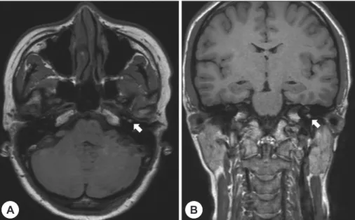
Magnetic resonance imaging (MRI) revealed a small, nodular soft tissue lesion of high T1 signal intensity but low T2 signal intensity in the medial aspect of the left mesotympanum (Fig. 3).
Although we proposed surgical exploration, the pa-
A Case of Benign Blue Nevus Located in Middle Ear Mucosa
Jae-mahn Cho, MD and Moo-jin Baek, MD, PhD
Department of Otolaryngology-Head and Neck Surgery, Inje University College of Medicine, Haeundae Paik Hospital, Busan, Korea
- ABSTRACT -
A blue nevus, characterized by the proliferation of dermal dendritic melanocytes, usually appears as a solitary, small, asymptomatic blue-to-blue-black macule or papule on the dorsal aspect of the extremities, scalp, or but- tocks. Blue nevi have also been reported in several extracutaneous sites, including the vagina; prostate; and in- traoral, sinonasal, and colorectal mucosae. A 38-year-old female presenting with intermittent dizziness had a blue-black mass in the left middle ear, behind an intact tympanic membrane; the mass was evident on otoendo- scopic examination. The mass was surgically removed and identified histologically as a benign blue nevus. To date, two cases of blue nevi in the middle ear have been reported. Here, we describe another such rare case. (J Clinical Otolaryngol 2018;29:82-86)
KEY WORDS:Blue nevusㆍMiddle earㆍMelanocytes.
tient insisted on observation only because of sponta- neous symptom relief. After 4 months of loss to fol- low-up, the symptom recurred and the patient agreed to undergo surgery. She underwent diagnostic explor- atory tympanotomy. Under general anesthesia, we exposed the middle ear via an endaural approach us- ing Lempert I and II incisions. A dark-blue mass was evident anterior to the malleus handle. Otherwise, the middle ear was clear and the ossicular chain intact and freely mobile. The mass was grossly removed. A small blue lesion was evident around the opening of the Eustachian tube, but was not removable because of adhesion to the adjacent mucosa. The resected tis- sue was sent to the Department of Pathology for evaluation.
The specimen consisted of a fragment of brown- ish-gray soft tissue 0.4×0.3×0.3 cm in dimension (Fig. 4). A microscopic examination of histological sections revealed moderately sized spindle-to-epithe- lioid-shaped melanocytes with high levels of mela- nin. The melanocytes contained abundant cytoplasm but did not exhibit cytologic atypia or mitosis. We stained the tissue with HMB45 combined with AP- Red to rule out a melanoma. The histopathological data confirmed that the tissue was a benign blue ne- vus (Fig. 5).
Four months of follow-up have now elapsed with-
out any sign of recurrence.
Discussion
Melanocytes are found throughout the mucosal membranes of the head and neck. Embryologically, the middle ear mucosa originates from pharyngeal pouches that also develop into the oral and nasopha- ryngeal mucosae.8) A blue nevus is characterized by the proliferation of dermal dendritic melanocytes.
Generally, benign blue nevi are associated with a good prognosis because they tend to remain unchanged throughout life or flatten and fade in color. However, rarely, the nevi exhibit malignant changes, a sudden increase in size, or ulceration.9,10) Several blue nevi have been reported in the oral and nasopharyngeal
Fig. 1. Endoscopic finding of a blue-black color mass in the left middle ear.
Fig. 2. Preoperative temporal bone CT identifying a 5mm-sized small nodular soft tissue density lesion at me- dial aspect of the left mesotympanum in (A) axial view and (B) coronal view (yellow arrows).
A
B
mucosae. Only one blue nevus confined to the mid- dle ear has been reported and, recently, a blue nevus extending from the middle to the inner ear and into the nasopharynx through the Eustachian tube was described.11)
During the differential diagnosis of a melanocytic blue lesion in the middle ear, a melanoma, dermato- fibroma, neurofibroma, perineuroma, and other fi- broblastic proliferations, including scarring, must be considered.12) It is essential to exclude a malignant melanoma because such melanomas, reflecting the neoplastic proliferation of melanocytes in the epider-
mis, retina, and mucosa, are associated with a 5-year survival rate of only 31.7%, as revealed by a review of 84,836 melanomas of the head-and-neck mucosae from the American Cancer Database.13)
In our case, there were no clinical symptoms or signs suggestive of a malignant tumor in the middle ear.
Cases with malignant melanoma present with pain, otorrhea, or otitis media with effusion.14) On preoper- ative CT and MRI, the mass showed no evidence of invasion into surrounding bones. Similarly, no gross evidence of malignancy was apparent. A histological examination revealed the proliferation of spindled melanocytes without any evidence of cellular atypia, mitotic activity, or necrosis. A histopathological ex- amination confirmed that the tissue was a benign blue nevus.
Long-term follow-up data are lacking for both of the two prior cases; neither recurrence nor a malig- nant change was addressed. As the melanocytes of the middle ear and sinonasal tract are presumed to have the same origin, we reviewed the literature on solitary blue nevi of the sinonasal tract. In the five re- ported cases, no recurrence after complete excision was evident during 13 months to 20 years of follow-
Fig. 4. Blue black soft tissue specimen measuring 0.4×
0.3×0.3 cm.
Fig. 3. Preoperative T1 weighted MRI images of small nodular mass at medial aspect of mesotympanum in (A) axial view and (B) coronal view (white arrows).
A B
up.3,15,16) Although blue nevi rarely become malignant, or transform into malignant melanomas, complete surgical excision with regular follow-up is required because the number of reported cases is small and blue nevi of the middle ear remain poorly characterized.
A blue nevus in the middle ear is rare; only two cas- es have been reported. We found no report from our country; indeed, no blue nevus in the head-and-neck region has been described. In the previous cases, the patients presented with hearing loss, with or without a bluish mass behind the tympanic membrane, and underwent complete excision if possible.12) A blue ne- vus per se is benign, but a malignant blue nevus and malignant melanoma must be considered during dif- ferential diagnosis; no established guidelines for the
management of symptomatic disease have appeared.
Additional studies on the prognosis of middle ear blue nevi are required.
REFERENCES
1) Phadke PA, Zembowicz A. Blue nevi and related tumors.
Clinics in Laboratory Medicine 2011;31(2):345-58.
2) Anderco D, Lazar E, Taban S, Miclea F, Dema A. Prostat- ic blue nevus. Romanian Journal of Morphology and Em- bryology = Revue Roumaine de Morphologie et Embry- ologie 2010;51(3):555-7.
3) Chuang WY, Hao SP, Yeh CJ, Jung SM. Blue nevi of the sinonasal mucosa: a report of two cases and review of the literature. The Laryngoscope 2007;117(2):371-2.
4) Fitzhugh VA, Houck K, Heller DS. Vaginal blue nevus: re- port of a case and review of the literature. Journal of Low- er Genital Tract Disease 2011;15(4):325-7.
5) Schreiber ZJ, Pal TR, Hwang SJ. Blue nevus of the colorec- Fig. 5. Pathologic finding (A) heavily pigmented 0.4cm sized dome shaped nodule, (B) proliferation of melanocytes and melanophages, (C) with melanin bleaching, diffuse proliferation of spindled melanocytes with abundant cyto- plasm but no cytologic atypia or mitoses are present and (D) positive for melanocytes. (A), (B), (C) and (D) are pathologic findings in HE*40, HE*100, HE*400 and HMB45*400 with AP red dye, respectively.
A
C
B
D
tal mucosa. Annals of Diagnostic Pathology 2011;15(2):
128-30.
6) Shumway BS, Rawal YB, Allen CM, Kalmar JR, Magro CM. Oral atypical cellular blue nevus: an infiltrative me- lanocytic proliferation. Head and Neck Pathology 2013;
7(2):171-7.
7) Babin RW, Benjamin SP. Pigmented lesion of the middle ear: report of a case. The Annals of Otology, Rhinology, and Laryngology 1976;85(3 pt 1):387-90.
8) Lin CS, Zak FG. Studies on melanocytes. VI. Melanocytes in the middle ear. Archives of Otolaryngology (Chicago, Ill : 1960) 1982;108(8):489-90.
9) Katz B, Rao B, Marghoob A. Blue nevus/combined nevus.
An Atlas of Dermoscopy 2004.
10) Ryu JY, Moon JH, Lee DH, Bang JH. Two cases of intra- dermal nevus in external auditory canal. J Clinical Otolar- yngol 2005;16:289-92.
11) Saito C, Ishikawa K, Nakamura KI, Fujita A, Shimizu M, Fukushima N, et al. A melanocytic lesion extending from
the right ear to the nasopharynx in a pediatric patient: a case report. The Annals of Otology, Rhinology, and Lar- yngology 2015;124(8):609-13.
12) Zembowicz A, Phadke PA. Blue nevi and variants: an up- date. Archives of Pathology & Laboratory Medicine 2011;
135(3):327-36.
13) Chang AE, Karnell LH, Menck HR. The National Cancer Database report on cutaneous and noncutaneous melano- ma: a summary of 84,836 cases from the past decade. The American College of Surgeons Commission on Cancer and the American Cancer Society. Cancer 1998;83(8):1664-78.
14) Uchida M, Matsunami T. Malignant amelanotic melano- ma of the middle ear. Archives of Otolaryngology--Head
& Neck Surgery 2001;127(9):1126-8.
15) Lim LM, Tan KB, Petersson F, Thong M. Sinonasal blue naevus: case report and clinicopathological review. The Journal of Laryngology and Otology 2013;127(9):939-41.
16) Piana S, Asioli S. Blue nevus of the nasal mucosa. Virchows Arch 2005;446 (3):342-3.