Totally Laparoscopic Total Gastrectomy Using Intracorporeally Hand-Sewn Esophagojejunostomy
Kwang Oh So, and Jong-Min Park
Department of Surgery, National Medical Center, Seoul, Korea
Purpose: Laparoscopic total gastrectomy (LTG) for gastric cancer is still uncommon because of technical difficulties, especially in esoph- agojejunostomy (EJ). There are many reports for various laparoscopic procedures of EJ using linear or circular staplers. On the other hands, there has been no report for hand-sewn anastomosis. We report successfully performed intracorporeally hand-sewn EJ after LTG.
Materials and Methods: The clinicopathologic data and short-term surgical outcomes of 6 patients who underwent totally laparoscopic total gastrectomy for upper gastric cancer from December 2010 and July 2011 were retrospectively reviewed.
Results: The mean age was 66.5 years and mean body mass index (kg/m2) was 24.6. All patients had medical comorbidities. The mean patient ASA score was 2.17. Among the 6 patients, previous abdominal operation was performed for 2 patients and combined operation was performed for 3 patients. The mean blood loss, operation time, and EJ anastomosis time was 130 ml, 379.7 minutes, and 81.5 minutes, respectively. The mean time to first flatus, first oral intake, and postoperative hospital stay was 3.0, 3.0, and 12.5 days, respectively. There was no 30-day mortality case. Postoperative aspiration pneumonia and multiple periventricular lacunar infarc- tions developed in 1 patient. There were no anastomosis-related complications and other major surgical complications.
Conclusions: When the intracorporeal anastomotic technique becomes popular in LTG the intracorporeally hand-sewn EJ may be ac- cepted as one method among the various laparoscopic procedures of EJ.
Key Words: Stomach neoplasms; Laparoscopy; Gastrectomy; Reconstructive surgical procedures
Correspondence to: Jong-Min Park
Department of Surgery, National Medical Center, Euljiro 6-ga, Jung-gu, Seoul 100-799, Korea
Tel: +82-2-2260-4883, Fax: +82-2-2269-0750 E-mail: jmparkgs@gmail.com
Received September 30, 2011 Revised November 2, 2011 Accepted November 3, 2011
Copyrights © 2011 by The Korean Gastric Cancer Association www.jgc-online.org
This is an open-access article distributed under the terms of the Creative Commons Attribution Non-Commercial License (http://creativecommons.org/
licenses/by-nc/3.0) which permits unrestricted noncommercial use, distribution, and reproduction in any medium, provided the original work is properly cited.
Introduction
In recent years, laparoscopic distal gastrectomy (LDG) in the treatment for gastric cancer has been widely accepted as safe and feasible method. LDG has many advantages such as rapid recovery of gastrointestinal function, short hospital stay, lesser pain, bet- ter cosmesis, and acceptable oncologic outcomes.(1-5) However, laparoscopic total gastrectomy (LTG) has not been accepted wide- spreadly as LDG because of low incidence of gastric cancer requir-
ing LTG and its technical difficulties, especially the esophagojeju- nostomy (EJ) after total gastrectomy.(6,7)
In conventional open total gastrectomy with Roux-en-Y EJ, the standard reconstruction method was circular stapled EJ, it was most commonly performed by end-to-side fashion. Some surgeons used an extracorporeal approach using circular stapler through a minilaparotomy after LTG, which is similar to conventional open surgery.(8-12) However, in this technique, it is sometimes difficult to perform purse-string suture and anvil insertion through the minilaparotomy, especially for patients with obesity, large antero- posterior diameter, and acute subcostal angle. It may be required extension of the incision and could be associated with increasing pain and anastomotic ischemia due to extensive traction. Some alternative methods for intracorporeal EJ after LTG to overcome these technical problems have been reported.(13-20) However, unfortunately, these procedures still appear to be complicated and
have not been accepted widely.
Intracorporeal hand-sewn EJ using a needleholder after LTG is not a novel technique. If this technique could be performed by sur- geons familiar with intracorporeal hand-sewn suturing, it would be very simple and classic as the conventional open total gastrectomy.
Nevertheless, it is a more time-consuming and difficult procedure than other alternative methods for intracorporeal EJ using stapler after LTG. This study was our preliminary experience of intracor- poreal hand-sewn EJ after LTG and aimed to assess its technical feasibility.
Materials and Methods
1. Patients
We retrospectively reviewed 6 consecutive patients who under- went totally laparoscopic total gastrectomy (TLTG) using intracor- poreal hand-sewn EJ at our institution between December 2010 and July 2011. Written informed consent was obtained from every patient who agreed to undergo TLTG. Preoperative staging of all patients was based on endoscopic biopsy and abdominal computed tomography. At our institution, the indication for TLTG in gastric cancer is limited to preoperative stage T1~3N0~2. Patients with an ASA score 4 or more points, serosal invasion in laparoscopic view, extensive esophageal invasion, extensive lymph node metastasis, and patients suitable for endoscopic submucosal dissection were ex- cluded. The indication for endoscopic submucosal dissection was a well differentiated mucosal lesion smaller than 2 cm without ulcer- ation. Disease stage was classified according to the 7th UICC TNM classification. The characteristics of patients, such as age, gender, body mass index (kg/m2), history of abdominal surgery, ASA score, co-morbidities, and surgical outcomes (operative methods, com- bined operation, operative time, EJ anastomosis time, number of the used linear stapler, blood loss, complications, pathologic results, time-to-first flatus, time-to-first oral intake, and postoperative hospitalization period) were examined.
2. Surgical techniques
Patients were placed in the supine position, and the operator stood on the right side of the patient. The scopist was positioned on the right side of the operator and the first assistant on the left side of the patient. After general anesthesia, a 10 mm trocar was inserted at the sub-umbilical area, and a pneumoperitoneum was formed by insufflation of carbon dioxide. The patient was placed in reverse Trendelenburg position and four or five additional tro- cars were inserted. The intraperitoneal pressure was maintained as
11~13 mmHg. A 12 mm trocar was inserted on the right side of the umbilicus, and lateral to the rectus abdominis muscle. A 5 mm trocar was inserted in the right subcostal area on the lateral side of the rectus abdominis muscle for use by the surgeon. A 10 mm tro- car was inserted at the left subcostal area on the lateral side of the rectus abdominis muscle. A 5 mm trocar was inserted on the left side of the umbilicus and the lateral side of the rectus abdominis muscle for use by the assistant in retracting organs. A 10 mm trocar was inserted at the subxiphoid process area and used for insertion of a fan-shaped liver retractor and it was sometimes omitted (Fig. 1).
The EXERAⓇ laparo-thoraco videoscope (Olympus, Tokyo, Japan) was used. For dissection of tissues, an ultrasonic cautery Harmonic scalpelⓇ (Ethicon Endo Surgery, Cincinnati, OH, USA) was used.
Total gastrectomy with complete omentectomy and extended lymphadenectomy (D1+β or D2) was performed laparoscopically in all patients. After full mobilization of the abdominal esopha- gus, Two detachable Endo intestinal clipⓇ (Aesculap, Tuttlingen, Germany) was placed at the esophagus to avoid contamination and withdrawal of the transected esophagus into the mediasti- num after subsequent transection. The esophagus was transected perpendicularly with laparoscopic metzenbaum scissors between the two detachable Endo intestinal clipⓇs. And then we obtained proximal resection margin using laparoscopic metzenbaum scissors, frozen biopsy was performed. After we confirmed negative resec- tion margin, and then following EJ procedure was proceeded. The Jejunum was transected 20 cm distal to the ligament of Treitz using 45 mm linear stapler ECR45ⓇB (blue cartridge, Ethicon Endo Sur- gery, Cincinnati, OH, USA). The transected distal Roux limb was positioned gently in an antecolic or retrocolic fashion. A 2.5 cm
Fig. 1. Port placement and minilaparotomy for specimen removal.
sized enterotomy was made 5 cm distal to the stapler line on the antimesenteric side of the Roux limb.
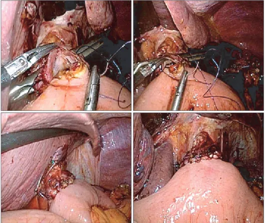
A braided absorbable suture was used for all suturing. Most of the suture could be done through the right sided trocar and sometimes left sided trocar was used due to the limited degree of freedom. At first, the suture was started from the left side of the esophageal stump and enterotomy of the Roux limb. After an- choring suture was performed at left end of anastomosis and then posterior wall of the EJ was closed by intracorporeally hand-sewn suture from left to right end of anastomosis with continuously run- ning suture, full-thickness stitch. After posterior wall closure was completed, a new thread was used for anterior wall closure. Ante- rior wall closure was also performed in the same way. Two threads were intracorporeally tied at the right end of the EJ anastomosis.
Reinforcement sutures were interruptedly performed 14 to 18 times at whole circumference after removing detachable Endo intestinal clipⓇ. Another two stitches were done to prevent the EJ anasto- mosis falling between the diaphragm and the anterior wall of Roux limb. Finally, intracorporeally hand-sewn EJ anastomosis was completely established (Fig. 2).
At 40 cm distal from EJ anastomosis site, side-to-side jejunoje- junostomy was performed using an ECR45ⓇB, and the common entry holes were also closed intracorporeally by using an ECR45ⓇB. Rein-
forcement sutures were also interruptedly performed at the stapler lines.
After abdominal cavity was checked and irrigation was done, then two closed suction drainage tubes were inserted through a port site in right subhepatic space and left subphrenic space, For speci- men removal, a 4 to 5 cm transverse or vertical minilaparotomy was made in intraumbilical port. The minilaparotomy was retracted and protected by ALEXISⓇ wound retractor (2.5~6 cm; Applied Medical, Rancho Santa Margarita, CA, USA), then the specimen was retrieved. The operation was finished by closing the skin of the trocar wounds.
3. Postoperative management
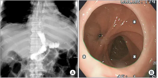
The preoperatively inserted nasogastric tube for air decompres- sion was removed at the end of operation. Gastrograffin study was performed to evaluate leakage and sips of water was started on postoperative day 3 (Fig. 3). Without further complications, patients were discharged on postoperative day 7 to 10 after tolerance of a soft diet for 2 more days.
Results
In our study, TLTG was underwent by one surgeon who ex-
Fig. 2. Laparoscopic view for intracor- poreally hand-sewn esophagojejunos- tomy.
perienced about 50 cases of laparoscopically assisted gastrectomy for gastric cancer. All patients were successfully performed without intraoperative complications or conversion to open surgery. Patient characteristics are summarized in Table 1. Mean follow up duration was 4.7 months (range, 2~10 months).
All patients were performed complete omentectomy and in- tracorporeally hand-sewn Roux-en-Y EJ. Antecolic type was performed in 3 patients and retrocolic type was performed in 3 patients. Combined operation was performed in 3 patients. The combined procedures were adhesiolysis for a patient with chronic pancreatitis; wedge biopsy of the liver for a patient with eosino- philic liver abscess; and lateral sectionectomy of the liver for pa- tient with extrahepatic cholangiectasis. The extent of lymph node dissection was D1+β in 5 patients and D2 in 1 patient. The mean size of minilaparotomy was 4.7 cm (range, 4~5 cm). The mean operating time was 376.7 min (range, 310~430 min) and the mean EJ anastomosis time was 81.5 min (range, 62~95 min). The mean
estimated blood loss was 130 ml (range, 80~200 ml). The mean number of used linear stapler was 4.2 (range, 4~5). The mean time to first flatus, time to first oral intake, and postoperative hospital stay were 3 days, 3 days, and 12.5 days, respectively. The mean postoperative hospital stay of 5 patients without complication was 9 days (range, 7~10 days).
On the pathologic examination, the location of the tumor was the upper body in 5 patients and the midbody in 1 patient. Esopha- geal invasion was detected in 3 patients and double lesions were detected in 1 patient who had a midbody cancer. The mean tumor size was 3.95 cm (range, 1.2~6 cm) and the mean proximal resec- tion margin was 1.92 cm (range, 1.5~4.0 cm). All resected speci- mens had tumor-free resection margins. The depth of invasion was submucosa in 3 patients, proper muscle in 1 patient, and serosa in 2 patients. In 2 patients with serosal invasion, the serosal invasion was not detected in the operative field, but diagnosed as focal serosal invasion based on pathologic evaluation. The histologic type was Fig. 3. Postoperative contrast study and esophagogastroduodenoscopy of the first patient. (A) Constrast study was performed on postoperative day 3.
It shows no anastomotic leakage. (B) It is endoscopic finding of the esoph- agojejunostomy site on postoperative 6 months. *It shows no anastomotic stenosis.
Table 1. Characteristics of patients
Case Age Gender BMI (kg/m2) Comorbidity ASA score Previous abdominal operation
1 78 Male 26.7 HTN, CHF, old CVA 3 Appendectomy
2 68 Male 23.2 HTN, DM, old CVA 3 No
3 69 Male 27.1 HTN 2 No
4 71 Male 26.4 Chronic pancreatitis 1 Laparoscopic cholecystectomy
5 66 Male 19.6 HTN,
atrophy of lateral segment, old CVA
2 No
6 47 Male 24.2 HBV hepatitis,
mediastinal abscess
2 No
Mean 66.5 - 24.6 - 2.2 -
BMI = body mass index; ASA = American Society of Anesthesiologists; HTN = hypertension; CHF = congestive heart failure; CVA = cerebrovascular attack; DM = diabetes mellitus; HBV = hepatitis B virus.
poorly differentiated carcinoma in 5 patients and signet ring cell carcinoma in 1 patient. The mean number of retrieved lymph nodes was 32.2 (range, 14~47). Lymph node metastasis was detected in 3 patients. The final pathologic staging according to the 7th UICC TNM staging system was stage IA in 3 patients, stage IIB in 1 pa- tient, stage IIIA in 1 patient, and stage IIIB in 1 patient. Adjuvant chemotherapy has been administered in 3 patients over stage II.
In the first patient, comorbidities were hypertension, well- controlled congestive heart failure, old cerebral infarction, and gall- bladder stone. The patient had suffered from medical complications such as pneumonia and multiple periventricular lacunar infarctions on postoperative day 9. The patient transferred to neurologic de- partment for anticoagulation therapy and discharged on postopera- tive day 30 without sequelae. In all patients, there were no mortali- ties and anastomosis-related complications, such as stenosis, minor and major leakge.
Discussion
The optimal method for EJ after LTG has not been established yet. Roux-en-Y EJ by extracorporeal anastomosis using circular stapler through a minilaparotomy is the most commonly reported because this method is simple and similar to conventional open surgery.(11,12) However, this method sometimes encountered dif- ficulties to perform purse-string suture and anvil insertion through the minilaparotomy, especially in patients with obesity, large an- teroposterior diameter, and narrow subcostal angle. Therefore, some alternative methods for intracorporeal EJ techniques after LTG to overcome these problems were reported. Usui et al.(17) introduced LTG using endoscopic purse-string suture instrument (Endo PSI, Hope Electronics, Chiba, Japan) and circular stapler. Jeong and Park (18) and Kunisaki et al.(19) introduced intracorporeal circu- lar stapling EJ using transorally inserted anvil (OrVilTM, Coviden, Mansfield, MA, USA). These devices are all excellent but have not become widely available. In transorally inserted anvil method, it has the risk of injury from the pharyx to the esophagus by passage of the anvil and has not gained favor as a common procedure. Also, Kinoshita et al.(20) reported intracorporeal circular-stapled EJ using hand-sewn purse-string suture after LTG. However, these circular-stapled methods are sometimes cumbersome and have some demerits such as reestablishment of pneumoperitoneum and placement of minilaparotomy in supraumbilical area for insertion of the circular-stapled instruments. Several groups reported vari- ous intracorporeal linear stapling EJ methods to overcome demerits
of circular-stapled method, such as side-to-side, functional end- to-end, and end-to-side.(13-16) Nevertherless, other problems appeared in these techniques. Extensive mobilization of the distal esophagus and the Roux limb is required to reduce the tension at the anastomosis site. Withdrawal of the transected esophagus into mediastinum can be developed and the closure of the common en- try hole is occansionally difficult because of the limited hiatal field.
Our presented technique has some merits. It is very simple, safe, and cost-efffective. Furthermore, it requires no excessive mobiliza- tion of the esophagus and Roux limb. Also, it has the least tension and tissue injury of anastomosis site than other stapled methods because there is no use of inserting circular or linear stapler as conventional open surgey. Another merit in our technique can be the lowest location of the minilaparotomy that is created after all procedures have been completed. Consequently, it can contribute to minimize the wound pain. Hur et al.(21) previously reported the excellent outcomes and the technical feasibility of robot-sewn anastomosis. Our technique is more applicable to robot system since it is very clear that robot system tends to have higher degree of the freedom in the limited field. However, the robot system is highly expensive, hard to own, and difficult to maintain in small institutes.
Intracorporeal hand-sewn EJ using a needleholder after LTG is not a novel technique. In our study, this technique shows acceptable surgical outcomes in terms of anastomosis-related complication and early postoperative course. However, the number of patients was very small in this study. It will be necessary to confirm these results in a large number of patients to show the validity of this procedure. If this technique could be performed by surgeons fa- miliar with intracorporeal hand-sewn suturing, it would be very simple as the conventional open total gastrectomy. Nevertheless, it is a more time-consuming and difficult procedure than other al- ternative methods for intracorporeal EJ using stapler after LTG due to the limited field. When the intracorporeal anastomotic technique becomes popular in LTG, the intracorporeally hand-sewn EJ may be accepted as one method among the various laparoscopic proce- dures of EJ. It can be performed safely by experienced laparoscopic surgeons.
References
1. Azagra JS, Goergen M, De Simone P, Ibanez-Aguirre J. The current role of laparoscopic surgery in the treatment of benign gastroduodenal diseases. Hepatogastroenterology
1999;46:1522-1526.
2. Adachi Y, Suematsu T, Shiraishi N, Katsuta T, Morimoto A, Kitano S, et al. Quality of life aft er laparoscopy-assisted Billroth I gastrectomy. Ann Surg 1999;229:49-54.
3. Kim MC, Kim KH, Kim HH, Jung GJ. Comparison of lapa- roscopy-assisted by conventional open distal gastrectomy and extraperigastric lymph node dissection in early gastric cancer. J Surg Oncol 2005;91:90-94.
4. Kitano S, Shiraishi N, Uyama I, Sugihara K, Tanigawa N; Japa- nese Laparoscopic Surgery Study Group. A multicenter study on oncologic outcome of laparoscopic gastrectomy for early cancer in Japan. Ann Surg 2007;245:68-72.
5. Huscher CG, Mingoli A, Sgarzini G, Sansonetti A, Di Paola M, Recher A, et al. Laparoscopic versus open subtotal gastrectomy for distal gastric cancer: fi ve-year results of a randomized pro- spective trial. Ann Surg 2005;241:232-237.
6. Huscher CG, Mingoli A, Sgarzini G, Brachini G, Binda B, Di Paola M, et al. Totally laparoscopic total and subtotal gastrec- tomy with extended lymph node dissection for early and ad- vanced gastric cancer: early and long-term results of a 100-pa- tient series. Am J Surg 2007;194:839-844.
7. Tanimura S, Higashino M, Fukunaga Y, Kishida S, Ogata A, Fujiwara Y, et al. Laparoscopic gastrectomy with regional lymph node dissection for upper gastric cancer. Br J Surg 2007;94:204-207.
8. Kim SG, Lee YJ, Ha WS, Jung EJ, Ju YT, Jeong CY, et al. LATG with extracorporeal esophagojejunostomy: is this minimal invasive surgery for gastric cancer? J Laparoendosc Adv Surg Tech A 2008;18:572-578.
9. Usui S, Yoshida T, Ito K, Hiranuma S, Kudo SE, Iwai T. Lap- aroscopy-assisted total gastrectomy for early gastric cancer:
comparison with conventional open total gastrectomy. Surg Laparosc Endosc Percutan Tech 2005;15:309-314.
10. Mochiki E, Toyomasu Y, Ogata K, Andoh H, Ohno T, Aihara R, et al. Laparoscopically assisted total gastrectomy with lymph node dissection for upper and middle gastric cancer. Surg En- dosc 2008;22:1997-2002.
11. Okabe H, Satoh S, Inoue H, Kondo M, Kawamura J, Nomura A, et al. Esophagojejunostomy through minilaparotomy aft er laparoscopic total gastrectomy. Gastric Cancer 2007;10:176- 180.
12. Cheong O, Kim BS, Yook JH, Oh ST, Lim JT, Kim KJ, et al.
Laparoscopic assisted total gastrectomy (LATG) with extra- corporeal anastomosis and using circular Stapler for middle or upper early gastric carcinoma: reviews of single Surgeon’s experience of 48 consecutive patients. J Korean Gastric Cancer Assoc 2008;8:27-34.
13. Uyama I, Sugioka A, Fujita J, Komori Y, Matsui H, Hasumi A.
Laparoscopic total gastrectomy with distal pancreatosplenec- tomy and D2 lymphadenectomy for advanced gastric cancer.
Gastric Cancer 1999;2:230-234.
14. Inaba K, Satoh S, Ishida Y, Taniguchi K, Isogaki J, Kanaya S, et al. Overlap method: novel intracorporeal esophagojeju- nostomy aft er laparoscopic total gastrectomy. J Am Coll Surg 2010;211:e25-e29.
15. Okabe H, Obama K, Tanaka E, Nomura A, Kawamura J, Na- gayama S, et al. Intracorporeal esophagojejunal anastomosis after laparoscopic total gastrectomy for patients with gastric cancer. Surg Endosc 2009;23:2167-2171.
16. Bracale U, Marzano E, Nastro P, Barone M, Cuccurullo D, Cu- tini G, et al. Side-to-side esophagojejunostomy during totally laparoscopic total gastrectomy for malignant disease: a multi- center study. Surg Endosc 2010;24:2475-2479.
17. Usui S, Nagai K, Hiranuma S, Takiguchi N, Matsumoto A, Sanada K. Laparoscopy-assisted esophagoenteral anastomosis using endoscopic purse-string suture instrument “Endo-PSI (II)” and circular stapler. Gastric Cancer 2008;11:233-237.
18. Jeong O, Park YK. Intracorporeal circular stapling esophago- jejunostomy using the transorally inserted anvil (OrVil) aft er laparoscopic total gastrectomy. Surg Endosc 2009;23:2624- 2630.
19. Kunisaki C, Makino H, Oshima T, Fujii S, Kimura J, Takagawa R, et al. Application of the transorally inserted anvil (OrVil) after laparoscopy-assisted total gastrectomy. Surg Endosc 2011;25:1300-1305.
20. Kinoshita T, Oshiro T, Ito K, Shibasaki H, Okazumi S, Katoh R. Intracorporeal circular-stapled esophagojejunostomy using hand-sewn purse-string suture aft er laparoscopic total gastrec- tomy. Surg Endosc 2010;24:2908-2912.
21. Hur H, Kim JY, Cho YK, Han SU. Technical feasibility of robot-sewn anastomosis in robotic surgery for gastric cancer. J Laparoendosc Adv Surg Tech A 2010;20:693-697.