687 대한안과학회지 2018년 제 59 권 제 7 호
J Korean Ophthalmol Soc 2018;59(7):687-690 ISSN 0378-6471 (Print)⋅ISSN 2092-9374 (Online)
https://doi.org/10.3341/jkos.2018.59.7.687
Case Report
Nail-patella 증후군에 동반된 선천녹내장 1예
A Case of Congenital Glaucoma in Associated with Nail-patella Syndrome
이수민⋅한종철⋅기창원
Soomin Lee, MD, Jong Chul Han, MD, Chang Won Kee, MD, PhD
성균관대학교 의과대학 삼성서울병원 안과학교실
Department of Ophthalmology, Samsung Medical Center, Sungkyunkwan University School of Medicine, Seoul, Korea
Purpose: To report a case of congenital glaucoma associated with nail-patella syndrome.
Case summary: A 20-day-old female was referred to our clinic for bilateral intraocular pressure (IOP) elevation and treatment of corneal opacities. Her IOP was 25 mmHg and 30 mmHg in the right and left eyes, respectively. After a diagnosis of congenital glaucoma, bilateral trabeculotomy was performed under general anesthesia. On the first postoperative day, the IOP was 12 mmHg in the right eye and 10 mmHg in the left eye, and remained stable thereafter. The infant was the second of fraternal twins (birth weight of 2.42 kg) and had no family history of any particular disease. During the regular checkup, she was referred to an orthopedic clinic for disorders of the elbow and knee. She presented with a dystrophic thumbnail, patella hypoplasia, elbow hy- poplasia, and bilateral triangular protrusions of the lateral iliac crest (iliac horn). Based on the above findings, typical nail-patella syndrome was diagnosed and a mutation in the LMX1B gene was detected.
Conclusions: If glaucoma patients have nail deformities or musculoskeletal abnormalities, nail-patella syndrome should be sus- pected and a multidisciplinary approach should be conducted.
J Korean Ophthalmol Soc 2018;59(7):687-690
Keywords: Congenital glaucoma, Nail dysplasia, Nail-patella syndrome
■Received: 2017. 9. 7. ■ Revised: 2017. 10. 15.
■Accepted: 2017. 11. 24.
■Address reprint requests to Chang Won Kee, MD, PhD Department of Ophthalmology, Samsung Medical Center, #81 Irwon-ro, Gangnam-gu, Seoul 06351, Korea
Tel: 82-2-3410-3548, Fax: 82-2-3410-0074 E-mail: [email protected]
* Conflicts of Interest: The authors have no conflicts to disclose.
ⓒ2018 The Korean Ophthalmological Society
This is an Open Access article distributed under the terms of the Creative Commons Attribution Non-Commercial License (http://creativecommons.org/licenses/by-nc/3.0/) which permits unrestricted non-commercial use, distribution, and reproduction in any medium, provided the original work is properly cited.
Nail-patella syndrome은 상염색체 우성으로 유전되는 매 우 드문 질환으로 약 5만 명 중 1명에서 발생하는 것으로 알려져 있다.1 Hereditary onycho-osteodysplasia, Turner-Keiser syndrome, Fong disease라고도 알려진 이 질환은 손톱 이형 성 및 무릎과 팔꿈치 이형성을 나타내는 질환으로 1820년 Chatelain에 의해 처음 보고되었다.2 1883년과 1897년 Pye- Smith와 Little이 nail-patella syndrome의 유전에 대해 보고하
였고,3 1946년 Fong은 iliac horn과 관련이 있음을 보고하였 다.3,4 이후 다양한 임상 양상을 가진 환자들이 보고되었고 현재는 손톱 이형성(nail dysplasia), 슬개골의 이형성 혹은 무형성(patella and elbow dysplasia or hypoplasia), 주관절 형 성 이상 혹은 탈구(elbow dysplasia or dislocation), 장골 돌기 (iliac horn)를 기준으로 임상적 진단을 내리고 있고, 신장과 눈에 영향을 주는 것으로 알려져 있다. 안과적으로 녹내장 이나 고안압증과 연관이 있다고 알려져 있는데,1,5 주로 40 대가 넘어서 안과적 이상이 발견되고 아주 드물게 소아에 서 발생하는 안압 상승이 보고되었다.2,3
한편, nail-patella syndrome에 대한 기존의 국내 보고들은 정형외과적 임상 양상 및 손톱의 피부과적 임상 소견, 신장 질환에 한해 보고되었고, 안과적 이상을 동반한 nail-patella syndrome은 국내에서 문헌 보고된 예가 없는 바이다. 이에 저자들은 선천 녹내장과 연관된 전형적인 nail-patella syn-
688
- 대한안과학회지 2018년 제 59 권 제 7 호 -
A
B
C
Figure 1. Ophthalmologic findings of the patient diagnosed
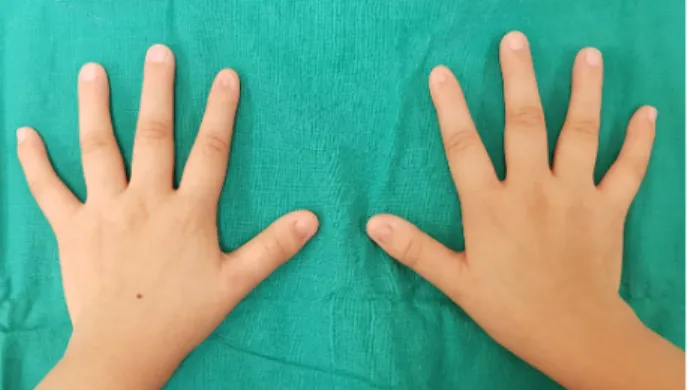
with congenital glaucoma associated with nail-patella syn- drome before and after trabeculotomy. (A) Anterior segment photos, 1 day before trabeculotomy show bilateral corneal edema. (B) Anterior segment photos, 1 year after trabeculot- omy show clear cornea and deep anterior chamber. (C) Fundus photographs, 2 years after trabeculotomy show myopic fundus and tilted disc.Figure 2. Clinical manifestation of the patient's hands. Dystrophic
nails of the thumb and index finger, common findings in the nail-patella syndrome.drome으로 진단된 1예를 경험하였기에 이를 문헌 고찰과 함께 보고하고자 하는 바이다.
증례보고
생후 20일 된 여아가 양안 선천 녹내장이 의심되어 본원 응급실에 내원하였다. 환아는 만삭, 2.42 kg으로 건강하게 태어난 이란성 쌍둥이 중 둘째인 환아로 특이 가족력은 없 었다. 생후 4일경 눈 색깔이 이상해 보여 안과 진료를 시행 하였고, 양안 선천 녹내장 진단하에 본원에 의뢰되었다. 내 원 당시 안압은 퍼킨스 압평안압계로 우안 25 mmHg, 좌안 30 mmHg로 측정되었고, 양측에 심한 각막 부종 소견이 관 찰되었으며 심한 각막 부종 및 혼탁(Fig. 1A)으로 시신경은 확인할 수 없었다. 각막 직경은 우안 10.5 mm, 좌안 10.0 mm 로 측정되었다. 양안 선천 녹내장 진단하에 환아는 전신마 취하 양안 섬유주절개술을 시행받았고 수술 후 1일째 안압 은 우안 12 mmHg, 좌안 10 mmHg로 안정되어 퇴원하였다.
수술 후 안압은 양안에서 12-15 mmHg로 잘 유지되었고 각막 부종도 호전되었다(Fig. 1B). 수술 1년 후 시행한 안저 및 시신경검사상 양안 범무늬 안저, 기울어진 유두(Fig. 1C) 이외에 특별한 이상은 관찰되지 않았고, 안축장 길이는 우 안 26.30 mm, 좌안 25.86 mm로 측정되었다. 홍채 및 수정체 에는 이상 소견을 보이지 않았다.
환아는 만 3세경 시행한 조절마비굴절교정검사상 우안 -10.0 Dsph = -1.0 Dcyl × 180º, 좌안 -13.5 Dsph의 심한 근시 소견을 보여 안경을 처방 받았고 6 PD 내사시가 관찰되었 지만 안구 운동의 이상은 없어 정기적인 안과 검진을 받으 면서 경과관찰하였다.
환아가 만 8세경 내원했을 때, 출생 시부터 양 팔꿈치가 잘 펴지지 않았다는 것을 호소하였고 양측 엄지 손가락 및 검지 손가락의 손톱 형성 이상이 관찰되었다(Fig. 2). 본원 정형외과에 의뢰하여 슬관절, 주관절 및 골반의 단순 방사선 촬영을 시행하였고 양측 슬개골 형성저하(Fig. 3A) 및 외반 슬(Fig. 3B), 양측 주관절 형성저하(Fig. 3C, D), 양측 장골 후외측의 삼각뿔 모양의 장골 돌기(iliac horn) (Fig. 3E)가 관 찰되었다. 위의 소견을 바탕으로 전형적인 nail-patella syn- drome으로 진단하였고 정형외과에서는 수술 등의 특별한 치 료 없이 1년 후 경과관찰하기로 하였다. 임상적 소견을 바탕 으로 nail-patella syndrome를 유발하는 유전자인 LMX1B의 변이 여부를 확인하기 위해 검사 의뢰하였고 LMX1B 유전자 변이(NM_002316.3:c.819+2T>G)가 발견되어 nail-patella syn- drome을 확실히 진단할 수 있었다. 환아의 어머니에서는 유 전자검사상 돌연변이가 발견되지 않았다. 병력 청취를 통해 환아의 부모, 조부모 및 외조부모는 임상적으로 nail-patella
syndrome을 의심할 만한 소견은 없었다는 것을 확인할 수 있었다. 가장 최근 내원인 만 9세경 시행한 안과검사상 최대 교정시력 우안 0.2, 좌안 0.3, 골드만 압평 안압계로 측정한 안압은 양안 모두 12 mmHg, 굴절교정검사상 우안 -19.0 Dsph, 좌안 -17.5 Dsph = -0.5 Dcyl × 180º로 측정되었다. 초음 파 각막 두께 측정법(ultrasound pachymetry)을 이용한 중심 각막 두께는 우안 496 µm, 좌안 474 µm로 측정되었고, 안축 장 길이는 우안 29.45 mm, 좌안 28.30mm로 측정되었다.
689 - 이수민 외 : 국내에서 발생한 Nail-patella 증후군 -
A B C D
E
Figure 3. Radiographic findings of the bilateral femurs, elbows, pelvis com patible with the diagnosis of nail-patella syndrome.
(A ) H ypoplastic patellae. (B) G enu valgum . (C , D ) H ypoplasia of the radial head, capitellum , olecranon and coronoid fossa.
(E) Presence of bilateral triangular osseous excrescences from the posterior aspect of ilia, known as iliac horns (arrows).
4 PD 내사시가 관찰되었고 사시각의 큰 변화 없는 상태로 정기 경과관찰하기로 하였다.
고 찰
Nail-patella syndrome은 매우 드문 상염색체 우성 유전질 환으로 대부분의 경우 9번 염색체의 장완(9q34)에 위치한 LMX1B 유전자의 돌연변이에 의해 발생한다. 88%의 환자는 질환에 이환된 부모로부터 돌연변이 유전자를 받지만, 12%
의 환자에서는 새롭게 돌연변이가 발생한다(de novo muta- tion).3 본 증후군 환자는 부모와 조부모에서 nail-patella syn- drome을 의심할 만한 이상 소견이 발견되지 않아 sporadic case로 생각된다. LMX1B 유전자는 사지 근골격계 발달과 콩팥 발달, 중추신경계의 분화, 안구의 전방 발달을 담당하 는 것으로 알려져 있다.6 150 가지가 넘는 다양한 유전자 변 형이 보고되고 있으나 nail-patella syndrome의 진단은 임상
적 진단을 기본으로 한다. 본 증후군 환자는 매우 다양한 임 상 양상을 나타낼 수 있는데, 손톱 형성 이상은 약 80-90%
의 환자들에서 나타난다. 출생 시부터 진단되는 경우가 많 고 주로 엄지손가락 및 검지손가락에 나타나며, 손톱 압흔 (nail ridging), 세로 갈림(splitting) 등이 나타날 수 있다.7 슬 개골 형성저하는 약 74-93%의 환자에서 나타나며 반복적인 탈구 및 관절염의 원인이 될 수 있다. 주관절 형성저하는 약 93%의 환자에서 나타나며 반복적인 탈구의 원인이 될 수 있다.7 장골 돌기는 무증상으로 치료가 필요하지는 않으나 질병의 특징적 소견으로 30-70%의 환자에서 발생한다.8 약 50%의 환자에서 단백뇨, 혈뇨, 신기능 부전 등의 신장 침범 소견을 나타내는데, 5-10%의 환자는 유년기부터 신장기능 이상이 발생할 수 있고 5%의 환자에서 신장 이식이 필요하 므로7 nail-patella syndrome이 의심되는 환자에 있어 신장 기능 검사가 반드시 시행되어야 한다.
안과적으로는 개방각 녹내장, 고안압증, 각막 두께 증가,
690
= 국문초록 =
Nail-patella 증후군에 동반된 선천녹내장 1예
목적: Nail-patella syndrome과 연관된 선천녹내장 1예를 보고하고자 한다.
증례요약: 생후 20일된 여자 환아가 양안 각막혼탁을 주소로 내원하였다. 내원 당시 양안에 심한 각막 부종이 나타났고 퍼킨스 압평안 압계로 우안 안압 25 mmHg, 좌안 안압 30 mmHg로 측정되었다. 양안 선천 녹내장 진단하에 전신마취하 양안 섬유주 절개술을 시행 하였다. 수술 후 1일째 안압은 우안 12 mmHg, 좌안 10 mmHg로 측정되었고 이후 잘 조절되어 특별한 소견 없이 정기 경과 관찰하였 다. 환아는 이란성 쌍둥이 중 둘째로, 만삭, 2.42 kg으로 태어났으며 특이 가족력은 없었다. 정기적인 경과 관찰 중 환아는 출생 시부 터 양 팔꿈치가 잘 펴지지 않는 증상과 양측 엄지 손가락 손톱 형성 이상 소견으로 정형외과로 의뢰되었다. 환아는 양측 슬개골 형성 저하, 양측주관절 형성저하, 양측 장골 후외측의 삼각뿔 모양의 장골 돌기(iliac horn) 소견을 바탕으로 전형적인 nail-patella syndrome을 진단받았고 nail-patella syndrome을 유발하는 유전자인 LMX1B 유전자 변이가 관찰되었다.
결론: 선천 녹내장 환자에서 손톱 형성 이상 또는 근골격 이상이 동반되었을 경우 nail-patella syndrome을 의심하여 다학제적인 접근이 필요할 것으로 생각된다.
<대한안과학회지 2018;59(7):687-690>
- 대한안과학회지 2018년 제 59 권 제 7 호 -
홍채 중심부 색소 침착(Lester sign), 선천 백내장, 선천 녹내 장, 소각막(microcornea) 등을 나타낼 수 있다.1,3 Mail-patella syndrome 환자의 약 7%에서 고안압증을 나타내고, 10%에 서 원발 개방각 녹내장을 나타낸다고 보고되었다. Lester sign은 정상인에서도 나타날 수 있지만 nail-patella syndrome 환자는 약 50%로 훨씬 높은 빈도로 나타난다.3,9,10
123명의 nail-patella syndrome 환자를 연구한 대규모 연구 에 따르면 nail-patella syndrome 환자에서 고안압증이나 개 방각 녹내장의 평균 진단 나이는 48세로 22세 이하 환자는
없었다.1,3,9 여러 세대에 걸쳐 이 두 질환이 동시에 발생한
두 가계도를 분석한 한 연구에 따르면 개방각 녹내장과 nail-patella syndrome이 모두 특정 유전자의 변화와 연관이 있고, 이 가계의 모든 녹내장 환자는 nail-patella syndrome으 로 진단되어 nail-patella syndrome이 진단된 환자에서 정기 안과적 검진이 필요하다고 하였다. 또한 이 연구에서는 2명 의 선천 녹내장 환자가 수년 후 nail patella syndrome으로 진단되기도 하였다.1 국내에서는 신기능 손상을 동반한 소 아 환자의 nail-patella syndrome 증례가 보고된 바 있다.11
본 증례는 선천 녹내장 환자에서 전형적인 nail-patella syn- drome이 진단되고 장기간 추적 관찰한 예로, 아직 국내에서는 보고된 적이 없는 증례였다. 본 증례 및 기존 연구들의 결과를 보았을 때 nail-patella syndrome으로 진단된 환자의 경우 정기적 안과 진료가 필요하고, 선천 녹내장으로 진단된 소아 환자의 경우에도 손톱 형성 이상 및 관절 이상을 동반하고 있을 경우 nail-patella syndrome를 의심하여 정형외과와 소아과와의 협진 을 통한 다각적 접근이 필요할 것으로 생각된다. 또한 환자와
보호자에게 충분한 유전 상담도 필요할 것으로 사료된다.
REFERENCES
1) Lichter PR, Richards JE, Downs CA, et al. Cosegregation of open-angle glaucoma and the nail-patella syndrome. Am J Ophthalmol 1997;124:506-15.
2) Thompson EA, Walker ET, Weens HS. Iliac horns; an osseous manifestation of hereditary arthrodysplasia associated with dys- trophy of the fingernails. Radiology 1949;53:88-92.
3) Bongers EM, Gubler MC, Knoers NV. Nail-patella syndrome.
Overview on clinical and molecular findings. Pediatr Nephrol 2002;17:703-12.
4) Fong EE. Iliac horns (symmetrical bilateral central posterior iliac processes). Radiology 1946;47:517.
5) Mimiwati Z, Mackey DA, Craig JE, et al. Nail-patella syndrome and its association with glaucoma: a review of eight families. Br J Ophthalmol 2006;90:1505-9.
6) Chen H, Lun Y, Ovchinnikov D, et al. Limb and kidney defects in Lmx1b mutant mice suggest an involvement of LMX1B in human nail patella syndrome. Nat Genet 1998;19:51-5.
7) Lemley KV. Kidney disease in nail-patella syndrome. Pediatr Nephrol 2009;24:2345-54.
8) Karabulut N, Ariyurek M, Erol C, et al. Imaging of "iliac horns" in nail-patella syndrome. J Comput Assist Tomogr 1996;20:530-1.
9) Sweeney E, Fryer A, Mountford R, et al. Nail patella syndrome: a review of the phenotype aided by developmental biology. J Med Genet 2003;40:153-62.
10) Millá E, Hernan I, Gamundi MJ, et al. Novel LMX1B mutation in familial nail-patella syndrome with variable expression of open an- gle glaucoma. Mol Vis 2007;13:639-48.
11) Lee BH, Cho TJ, Choi HJ, et al. Clinico-genetic study of nail-pa- tella syndrome. J Korean Med Sci 2009;24 Suppl:S82-6.