The Effect of Gaze Directions and Pressure Levels on longus colli and Sternocleidomastoid Thickness during Cranio-cervical flexor
Exercise in Young Adults
Ha-ri Cha, Byoung-Kwon Lee, Dong-Kwon Seo*
Department of Physical Therapy, Konyang University
젊은 성인에서 머리-목 굽힘근 운동 시 시선과 압력이 목긴근과 목빗근의 근두께에 미치는 영향
차하리, 이병권, 서동권*
건양대학교 물리치료학과
Abstract This study aimed to investigate the effect of changes in pressure levels and gaze directions on deep neck flexor muscle thickness. Twenty-seven subjects participated in this study. Ultrasound imaging of the longus colli (LC) and sternocleidomastoid (SCM) were measured in four gaze directions (0°, 20°, 40°, 60°) and five pressure levels (20 mmHg, 22 mmHg, 24 mmHg, 26 mmHg, 28 mmHg) during cranial-cervical flexor (CCF) exercises. Repeated ANOVA was performed for analysis of muscle thickness difference according to gaze direction and pressure levels in LC and SCM. Results: LC showed a significant difference between 0° and 20°, 0° and 40°, and 0° and 60° at pressures of 20 mmHg and 22 mmHg (p<.05). SCM displayed a significant difference between 0° and 20°, 20° and 40°, and 40° and 60°
at 28 mmHg (p<.05). In this study, it was found that setting the gaze direction to 20° for the CCF exercise can increase the activation of LC and lower the activity of SCM to obtain the effect of exercise. Based on the results of this study, it is hoped that the beneficial effects of the CCF exercise can be increased by setting an optimal gaze direction in a clinical environment.
요 약 이 연구는 정상 성인에서 머리-목 굽힘 운동 시 시선의 각도와 압력이 깊은목굽힘근의 두께변화에 미치는 영향을 알아보고자 하였다. 본 연구에는 27명이 참여하였다. 머리-목굽힘 운동하는 동안 4가지(0°, 20°, 40°, 60°)의 시선과 5가지(20mmHg, 22mmHg, 24mmHg, 26mmHg, 28mmHg)의 압력에서 목긴근과 목빗근의 근 두께는 초음파 영상 을 이용해 측정하였다. 시선 각도 및 압력변화에 따른 목긴근과 목빗근의 두께 변화를 비교 분석하기 위하여 반복측정분 산분석을 수행하였다. 본 연구결과, 목긴근은 20과 22mmHg에서 0°와 20°, 0°와 40°, 0°와 60°은 유의한 차이를 보였 으며, 20°에서 가장 근활성도가 높았다(p<.05). 목빗근은 28mmHg에서 0°와 20°, 20°와 40°, 40°와 60°은 유의한 차 이를 보였으며, 20°에서 가장 낮은 근활성도를 보였다(p<.05).
본 연구에서 머리-목 굽힘 운동 시에는 시선의 방향을 20°로 설정하는 것이 목긴근의 활성화를 높이고, 목빗근의 근활성 도를 낮추어 운동의 효과를 얻을 수 있다는 것을 찾았다. 본 연구의 결과를 바탕으로 임상환경에서 머리-목 굽힘 운동 시에 시선 방향을 설정하여 운동의 효과를 증대시키길 바란다.
Keywords : Longus colli, Sternocleidomastoid, Cranio-cervical flexor exercise, Ultrasonography, Gaze direction
*Corresponding Author : Dong-Kwon Seo(Konyang University) email: [email protected]
Received September 28, 2020 Revised October 20, 2020 Accepted February 5, 2021 Published February 28, 2021
1. Introduction
Among musculoskeletal disorders, neck pain is the most common[1,2]. With computers and mobile devices in widespread use in South Korea, the age group of complainants for cervical pain has gradually decreased, skewing towards younger generations[3]. In Korea, the internet usage rate is 78.4%, and the average computer usage time per week is 16.3 hours[4], which is reported to have an adverse effect on physical health[5]. Frequent use of electronic devices is one of the causes of neck pain, which is so common that 7 out of 10 adults experience it more than once in their lifetime, and more than one-third of them are chronically recurrent[6-8].
As such, neck pain causes restrictions and disabilities in daily activities, and increases medical costs such as hospital treatment.
Efficient management of neck pain relieves symptoms, reduces relapse rate, and reduces medical costs[9].
The most important structures for muscle activity around the neck are the longus colli(LC) and longus capitis, which are located deep in the neck. These muscles perform similar functions dynamically to provide vertical stability of the neck and to support and hold the front bend of the cervical spine and each joint[10]. In chronic neck pain patients, there is high muscle fatigue of the neck flexor, high muscle activity of surface muscles, and atrophy of the deep neck flexor(DNF)[11,12]. Cranio-cervical flexor(CCF) exercise is said to be suitable for activating DNF muscles, such as the LC and longus capitis, and the activity of the neck surface muscles[13]. CCF is the most representative exercise that trains the muscle strength of the DNF[14-16], to precipitate the action of DNF, and to suppress the action of the surface neck bending muscles. Increasing the mobilization of the DNF reduces chronic neck pain and disability levels, and improves posture control[17-19].
Much attention has been paid to the analysis of deep neck muscles recently, it is still difficult to analyze the muscles of the spine located in the deep part. Although it is challenging to directly promote DNF or apply surface electromyography, ultrasonography can measure muscle contraction in real time as line-of-sight angle feedback.
Various studies have recently been presented on ultrasound images of the Army near the neck of the heart. Greg et al(2005) observed that the activity of neck muscles varies depending on the height of the information medium[20], while the U.S. National Institute of Occupational Safety and Health and Canada's National Standards Association state that when the computer monitor is placed in a position that is tilted 2-15° vertically from the line of sight, it can reduce the posture load of neck muscles[21,22].
CCF exercises increase pain control and function in the daily lives of neck pain patients.
However, studies that identify a better degree of activation of DNF are insufficient, especially studies with an ocular focus, which are not actively conducted. The normal human field of vision is 60° to the nose, 90-95° to the ear, 50°
to the top, and 70-75° to the bottom[10].
Turville(1998) compared the angles of 15° and 40° of the line of sight from the horizontal of the eye to the center of the eye, because the range of eyes for each subject is different, In this experiment, the angle of the downward line of vision was set at 0°, 20°, 40°, and 60° with a maximum of 60° towards the ear. In this study, during CCF exercises using pressure bio-feedback equipment, the muscle thickness of the muscle groups involved in DNF changed depending on the direction, angle and pressure of the line of sight[22].
2. Method
2.1 SubjectsA total of 30 participants(20 males, 10 females) were enrolled in this study. Of these, 3 were excluded. All subjects received a full explanation of the CCF exercise and then signed the agreement directly. The number of subject was set up through the G-power program. When we set the effect size to 0.6, the power to 0.95, and the significance level (α) to 0.05, the sample size for obtaining the difference between the two groups was calculated as 8/group. And the total number of participants was 27.
Spec. Age
(year) Height
(cm) Weight
(kg) BMI
(kg/㎡) Neck girth (cm) (n=18)Male 24.77
±1.86 174.66
±5.19 73.01
±9.88 23.89
±2.78 38.35
±1.59 Female(n=
9) 24.11
±2.57 161.55
±4.30 52.60
±3.03 20.16
±.97 31.57
±1.00 Entire
sample (n=27)
±2.1024 170
±7.93 66.21
±12.76 22.65
±2.92 36.09
±3.54 Table 1. Anthropometric data of the subjects
2.2 Experimental procedures
2.2.1 CCF exercise of gaze directions and pressure levels
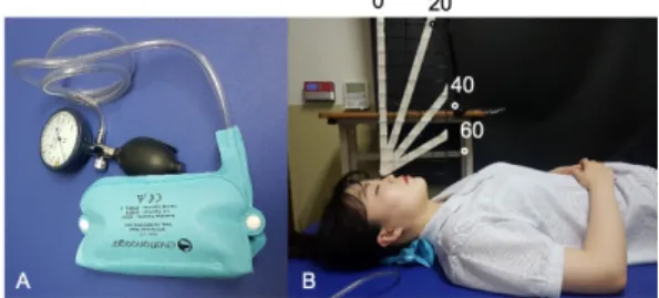
Subjects were placed in a neutral position so that the forehead and chin could be horizontal in the reclined position. The pressure biofeedback unit(Chattanooga, USA) was placed at the back of the target's neck and the pressure gauge was set to maintain a reading of 20 mmHg with the target sufficiently relaxed. There were a gaze to inferior, and the angles were fixed to 0°, 20°, 40°, and 60°. At this time, the angles were set by using a goniometer and a preformed angle plate(Fig. 1). The pressure feedback mechanism started at 20 mmHg and set a target of 28 mmHg.
Each step was held for 10 seconds, and the pressure was sequentially increased by 2 mmHg, for a total of five steps, and an ultrasound imaging was performed. At this time, the subject pulled the jaw and pressed the pressure device
under the neck. During each stage of muscle contraction, a 10-second rest period was provided so that the subject did not induce muscle tone. The angle of the line of sight was selected by using a support to fix the angle so that the subject could adjust the line of sight. At this time, the movement of the neck joint was restricted to allow only the movement of the gaze, and the measurer confirmed that the scale of the pressure biofeedback unit did not move.
Fig. 1. Pressure biofeedback unit(A) and CCF(B)
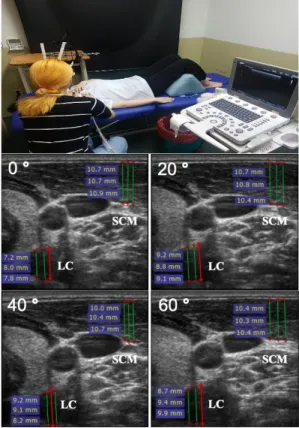
2.2.2 Measurement of muscle thickness Ultrasound imaging was measured of LC and sternocleidomastoid(SCM) using ultrasonography device(SAMSUNG, South of Korea, 2017), according to previous studies[23]. The probes were linear type B and a 7.5 MHz transducer was used. The measurement level was adapted to the fifth cervical spine(C5)[24]. At this level, DNF, including the LC, were visible. For this purpose, the second and third fingers of the subject were stretched and placed on the upper part of the laryngeal prominence and the third fingertip was marked with an oil pen. This point is 1.5 cm below the laryngeal projection and corresponds to the fifth cervical vertebra. This point was placed at the centerline of the transducer and the transducer was placed at a right angle to the longitudinal axis of the subject’s neck to obtain across-sectional image. The muscle in the upper part of the image is the SCM, the lower muscle is the LC, the carotid artery is in the upper border of the DNF. There is a vertebral lamina at the lower border of DNF. When measuring the
anteroposterior distance(APD) of the LC and SCM, the muscle furthest away from the anteroposterior distance of the muscle was analyzed, based on the inside of the muscle fascia. Thickness was measured 3 times, and the mean value was used for data analysis(Fig. 2).
Since the thickness of the muscle is different between the individual incisors, the muscle thickness of the resting muscle was determined[(muscle thickness of the contraction - muscle thickness at rest)/muscle thickness at rest] to analyze the state of the muscle through ultrasonic waves[25-27].
Fig. 2. Ultrasound imaging of LC and SCM thickness during Deep Cervical Flexor exercise. LC:
longus colli, SCM: sternocleidomastoid.
2.3 Analysis method
In this study were analyzed using SPSS ver.
20.0 program for data collected. Muscle thickness differences, according to gaze angle and pressure
level in the LC and SCM, were compared using repeated measures ANOVA. The group - intervening factor was set as the angle, the grouping factor was set as the independent variable, and the thickness of the muscle was set as the dependent variable. Least Significance Difference(LSD) was used for post-test analysis. Corresponding T-tests were used to compare the difference between the lower and the lower gaze. The statistical significance level was set at .05 or less.
3. Results
3.1 Thickness variation of LC according to gaze direction and pressure levels LC on 20 mmHg was going thicker when the subjects inferior gaze angle is going higher.
pressure on 22 and 24 mmHg, LC shown two peak value on 20° and the 40°. When the pressure on 26 and 28 mmHg, the highest thickness of LC was on gaze angle 20° and after that if the angle goes higher the muscle thickness was changed to thinner the before(Table 2).
There was no difference in the between-subjects effect of gaze angles. But, there was a statistically significant difference in the within-subjects effect within the group of pressure levels(p<.05).
Spec.
pressure levels (n=27) mmHg20 22
mmHg 24
mmHg 26
mmHg 28
mmHg
gaze
0° 0.22
±0.13 0.23
±0.13 0.31
±0.13 0.27
±0.13 0.28
±0.13 20° 0.35
±0.18 0.38
±0.18 0.37
±0.17 0.35
±0.18 0.30
±0.14 40° 0.36
±0.15 0.36
±0.17 0.32
±0.13 0.32
±0.12 0.30
±0.14 60° 0.37
±0.16 0.39
±0.16 0.34
±0.19 0.30
±0.13 0.28
±0.14 p-
value
Between-subjects effect F=681.026, p=.60 Within-subjects effect F=10.218* p=.002 Interaction effect F=6.956**, p=.001
*p<.05, **p<.01
Table 2. Comparison of LC muscle thickness in four gaze directions and pressure levels
The interaction effect of angle and pressure was statistically significant(p<.01). When we compared the angles and pressure between pressure groups, in the case of 20 mmHg and 22 mmHg, there was a significant difference between 0° and 20°, 0° and 40°, and 0° and 60°(p<.05; Fig. 3). When the pressure was 24 mmHg and 26 mmHg, there was a significant difference when the angle of inferior gaze was 0°
and 20°(p<.05; Fig. 3). There was no significant difference in pressure at 28 mmHg.
Fig. 3. Comparison of LC thickness by gaze direction and pressure levels
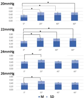
3.2 Thickness variation of SCM according to gaze direction and pressure levels Variation of the SCM at 20 mmHg pressure the thickness of the muscle was going thicker when the subjects inferior gaze angle is going higher.
but on pressure 22 to 28 mmHg, the thickness of SCM was thinner on 20° and after that if the angle goes higher the muscle was changed to thicker(Table 3). There was no difference in the between-subjects effect of gaze angle. There was
a statistically significant difference in the within-subjects effect within pressure level groups(p<.01). There was no statistically significant difference in the interaction effect between angle and pressure. When we compared the angles and the pressure between pressure groups, the pressure difference at 28 mmHg was significant between 0° and 20°, 20° and 40°, and 40° and 60°(p<.05; Fig. 4). There was no significant difference in the remaining pressures.
Spec.
Level of neck pressure mmHg20 22
mmHg 24
mmHg 26
mmHg 28
mmHg
gaze
0° 0.24
±0.15 0.29
±0.15 0.30
±0.13 0.32
±0.13 0.35
±0.12 20° 0.25
±0.12 0.25
±0.12 0.27
±0.12 0.30
±0.13 0.31
±0.12 40° 0.26
±0.12 0.29
±0.13 0.31
±0.17 0.31
±0.15 0.33
±0.15 60° 0.26
±0.12 0.29
±0.11 0.30
±0.13 0.34
±0.11 0.38
±0.14 valueP-
Between-subjects effect F=.544, p=.653 Within-subjects effect F=42.336
p=.001**
Interaction effect F=745, p=.523
*p<.05, **p<.01
Table 3. Comparison of SCM muscle thickness in levels of cervical pressure according to four different eye directions
(n=27)
Fig. 4. Comparison of SCM muscle thickness variation by gaze direction on 28 mmHg.
4. Discussion
CCF exercise promotes DNF activation, inhibits SCM activation, and is effective in restoring function[12]. CCFT is generally
performed while looking slightly downward[15].
In this study, we tried to find the most appropriate gaze direction and pressure levels to increase the efficiency of CCF exercise.
In this study, it was confirmed that LC and SCM activation changed according to the gaze direction and pressure levels during CCF exercise. LC on 20 mmHg and 22 mmHg showed that thickness change was gradually increased as gaze direction increased. At 20 mmHg and 22 mmHg, values of 20°, 40°, and 60° were significantly increased compared to that at 0°.
And LC showed the highest value at 20° on 22, 24, 26, and 28mmHg pressure levels except thickness at 60° on 22mmHg. As a result, LC showed the highest value at 20° regardless of pressure in asymptomatic individuals. This is consistent with the report that computer monitors placed in a position 17-20° vertical to the line of sight, can reduce the postural load of the neck muscles[28]. And LC and eye movement muscles maintain the head position and adjust the postural balance in real time[31]. These studies support the results of this study.
On the other hand, SCM showed the smallest value at 20° and the highest value at 60° on all pressure levels. At 28 mmHg, the mean value was higher at all angles and there was a significant difference between 0° and 20°, 20° and 40°, and 40° and 60°. Only at 20° was the mean value of muscle thickness lower compared to other angles. This may be supported by the results of a study showing that the muscle contraction capacity of the DNF muscle decreases in the post-pressure stage of CCF exercises[29]. And in the previous study, when the computer monitor is in the gaze direction below 40°, the activity of SCM is increased caused by the head tilt angles are increased compared to when the computer monitor is below 15° [22]. In addition, in a study comparing the gaze directions above 45°, 90°
and below 45° for SCM activation during CCFT, below 45° showed the lowest activity [32]. These
studies support the results of this study.
Based on these results and the results of this study, the surface muscles cause neck pain under excessive tension[30]. When the CCF exercise is accompanied by moderate pressure, placing the gaze direction 20° the degree of tension in the surface muscle, which may relieve neck pain, and increases the thickness of the DNF.
In the present study, it was found that the change of gaze direction can activate the DNF muscles and limit the activation of the SCM. It was possible to cause muscle contraction without much pressure depending on the direction of the muscle, gaze direction and head-neck movements in healthy individuals. In patients with neck pain or difficulty adjusting head-neck movements, the angle and direction of gaze can be set during treatment to prevent neck muscle damage. As a limitation, this study was conducted in asymptomatic individuals. The ccf exercise is for people with neck pain in clinical environments.
In a future study, I hope that the study will be conducted on patients with neck pain.
5. Conclusion
This study compared the changes in muscle thickness of LC and SCM according gaze direction and pressure levels during CCF exercises. LC showed the highest value and SCM smallest at 20° all pressure levels. Therefore we would be recommended to set the gaze direction 20° for CCF exercise in a clinical environment, and future studies should be conducted to investigate the effects of CCF exercise for neck pain.
References
[1] H. Vernon, K. Humphreys, C. Hagino, “Chronic mechanical neck pain in adults treated by manual therapy: a systematic review of change scores in randomized clinical trials”, Journal of Manipulative
Physiological Therapeutics. Vol.30, No.3, pp.215-227, Mar. 2007.
DOI:https://doi.org/10.1016/j.jmpt.2007.01.014 [2] S. Haldeman, L. J. Carroll, J. D. Cassidy,
“Introduction/Mandate: the empowerment of people with neck pain. The Bone and Joint Decade 2000–
2010 Task Force on Neck Pain and Its Associated Disorders”, Spine. Vol.33, No.supplemet pp.S8-S13, Feb. 2008.
DOI:https://doi.org/10.1097/brs.0b013e3181643f51 [3] Y. H. Chung, T. W. Moon, T. W. Eom et al, “A study
of neck pain and its related factors among highschool students in the vicinity of Seoul and Kyung Ki province”, Journal of Oriental Rehabilitation Medicine. Vol.17, No.4, pp.185-198, Oct. 2007.
UCI:G704-001658.2007.17.4.011
[4] Korea Internet & Security Agency, 2012 Survey on the Internet Usage, Korea Internet & Security Agency, 2012, Available From:
https://www.kisa.or.kr/eng/usefulreport/surveyReport (accessed Feb. 22, 2013)
[5] P. Phillastrini, C. Farneti, R. Mugnai, et al, “Evaluation of two preventive interventions for reducing musculoskeletal complaints in operators of video display terminals”, Physical Therapy. Vol.87, No.5, pp.536-544, May. 2007.
DOI:https://doi.org/10.2522/ptj.20060092
[6] P. Côté, J. D. Cassidy, L. J. Carroll, “The Saskatchewan health and back pain survey. The prevalence of neck pain and related disability in Saskatchewan adults”, Spine. Vol.23, No.15, pp.1689-1698, Aug. 1998.
DOI:https://doi.org/10.1097/00007632-199808010-00015 [7] P. Côté, J. D. Cassidy, L. J. Carroll, et al, “The annual
incidence and course of neck pain in the general population: A population based cohort study”, Pain. Vol.112, No.3, pp.267-273, Dec. 2004.
DOI:https://doi.org/10.1016/j.pain.2004.09.004 [8] S. Hogg-Johnsons, G. van der Velde, L. J. Carroll, et al,
“The burden and determinants of neck pain in the general population: Results of the bone and joint decade 2000-2010 task force on neck pain and its associated disorders”, Spine. Vol.33, No.4, pp.S39-S51, Feb. 2008.
DOI:https://doi.org/10.1097/brs.0b013e31816454c8 [9] Borghouts, A. J. Jeroen, B. W. Koes, et al,, “Cost of
illness of neck pain in the Netherlands in 1996”, Pain. Vol.80, No.3, pp.629-636, Apr. 1999.
DOI:https://doi.org/10.1016/s0304-3959(98)00268-1 [10] J. H. Park, D. H. K, J. G. Her, J. H. Woo, “What is a
more effective method of cranio-cervical flexion exercises?”, Journal of Back and Musculoskeletal Rehabilitation. Vol.31, No.3, pp.415-423, Jun. 2018.
DOI:https://doi.org/10.3233/bmr-170860
[11] D. L. Falla, G. A. Jull, P. W. Hodges, “Patients with neck pain demonstrate reduced electromyographic activity of the deep cervical flexor muscles during
performance of the craniocervical flexion test”, Spine. Vol.29, No.19, pp.2108-2114, Oct. 2004.
DOI:https://doi.org/10.1097/01.brs.0000141170.89317.0e [12] K. Javanshir, M. A. Mohseni-Bandpei, A. Rezasoltani,
M. Amiri, M. Rahgozar, “Ultrasonography oflongus colli muscle: A relability study on healthy subjects and patients withchronic neck pain”, Journal of Bodywork and Movement Therapies. Vol.15, No.1, pp.50-56, Jan.
2011. DOI:https://doi.org/10.1016/j.jbmt.2009.07.005 [13] M. A. Mayoux-Benhamou, C. Vallée, M. Revel, et al,
“Longus colli has a postural function on cervical curvature”, Surgical and Radiologic Anatomy. Vol.16, No.4, pp.367-371, Dec. 1994.
DOI:https://doi.org/10.1007/bf01627655
[14] G. Jull, “Management of cervical headache”, Manual Therapy. Vol.2, No.4, pp.182-190, Nov. 1997.
DOI:https://doi.org/10.1054/math.1997.0298
[15] G. A. Jull, S. P. O’Leary, D. L. Falla, “Clinical assessment of the deep cervical flexor muscles: The craniocervical flexion test”, Journal of Manipulative and Physiological Therapeutics. Vol.31, No.7, pp.525-533, Sep. 2008.
DOI:https://doi.org/10.1016/j.jmpt.2008.08.003 [16] S. O’Leary, D. Falla, G. Jull, B. Vicenzino, “Muscle
specificity in tests of cervical flexor muscle performance”, Journal of Electromyography and Kinesiology. Vol.17, No.1, pp.35-40, Feb. 2007.
DOI: https://doi.org/10.1016/j.jelekin.2005.10.006 [17] Y. Dusunceli, C. Ozturk, F. Atamaz, et al, “Efficacy of
neck stabilization exercises for neck pain: A randomized controlled study”, Journal of Rehabilitation Medicine. Vol.41, No.8, pp.626-631, Apr. 2009.
DOI: https://doi.org/10.2340/16501977-0392
[18] D. Falla, T. Russell, G. Jull, B. Vicenzino, P. Hodges,
“Effect of neck exercise on sitting posture in patients with chronic neck pain”, Physical Therapy. Vol.87, No.4, pp.408-417, Apr. 2007.
DOI: https://doi.org/10.2522/ptj.20060009
[19] S. O’Leary, D. Falla, P. W. Hodges, G. Jull, B.
Vicenzino, “Specific therapeutic exercise of the neck induces immediate local hypoalgesia”, Journal of Pain. Vol.8, No.11, pp.832-839, Jul. 2007.
DOI: https://doi.org/10.1016/j.jpain.2007.05.014 [20] A. M. Greig, L. M. Straker, A. M. Briggs, “Cervical
erector spinae and upper trapezius muscleactivity in children using different information technologies”, Physiotherapy. Vol.91, No.2, pp.119-126, Jun. 2005.
DOI:https://doi.org/10.1016/j.physio.2004.10.004 [21] S. A. Kumar, “A computer desk for bifocal lens
wearers, with special emphasis on selected telecommunication tasks”, Ergonomics. Vol.37, No.10, pp.1669-1678, Oct. 1994.
DOI:https://doi.org/10.1080/00140139408964944 [22] K. L. Turville, J. P. Psihogios, T. R. Ulmer, F. A. Mirka,
“The effects of video display terminal height on the operator: A comparison of the 15 degree and 40 degree recommendations”, Applied Ergonomics. Vol.29, No.4, pp.239-246, Aug. 1998.
DOI: https://doi.org/10.1016/s0003-6870(97)00048-3 [23] H. Ishida, T. Suehiro, K. Ono, C. Kurozumi, S.
Watanabe, “Correlation between deep cervical flexor muscle thickness at rest and sternocleidomastoid activity during the craniocervical flexion test”, Journal of Bodywork and Movement Therapies. Vol.20, No.1, pp.208-213, Jan. 2016.
DOI:https://doi.org/10.1016/j.jbmt.2015.06.005 [24] B. Cagnie, L. Vandamme, E. Derese, et al, “Validity and
reliability of ultrasonography for the longus colli in asymptomatic subjects”, Manual Therapy. Vol.14, No.4, pp.421-426, Aug. 2009.
DOI: https://doi.org/10.1016/j.math.2008.07.007 [25] P. W. Hodges, L. H. Pengel, R. D. Herbert, S. C.
Gandevia, “Measurement of muscle contraction with ultrasound imaging”, Muscle & Nerve. Vol.27, No.6, pp.682-692, Jun. 2003.
DOI: https://doi.org/10.1002/mus.10375
[26] F. M. Jesus, P. H. Ferreira, M. L. Ferreira,
“Ultrasonographic measurement of neck muscle recruitment: a preliminary investigation”, Journal of Manual & Manipulative Therapy. Vol.16, No.2, pp.89-92, Apr. 2008.
DOI: https://doi.org/10.1179/106698108790818486 [27] F. R. Jesus-Moraleida, P. H. Ferreira, L. S. Pereira, C.
M. Vasconcelos, M. L. Ferreira, “Ultrasonographic analysis of the neck flexor muscles in patients with chronic neck pain and changes after cervical spine mobilization”, Journal of Manipulative and Physiological Therapeutics. Vol.34, No.8, pp.514-524, Oct. 2011.
DOI: https://doi.org/10.1016/j.jmpt.2011.08.006 [28] D. R. Ankrum, K. J. Nemeth, “Head and neck posture
at computer workstations-what’s neutral?”, Proceedings of the Human Factors and Ergonomics Society Annual Meeting. Vol.44, No.30, Jul. pp.565-568. 2000.
DOI:https://doi.org/10.1177/154193120004403046 [29] T. T. Chiu, E. Y. Law, T. H. Chiu TH. “Performance of
the craniocervical flexion test in subjects with and without chronic neck pain”, Journal of Orthopaedic and Sports Physical Therapy. Vol.35, No.9, pp.567-571, Sep. 2005.
DOI:https://doi.org/10.2519/jospt.2005.35.9.567 [30] G. Johnson, N. Bogduk, A. Nowitzke, D. House, “Anatomy
and actions of the trapezius muscle”, Clinical Biomechanics. Vol.9, No.1, pp.44-50, Jan. 1994.
DOI: https://doi.org/10.1016/0268-0033(94)90057-4 [31] J. A. Cromer, D. M. Waitzman. “Neurones associated with saccade metrics in the monkey central mesencephalic reticular formation.” Journal of physiology. Vol.570, No.3, 2006. pp. 507-23, Feb. 2006.
DOI: https://doi.org/10.1113/jphysiol.2005.096834.
[32] J. Park, D. Ko, J. Her, et al., “What is a more effective
method of cranio-cervical flexion exercises?” Journal of Back Musculoskelet Rehabilitation. Vol.31, No.3, 2018. pp. 415-423.
DOI: https://doi.org/10.3233/BMR-170860.
차 하 리(Ha-ri Lee) [정회원]
• 2019년 8월 : 건양대학교 물리치 료학과 (이학석사)
• 2015년 2월 ~ 2016년 2월 : 대전 웰니스요양병원
• 2016년 3월 ~ 2020년 6월 : 대전 IAME트레이닝센터
• 2020년 7월 ~ 현재 : 서초리더스 정형외과재활의학과의원
<관심분야>
근골격계물리치료학, 스포츠물리치료학
이 병 권(Byoung-kwon Lee) [정회원]
• 2010년 10월 : 국립독일체육대학 교 재활 및 예방학 (이학박사)
• 2011년 3월 ~ 현재 : 건양대학교 물리치료학과 교수
<관심분야>
스포츠물리치료학
서 동 권(Dong-kwon Seo) [정회원]
• 2011년 2월 : 대전대학교 물리치 료학과 (이학석사)
• 2014년 3월 ~ 현재 : 건양대학교 물리치료학과 교수
<관심분야>
근골격계물리치료학, 신경계물리치료학