大韓放射線醫學會誌 Vol. XVI, No. 2, 1980
黃煩의 超音波所見에 關한 冊究
서울大學校 醫科大學 放射線科學敎室
훌훌炳濟 • 裵‘뼈勳 · 李承훌 • 金周完
- Abstract-
Ultrasonography of Jaundice
Byung )ae Cho, M.D., Sang Hoon Bae, M.D., Seung Ro Lee, M.D., Chu Wan Kim, M.D Dept. of Radiology
,
Seoul National University,
Medical Col/egeThe importance of ultrasonography in the evaluation of jaundice is stressed with an analysis of 47 cases of jaundice.
31 cases proved to be obstructive and 16 non.obstructive jaundice. Obstructive jaundice could be differen.
tiated from non-obstructive jaundice in all but 2 cases, (96%). The site of obstruction in 31 cases of obstructive jaundice could be predicted correctly in 23 cases of 31, (90%), and cause of obstruction with an accuracy of 35% as well.
One can certainly recommend ultrasonography as an initial procedure of choice in jaundice patients.
1 . 精 論
黃;효愚者에 있어 手術을 강용하는 !펌銷{生黃훤(外科的 黃흉)과 R千細뼈{生 原因에 依한 非開銷性 黃펄(內科的 黃훨)을 강렬하는데는 무엇보다도 安全하고 非홉觀{生 該斷方法으로 그레 이 스케 일 (gray scale) 超音波檢홀 의 重떻↑生이 認識되어 왔다 1-20 ,23 , 21)
經皮經R千廳道造影術21)이나 內視鏡的 않行 f生 觸· 牌 管造影術22) 도 m쩔管을 造影該斷에 는 重要한 檢흉方法。1 기 는 하나 슴없효과 失敗率도 無視할 수 없 다. 전산화단
층촬영(C-T) 도 經濟的인 理由等으로 除限펀 경우外는
施行하기가 어렵다하겠다.
最近 그레 이 스케 일 (gray scale) 方式의 超훌波該斷 機의 짧達도 黃훨에 관한 超音波의 양斷的 價{直를 높 은 位置를 차지 하게 되었다.
著者들은 超音波로서
첫 째 : 閒銷性 또는 非봐l 銷{生 黃훨의 강별 둘째 · 外科的 黃훨에 있어서 閒銷된 部{立의 正確
位置
셋째 : 까科的 黃훨에 있어 病因學的 談斷 (etiolo
gical diagnosis) 等을 규영 할려 고 하였 다.
n.
對象 및 方法1) 對 훌
1980年 2月부터 5月까지 黃훨을 主訴로 入院하여 右 上題部 超音波檢훌를 行한 思者에서 臨 VR所見. 生化學 檢훌, 生檢 빛 追없조사와 經皮經 IlH뿔道造影術, 內視 鏡的 않行{生 廳· 降管造影術파 手術및 病理學的 所見 結果과 確認된 47例을 對象으로 하였 다 (르, 觸훌結石 을 主訴로 超音波檢흉를 行한 것 은 除外하였 다. )
2) 方 法
音波檢훌는 Picker 80L 스켄너 (scanner) 를 使用하 였 고, 2.25MHz, 19mm, medium internal focused 트란스듀서 (transducer) 를 主로 使用하였 다.
- 370-
連結的인 스켄 (scan) 은 可能한 限 深吸入狀態에서 維斷, 橫斷스켄을 했고, 總願管과 門服 (portal tein) 을 쉽게 區分하기 위해 右測을 들어올려 左後짧位 (LPO position) 로 한後, 維斷스켄을 行하였다 1) 遠{立總廳管 을 잘 보기위해셔 右예을 더욱 들어올려서 十二指陽球 빛 大陽·깨스 (gas) 로 因한 흉쩔陰影 (acoustic shadow) 으로부터 遠位總願管올 分離시 킬 수 있었 다. 또한 超 音波檢홉로서 nf寶質 .ßf 內廳管. ij주外廳管, 죽 總R주脫 管 (commun hepatic duct). 總廳管 (common bile
duct) 와 g¥頭部둥을 스켄하였다.
不確賢한 所見을 보인 f71J 에 서 는 可能한 限 다음날 또는 수일 後에 다시 施行하였 다.
nI.
成 짧確양된 47f71J 中 內科的 黃펄이 16例 外科的 黃훨이 31 例였고 內科的 黃훨은 ßfß{ 6f71J. 擔管씻 5f71J. ß千硬 變 3f71J. 原發{生 府癡 l{정U. ßf轉移를 同伴한 몹찮 1 例。 1 며 , 外科的 黃훨은 總廳管結石 12例, 牌頭部癡 7f71J.
總觸管찮 6例. Æ웹훌훌 j품및 주위흡題 1 f71J. ¥L頭癡3f71J. 府 內및 廳훌훌結石 1 f71J. 廳管內 破앓을 同伴한 原發{生 府 癡 1 {7lj였 t:-J.(Table 1).
正힘人에 있어서는 超音波檢훌로서 ßf 內 8홈管分技는 찾기 가 어 렵 다. 萬若 門服의 分技와 하行하는 觸道의 分技가 함께 나타냐연 이 는 二重曉所見. àou ble chan nel sign. 이 라하여 觸道閒銷의 主要所見의 하나안데 2
,
이 :重隨所見等 R千內觸管의 據張을 보인 例를 分析 하연 內科的 黃훨 16f71J 中 原發{生 8千훨 1 f71J 따轉移를 同伴한 뿜 癡 1 f71J로 近位據張 (proximaldila ttion)을 보
Table 1. Pathoanatomic diagnosis of jaundice patients
Medical jaundice(I6 pts) Surgical jaundice(31pts)
Hepatitis 6
Cholangitis 5 Cirrhosis 3
Hepatoma l
Stomach cancer ‘ 1 with iver
metastasls
C.B.C. stone 12 Pancreatic carcinoma 7 C.B.D. cancer 6 Ampulla cancer
G.B. cancer with regional invasion
Intrahepatic stone with 1 G.B. stone
Hepatoma with rupture 1 into biliary tree
Table 2. Correlation between intrahepatic biliary dila ta tion and jaundice
medical jaundice surgical jaundice Dilatation
No dilatation
2* 29
2 14
Total 16 31
... proximal dilatation due to hepatoma and metas- tasis from stomach
인 2例를 除外하연 모두 표常所見을 보였고 外科的 黃 훨 31 例는 29f71J에서 9千內廳管廳張을 보였고 2f71J에서만 나타나지 않았다 (Table 2).
m훌管의 閒짧部位를 該斷하려 연 .ßf 外廳管의 據張有 無를 알아야 하는데 外科的 黃훨 31 f71J를 分析해 보연 總R꾸觸管 (common hepatic duct) 이 閒銷된 3 例에서 는 모두 싸外廳管의 據張이 없었으며 ?遠位總廳管閒銷 된 28f71J에서는 24 f71J는 據張된 總願管올 폴 수 있었고 4例에서는 나타나지 않았다. 이 4例中 1 例에서는 手術 後 據張이 없었음이 증명되어 3例의 假陰性 談斷이 있었어냐, 假陽性은 全無했다 (Table 3).
"Table 3. Obstructive Leyel of Surgical Jaundice
US Dilated CBD Not dilated CBD Obstrutive leveJ
Proximal C.B.D.
Distal C.B.D.
0 24
3 4*
*1 case is confirmed to be not dilated by opera- tlOn.
病因學的 談斷 (etiological diagnosis) 은 總願管結 石은 12例中 7例, 降頭部癡은 7 f71J 中 3例 • ß주內廳管및 廳훌結石은 1 例中 1 껴1 에서 ~斷이 可能했고 그 外 總、
體管훨 6f71J. 觸활癡및 주의 홉꿇 1例, ¥L頭癡 3例와 廳
管內 破앓올 同伴한 原發{生Jlf癡 1 例는 不可能했 다. 總 31 例中 ll f71J( 35%) 에서 原因의 談斷。l 可能하였다 (Table 4).
3
1
N.
考 按黃훤愚者의 ä~斷方法에는 여러가지檢흉 方法이 있겠 는데 安全하고 非홉製性이 며 응?斷的 價{直가 커 야만 理想 的안 檢흩인 것 이 다. 밸 리 추빈 (bilirufin) 植가 4mg/dl
以上이 변 經口빚 經靜Il*n쩔管 4mg/dl 造影術을 行하기 가 困難하며 經皮經ßf願道造影術빛 內視鏡的 않行性 勝 윈
-
Table 4. Etiological Diagnosis of Surgical Jaundice
Etiological Diagnosis Correct Dx
C.B.D. stone 7/12
3/7 0/6 Pancrea tic carcinoma
C.B.D. cancer
Intrahepatic stone with G.B. stone 2/1 G.B. cancer with regional invasion 0/1
AmpuIla cancer 0/3
Hepatoma with rupture into biliary tree 0/1
Total 11/31(3 5%)
• 牌管造影術도 前者에 있어서는 정도의 차는 있으나
1îfJH!E을 유말할 수도 있고!!1) 後者의 경우에서도 相當 한 檢흉時間이 要하며 , 思者의 苦痛, 多少의 失敗率 (1 0"'30%)도 있다한다1 , 7 , 22) 이러한 단점을 보완하는 데 는 超音波檢홀가 좋은 位置에 있 다고 하겠 다 1-20)
解힘j學的으로 右 · 左빠內廳管이 府內에서 合쳐져있 다가 府門部에서 總Jlf廳管을 이루고 아래로 내혀와 4 cm 정 도에서 廳훌管과 合하여 總擔管을 이 루어 몹 8千 間體(lesser omenum) 의 右測을 따라 내려와 十一指 陽第-部位의 後뻐IJ을 달리 게 된다. 이 總觸管은 門!l!FC 의 앞쪽에 위 치 하고 下大靜!l!FC의 右前뼈Ij에서 비 스듬히 내려온다.
超音波檢훌에서 單純한 維, 橫斷스켄으로는 W쩔管의 據張이 어느정도 되지 않A연 쉰게 찾기가 어렵다. 뿐 안아니 라 real time 스켄으로도 門版과 總廳管이 크기 가 작고 또한 下大靜師, 題部大動!l!FC과 憐接해 있어 波 動 (pulsation) 만으로는 區別이 쉽지 않다 1) 그래서 右 {則을 들어 올려 左後힘位를 취 한다음 徵斷스켄을 함으 로써 Behan Ml) 은 96%에서 織張된 @홈管을 찾을 수가 있었다.
左後씹位(L. P.O Position) 스켄은 앞쪽에 위치한 擔管과 뒤 쪽의 門服을 쉽 게 區分할 수 있 A며 右8주葉 올 音짧 (acoustic window)로 이용하여 總觸管을 찰볼 수 있을 뿐만 아니라 十二指陽球 또는 大陽의 깨스로 因한 音響陰影으로 부터 遠位總體管을 合離시킬 수 있 는 장정 外에 @쩔훌훌, 總觸管과 아울러 際頭部도 함께 볼
수 있다 1.3,9, 11) (Fig. 1).
內科的 黃힘과 外科的 黃펄올 區分하는데 있어 Jlf內 廳管據張및 또는 總廳管의 었옮끓i을 중영 해야 하며 特히 外科的 黃훨에 는 觸管의 擬張올 各 部分別로, 따內廳 管, 總 IlH형管, 總廳管으로 區分하여 分析하는 것 이 閒
“‘
~
11UdllllluIlhlulllllllllllllllll1111‘”‘1"’,1‘”‘!
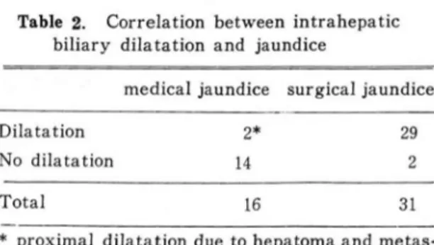
SNUH RAC、IOLOGν 50N02333 R 6. 5늑
Fig. 1. Normal CBD
Longitudinal scan in LPO : Sonolucent cystic structure is gaIIbladder (GB), not dilated, anterior located CBD(arrows),and posterior located portal
vein (P).
鎭된 正確한 位置와 病因學的 양斷에 도움을 줄 수 있
겠다 19)
Lang FC et aPO) 은 %內廳管의 據張과 Jlf內門!l!FC을 區分하는뎌1
CD
右門服파 主門服주위 의 解휩j學的 形態 의 變化,@ 擬張된 擔管의 불규칙한 벽 @ 擬張된 廳 管의 放射狀合流(steIIate confIuence)@
據張된 廳 管의 後面增꿇 (aconstic posterior enhancement) @ 據張펀 廳管의 周邊 ÜL置 (peripheral location) 等으로1IIIIIIIIIIIIIIIIIIIIdlllllllll
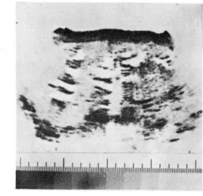
Fig. 2. Transverse scan:
Markedly dilated biIiary trees with tortuosity.
…
“
강별할 수 있으며 (Fig. 2) Conrad MR et a ]2>은 右,
左門~에 憐接해 있는 擬張된 擔管分技를 同時에 나타 내 는 “parallel channel sign" 으로 94%의 談斷率을 얻었다고 했고 또한 이것을 갱 uotgun sign"19) 로 말하 기 도 하벼 特히 左葉에 서 助骨下緣스켄 (snbcostal
scan) 을 할혜 잘 나다난다고 하였다 (Fig. 3).
보다 커진다고 한다. 이러한 所見들은 外科的 黃훤을 의미하게 된다. 造影齊l 를. 쭈IJ 用한 檢훌에서는 總廳管의 크기가 10mm以上이, 超홉波檢훌에서는 8mm1,3,4,15)
以上이연 意味있는 閒銷{生 黃훨을 냐타낸다. w함훌手術 을 行한 경 우에 종전어}는 總廳管의 據張이 온다고 했
£냐 Graham MF et aF) 이 보고한 바로는 願훌切除 術파 總擔管의 搬張과는 無關하며 據張된 總體管도 手 術後 正常으로 돌아온다고 했다.
病因學的 該斷은 擔道樓張外에 追加giJi‘見이 있억야“
可能한 것 이 다. Fig. 4와 강이 總廳管의 擬張이 있고 그 內部에 진한 에코가 있으연서 音쩔陰影이 있으연 總廳管結石을 :쉽 게 該斷할 수 있고, g¥頭部찮은 擬張 띈 總‘觸管과 體頭部에서 睡塊가 보일 경우에 可能하다
(Fig. 5). 그러나 (Fig.6) 에서와 같이 봐內觸管의 據 張만 보일뿐 體農과 總觸管의 據張이 찰보이 지 않을
-률 경 우에 는 總Rf觸管閒賴을 意味할뿐 擔管癡인지 轉移性
?휩인지 강별이 용이하지 않다.
閒銷{生 黃훨파 非閒銷{生 黃훤의 강별은 86%~97
% 5,8,11, l~ , 15, 17, 18) 에서 可能하여 , 閒銷된 部位의 正確한
位置는 85%~94%8 , 11) 에서. 病因學的 談斷은 58%~
Fig. 3. Parallel channel sign : 81%5,8) 에서 可能하다고 했으며 Sample WF et a1'5) Transverse scan : Anterior located, dilated left 은 超音波所見하나 만으로 22% 의 該斷率을 얻었다고 intrahepa tic biliary tree (arrow), posterior loca ted 한다. 그러 나 黃훨의 期間및 정 도와 廳管搬張의 크기
portal vein (arrows). 와는 판계 가 없‘다고 한다.
l펌銷{生 黃훨이 상당히 의심스러운데 超좁波所見이 正 總總觸管의 鍵張이 나타나연 外科的 黃훨을 意味하 常이 연 다른 홈製i生 檢흉를 行하기 前에 재 차 超홉波 게되는데 표常에서는 앞쪽에 위치한 總體管의 크기가 檢흉를 권하고 있다. 따外 總脫管은 平網節이 거의 없 뒷쪽의 門版보다 작다, 그러나 據張이 있으면 반대현 고 많은 結願下 彈{生組織혜문에 擬張된 廳管도 原因만 상으로 總廳管의 크기가 門1M과 같아지거냐 또는 門1M 除去되연 수일內 표常A로 되돌아 온다고 한다 16ì
.. ‘
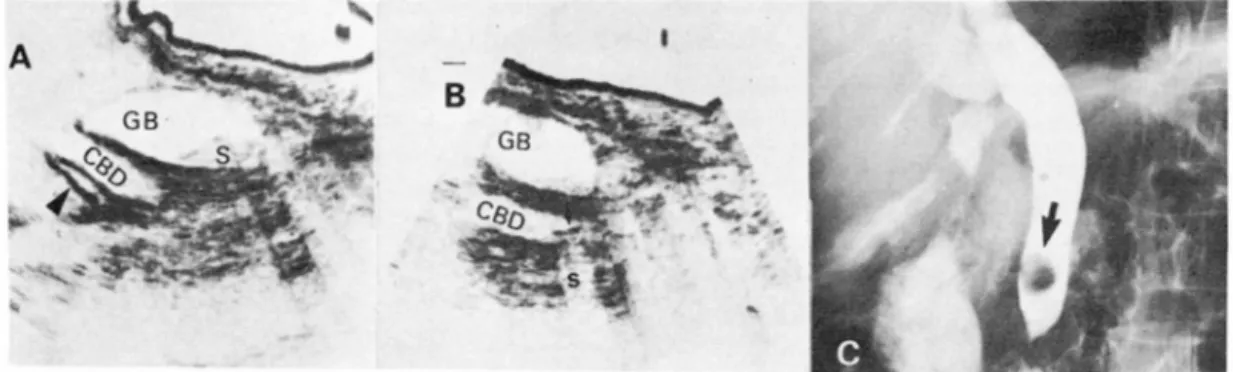
Fig. 4. Distal CBD stone
’(A) Longitudinal scan in LPO. Markedly dilated CBD, compressed protal vein(arrow head) and enlarged gallbladder with sludge (S).
<B) More elevated than A: Dense echo (arrow) within dilated CBD, with shadowing (S).
{C) ERCP : CBD stone within dilatated CBD (arrow).
… 꾀
Fig. 5. Pancreatic head cancer
~A) Longitudinal scan in LPO. Marked dilatation of CBD (C), comparing with portal vein (P).
{B) Transverse scan solid tumor mass in pancreatic head (arrows). Enlarged gallbladder (G).
‘(C) ERCP: Typical double duct sign suggests pancreat!c head cancer.
B
- -
‘-
낳?쓸예f ’
,~훌같
Fig. 6. Biliary cancer in CHD : .(A) Longitudinal scan: Dilated intrahepatic biliary ducts.
(B) Longitudinal scan in LPO: Not dilated CBD (arrows), Comparing with portal vein (P).
contracted gallbladder (g).
(C) ERCP: Biliary cancer at trifurcation of CHD (arrow).
더 군다냐 Weinstein DP et a P"'은 黃훨없 이 훨管의 搬랬이 올 수 있으며 이것은 部分的 觸管閒짧에서는 超音波檢흉가 生化學檢옳보다 더 욱 예 민하다고 할 수 있 A며 이때는 經皮經IltJl1쩔道造影術및 內視鏡的 i뽀行性 觸· 牌管造影~한올 行하여 原因규명 올 해 야한다고 하였 다.
以上 列學한 報告들을 보건티l 著者들의 經驗은 척으 나 合當한 意見들이라 할 수 있겠다.
V.
結 論1980年 2月부터 5月사이 에 서 울大學校 醫科大學 放
射線學科敎室에서 施行한 黃훨思者의 上陽部 超音波所 見과 臨tf:, 기 다檢훌, 手術및 病理所見과 比較함으로 써 다음과 같은 結果를 얻었다.
<D lì{Ht된 47例에 서 內科的 黃훨 16例, 外科的 黃훨
{7IJ 로 뭔分되 었 으며 外科的 黃훨 2例를 除外한 45
- 374-
9. Lee TG, Henderson SC, Ehrlich R : Ultrasoundi
diagηosis of commoη bile duct dilatation. Radi‘ ology 124:793-797, Sep 1977
10. Laing FC,London LA, Filly RA : Ultrasoηog
raPhic identicatioη 0/ dilated iη trahepatic bile ducts a η d their differeη tiation from portal venous structμ res. jCU 6 : 90-94, 1978
11. Malini S, Sabel J: Ultrasonography iη obstr- uctzve ;auηdice. Radiology 123 : 429-433, M ay 1977
12. Neiman HL, Mintzer RA : .4ccuracy 0/ biliary
dμ ct ultraso ηd; Comparisoη zνith cholaη giography.
REFERENCES Am j Roeηtgeη01 129 : 979-982, Dec 1977
13. Reid MH : Visualization of the bill ducts us떠g 1. Behan M, Kazam E : SoηograPhy 0/ the commoη focused ultrasound. Radiology 118: 155-158,
bile duct; Value of the right aηterior oblique Jan 1976
vtezν. Am j Roeηtgeη01 130: 701-709, APr 1978 14. Stone LB, Ferrucci JT, Warshaw AL, Witte- 2. Conrad MR, Landay MJ,Janes JO:SonograPhic nberg J, Slutsky M: Gray scale ultrasouηd
“parallel chaηηel sign" of biliary tree eηlar- diagηosis 0/ obstructive bilt'ary disease. Am j
gemeηt iη mild to moderate obstructive jaundice. Roeηtgenol 125 : 47-50, Sep 1975
Am j Roentgenol 130 : 279-286, Feb 1978 15. Sample WF, Sarti DA, Goldstein LI. Weiner 3. Cooperberg PL : High-resolutioη real time ult- M, Kadell BM : Gray scale ultrasonography of rasound in the evaluatioη of the normal aηd the jauηdiced patient. Radiology 128: 719-725, obstructed biliary tract. Radiology 129 : 477-480, Sep 1978
Nov 1978 16. Scheske GA, Cooperberg PL, Cohen MM,
4. Cooperberg PL, Li D, Wong p, Cohen MM, Burhenne HJ : Dyηamic chaηge in the caliber Burhenne HJ: Accμracy of commoη hepatic 0/ the major bile ducts, related to obstructioη.
dμct size iη the evaluatioη 0/ extrahepatic biliary Radiology 135 : 215-216, APr 1980
obstructioη. Radiology 135: 141-144, Apr 1980 17. Taylor KJ끼r , Rosenfield AT : Gray scale ult- 5. Dewbury KC, Joseph AEA, Hayes S, Murray rasoηography in the differeηtial diagηosis of
C : Ultrasolμ2d iη t he eval uatz‘0η and diagnosis jaundice. Arch Surg 112 : 820-825, Jμ1 1977 of jaundice. British j Radiology 52: 276-280, 18. Taylor KJW, Rosenfield AT, Spiro HM :
1979 Diagηostic accuracy of gray scale ultrasoηog-
6. Goldberg BB: Ultrasonic cholangiograþhY' .raþhy for the jaundiced patieηt. Arch 1:ηterη Radiology 118: 401-404, Feb 1976 Med 139: 60-63, jaη 1979
7. Graham MF, Cooperberg PL, Cohen MM, 19. Weill F, Eisencher A. Zeltner F: Ultrasoηic Burhenne HJ : The siz
例 (96%) 에서 內科的및 外科的 黃훤의 감별이 可 能했다.
@ 外科的 黃훨 3H쩨中 며]짧된 願管의 正確한 位置 를 나타낼 수 있었던 ØlJ는 28ØlJ( 90%) 에서 i'iJ能혔 다.
@ 病因學的 談斷이 可能한 率은 外科的 黃펄 31 ØlJ
中 ll ØlJ( 35%) 였다.
위와 같은 결과로 보아 黃훨,뽑者의 檢흉에 있어서 훔 觀性인 檢훌를 行하기 前에 安全하고 備便한 超音波檢 흉를 우선 行하는 것이 바랍직하다 하겠다.
얘 ω
22. 金仁元, 李承홉, 韓萬育, 朱東雲: 內視鏡的 않行 508 1979
{生 廳· 牌管造影術所見에 關한 冊究. 大韓放射線 24. 金周完, 徐廷守, 李寬世, 外 . 超音波該斷自驗 1018
醫學會誌 15 : 427-433. 1979 例에 關한 考察, 大韓放射線醫學會誌15 : 493-503.
23. 金周完, 李寬世, 趙秉濟. 徐廷守· 廳餐의 超音波 1979
嚴影에 關한 冊究. 大韓放射線醫學會誌 15 : 505-
- 376-