人~!( Ii'x:껴t*Jíl석 r}~씬ι Vol. 21, No. 2, 1985
R주硬化徒에 있어서 造影增彈後 電算化斷層擺影 所見의 意義
--血流力學的 觀點에서-
쏠山大學校 醫科大學 放射線科學敎室
李 錫 洪·金 炳 洙
- Abstract -
Significance of A Postenhancement Computed Tomography Finding in Liver Cirrhosis : In View
ofHemodynamics.
5uck Hong Lee, M.D. and Byung 500 Kim, M.D.
Department of Radiology, Co /lege of Medicine, Pusan National University
We observed a significant sign in postenhancement computed tomography of liver cirrhosis, that is visualization of portal venous branches
During postenhancement computed tomography scanning of liver, the portal vein can not be identified in liver parenchyme in 84% of patients without known cirrhosis (including chronic active hepatitis). The tw。
have the same hemodynamic changes in that there is diffuse fibrosis and resultant decrease in vascular bed Visualization of intrahepatic portal branches in postenhancement computed tomography is because of decreased diffusion ability and portal hypertension
1. 總 Eζi õll!!
지금까지 밝혀진 맑硬化徒에 對한 電算化斷뺨擾影 所 見은 크기의 變化, ~千변연부의 結節狀, 牌睡大尾形葉의 ll] 후, ~千門靜服 系統의 測副 血管의 擬張,題水, 뺨합質 의 部分的 밀도의 變化 等이다Il 또한 造影增彈前CT 스캔에서 8읍Jl1J~千과 혈색소증은 땀實質內 門靜 IW이 府實 質 自體 밀도의 變化와 비교되어 뚜렷이 보이는 所見으 로 正確히 장斷할 수 있다.
著者들은 뺨硬化효 愚者의 造影增彈後 CT스캔에서 R千
이 논문은 1984 년12월 22 일에 접수하여 1985 년 2 월 6 일에 채택되었음.
합質內 門靜 IW 分技들이 顯示(Visual ization) 되는 所見 을 觀察하였다 造影t曾彈方法은 主로 單純潤注法 (Drip infusion)이었지만 상당히 意義있는 것으로 나타났다 이 slgn 의 val idity을 確定짓기 위해 빠硬化효으로 確 定된 ,횡者와 比較對照、群의 一連의 C.T스캔을 比較觀察 하여 보았다
2. 對象 및 方法 1) 對象
1983 年 5 月부터 1984 年 7 月까지 15 個月동안 잃U 大學校 醫科大學附屬病院에 서 臨,*, 放射性同位元素檢훌,
組織生檢等에 의하여 듭?斷된 B千硬化효 훌、者중에서 上題 部
C.T.
스캔을 施行한 21 例로 하였다. 이에 對한 比較對照群으로서는 同期間中 上뼈部 C.T.스캔을 하고, 正 이었다 뽑痛 1例는 처음부터 간염 표식자를 위한 檢 常 또는 뺨硬化효과 相:互關係가 없다고 생각되는 훌愚 훌플 1훗施하지 않았다. 造影增彈後 C.T. 스캔에서 맑 의 훨、者 50 例플 選擇하였다 다만 땀찢의 파거력이나‘ 實質內 門靜服分校를 보인 1817U의 最終 談斷名을 分析
간엮 표식지가 發見되는 경우는 正람이라도 除外시켰다.
2) 方法
使用된 C.T. 기기는 GE CT/T 8 , 800 이었고, matrIX
320 x 320, 走훌時間 9.6 sec 였다. 擺影條件은 120 kVp, 250 - 300 mA, Pulse code width 3 이 었다. 造影 方法은 單純潤훈法 (Drip infusion) 이 였고, 使用된 造影 郵j는 Conray 로서 100 - 150 cc 정도 표入하였다. C.T.
펄픔의 zp:價는 情報를 갖지 옷한 放射線科 專門醫 2 名 이 判讀하였고, 必要한 경우는 wi ndow width와 level 을 여러가도에서 操作하였다.
3. 結 果
해 보면 R千硬化l'!E 7 例, 慢性活動i生 §千淡 2 例, 몹病을 同伴한 府硬{1:l'!E 1 t列, ~千1홉을 同{半한 g千硬{1:l'!E 7 例이 었다 CTable 1).
比較對照、群 50 例中에서 43 例에서 造影t曾彈後 C.T.
스캔에서 봐寶質과 門靜服分技와의 densi ty 差異를 分明 허 구별할 수 없었다. 니머지 8 例에서는 정도차이는 있으나 府뀔質과 門靜服分析와의 벌도差異를 분명허 구 별할 수 있었다‘ 이 差異를 認知할 수 있었던 比較對照 群 8 例의 臨tfZ~$;斷名을 살펴보면 쿠성효候群 3 例, 慢 性알코흘中毒l'!E 2 쩌1], 慢{生쩔굶뽑淡 1例 웨장염 1띠, f훨性담낭염 1例이었다. 上記 愚者 8 例에서 分明한 맑 機能 障댐 및 이 상소견은 臨tR的 觀察上에 서 짧見할 수 없었다 CTable2).
위 結果를 洗係的으로 처 리하연 感受性Csensitivity) 따硬化뾰 愚者 總 21 例中 18 例에서 造影增彈後 C.T 86 %, 特異性 ( specifici ty) 84 %, 正確性Caccurauy) 스캔에서 맑암質보다 높은 density를 보이는 빠門靜服 74% 플 얻을 수 있었다 CTable 3).
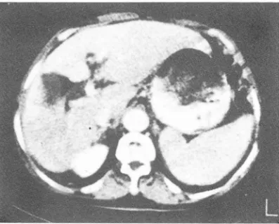
分技흘 確認할 수 있었다 (Fig.l). 냐머지 3 例에서는 맑實質과 門靜服分技의 densiψ 差異를 認知할 수 없었 다.
Fig. 1. There are evidences of 1iver cirrhosis, such as splenomegaly, & enlargement of left lobe, especially caudate lobe. Note visualization of portal veins on this postenhancement C.T.
scan.
8구î1r質內 門靜IW 分技플 確認할 수 있었던 18 例中 2 171에서는 심한 섬유성변화를 同伴한 헬性活때{生 8千찢 CC.A.H with fibrosis) 로 確앙되 었다. ~千硬化료中 뿜}끊」즈 로 確談된 1例를 除外한 全例에서 간염 표식자가 陽性
4. 考 按
一般的으로 C.T스캔 상 따內 血管構造는 造影뺑j의
Rapid intravenous infusion 이 나 i ntravenous bo 1- us i nj ectIOn에 의하여 觀察할 수도 있다 1) 그리고 따寶質內 閒防설潤때문에 용n똘質의 密度減Z↓로 인한 對 比交b果에 의 하여 j휠影前 스캔이 나 또는 통상의 단순적 주볍에 의해 따內 lÚL管構造가 보일 수도 있다1) 그러 나 통상의 단순적주법시 正常的얀 맑;寶質內에 서 괴연 正 常的£로 따門靜眼이 구별되어 보일 수 있는지 여부에 관한 文敵을 著者들은 찾아보지 못했 마. 그래서 著者들 은 맑實質 빛 機能에 臨tR的으로 異常이 없었던 50例 을 觀察해 본 結果 84 % C42 例 )에서 造影後 C.T.
스캔에서 Hî1r質과 門靜뼈分技시아의 밀도差異를 認知할 수없었다.
맑에 있어서 bolus injection실시 20 양後 즉 art - erial phase 동안에서 H.U ,는 70 정도이고약 45 쟁、 後 즉 Venous phase 동안에 H.U. 120 요로서 最高f直에 도달한다 3) 그러나 單純 적주법인 경우는 bolus inje-
ction과는 달리 대개 천천히 血中懷度가 最高個에 오 르고 이때 딴實質 density 도 最高個에 오른다 3) 般 的S로 組織과 血管內 造影협j의 平衝關係는 lfnf에서 血 管外 組織으로 30秋미만에 移動한다. 結局 注入된 造
- 263 -
影꼈에의 많은 해;5f은 注따後 따分이내에 Úll管內에서 除 去뭔디 9) 넓影뼈j의 Jfil해內 U꾀표는 用i"l1, ð:入速j풍품 排i'f!tjyßl흥, Úll쉽:外組級괴의 각;:W!Í16서係등.01/ 의해 結定된다
3) 즉 通常의 단순 적주법에 의폐서는 造影폈1 注射後 열마안되어 충분히 Jfll管內따양와 떠減內따度가 zp:術狀 態를 維持하여, bolus injection時와 김은 血管造影 t홉릿몇現像은 期待할 수 없음을 알 수 있다. 단지 時間經
j샘에 의한 월位j됨원當 造Jil3 Jlf1J Jfl iï\:의 짤化에 따븐 fff1
'r
質 全般的인 H.U. 의 핫化는 뼈찢할 수 있다 2) 本 iíff究의 9H힐化ilE 愚:1f 21 ørJ中 18ØlJ에 서 분명 한
造彩後 C.T. 스캔에서 따內 門원}派 分校가 보였다
IJ'F硬fUi능의 病웰組織學的 定義는 願爛f生 織維t쩔쩨과 0주tr 뀔의 再生結節의 形成이다4) Cf ig 2). 맘硬化뾰의 織 維增植이 始f'F도|떤 가장 먼저 그리고 가장 심한 破찮1를 Table 1. Patients with visualization of intrahepatic portal veins in postenhancement C.T. scans
Patients*
2 3 4 5
6
7t nxu ny
10 11 12
13 14 15 16
17 18
Clinical diagnosis Radionuclide scan Hepatitis marker Liver biopsy
Cirrhosis Marked hepatoceJ1ular HBsAg, HBcAb None Chronic active hepatitis dysfunction
Cirrhosis, ascites Marked hepatocellular HBeAb, HBcAb None dysfunction
Hepatitis Moderate hepatocellular HBsAg, HBcAb Chronic active hepatitis
dysfunction with fibrosis**
Cirrhosis Marked hepatoceJlular HBsAb Posthepatic cirrhosis Acute cholecystitis dysfunction
Cirrhosis Moderate hepatoceJ1ular HBsAg, HBcAb, Chronic active hepatitis
dysfunction with fibrosis
Cirrhosis Moderate hepato ceJ1ular HBsAb, HBcAb None dysfunction
Cirrhosis Marked hepatoceJ1ular HBsAb, HBcAb None
Stomach cancer None dysfunction None Macronodular cirrhosis
Hepatoma Multiple of focal defects HBsAb, HBcAb, HepatoceJ1ular carcinoma HBeAb and macronodular cirrhosis Hepatoma Single fo cal defect HBsAg, HBcAb, HepatocelIular carcinoma
HBsAg, HBcAb, and macronod ular cirrhosis
Hepatoma Single focal defect HBsAb Hepatocellular carcinoma
and macronodular cirrhosis
Hepatoma Single focal defect HBsAb Hepatocellular carcinoma
and macronod ular cirrhosis Hepatoma Single focal defect HBsAb, HBcAb HepatocelIular carcinoma
and macronodular cirrhosis
Hepatoma Sigle focal defect HBcAb Hepato cellular carcinoma
and macronodular cirrhosis Cirrhosis, ascites Marked hepato ceJ1ular HBsAg, HBcAb, None
dysfunction HBeAg
Hepatoma None HBsAg, HBcAb, Hepatocellular carcinoma
HBeAg and cirrhosis
Clonorchiasis None HBsAg, HBeAb Biliary cirrhosis
Cirrhosis Marked hepatocellular HBsAb, HBcAb None dysfunction
clide scans.
* In 3 cases with cirrhosis not included in this table, the intrahepatic portal veins were not enhanced on postenhancement C.T. scans. But they had clinical evidence of cirrhosis, hepatic markers, and diffuse hepatocellular changes on radionu-
**We include broadly simple fibrosis of liver into cirrhosis in the view of diffuse hemodynamic changes.
Table 3. Decision matrix analysis Table 2. Cases of ∞ ntrol group with visualization of
intrahepatic portal veins in postenhancement
C.T‘ scans Cirrhosis (+) Cirrhosis (-)
8 42 18
3 Test(+)
Test (-) Liver biopsy
None None None None None
Fatty infiltration None
Fatty infiltration
* AI1 cases above had no hepatitis markers.
;;7-18 x100=86(%) 21
Nosologic probalbilities Test abnormal Sensitivity=
Pathologic abnormal
~x100=84(%) 42 50
Test normal S pecificity=
Pathologic normal Clinical diagnosis
Cushing’s syndrome Chronic pyelonephritis Cushing’s syndrome Cushing’s syndrome Pancreatitis Alcoholism
Chronic cholecystitis Alcoholism
Cases*
1i
끼/-,、) A약
<
) /0
「l。。
~ 60 xlOO=74(%) 81
당하는 딴內血管構造는 洞樣血管이고 그 다음이 中心靜 服이다 5) 그러나 門靜服과 땀動派은 末期까지는 크 게 影響을 받지 않는다. 이 때 Ilf硬化훈의 構造的 變化에 익한 이차적인 血流의 變化를 보면 府細뼈 壞死後의 顧
~Ø1生 織維增꺼훌과 再生結節의 形成£로 인한 洞樣血管의 破壞 및 中心靜服의 破壞에 의 하여 lJf합質內 全般的인 vascular bed 의 減少와 더불어 中心靜服의 結節에 의 한 압박등의 要因이 합하여 門靜服壓이 上昇하게 된다
TP+TN Efficiency (Accuracy)=
TP+FP+FN+TN
Microscopic view of lh'er cirrhosis (H-E staining X100)
/
Fig.2.
Single plate of
꾀OR센& ABNORMAL
Abnormal relationship between ceIls and blood supply ir
,
cirrhotic Iiver.Fig.3.
3,4,5) Cfig .3). Porto-systemic collateral circula- tIOn 이 形成되고 더 심하면 府따11派 1[[l流뀔이 增加하여 Ilf lÚl流 供給原무로 代替된다. 이띠1 ij꾸門웹II@의 血流는
“
slow down" 되고“
to and fro" 現像을 일으키게 된다 3
,
4,
5) Cfig.4).R주硬ftJlE時 造影齊j를 注射하면 正常과 딸리 땀에 도 달하는 造影햄j의 是의 減少와 6千內 vascular bed 의 減少로 因한 확산障짧로 血管外組織內의 j훌影꼈~J 密度 도 떨어질 것이 다. 또한 문정맥의 高血壓으로 반한
m
流의 slow do빼 및 to and fro現像으로 인한, 造影 觸의 비교적 큰가지내의 停滿플 類推할 수 있다.
要約하면 著者들은 府硬化폼이 있을 경우 單純rr힘注法
£로 C.T.스캔상 땀內門靜服의 顯視現像을 (1) 府內 vasculer bed 의 減少에 의 한 府합-質內의 확산減少와 (2) 門靜服血趣上昇에 의한 血流停E뿜어l 基因된다고 생 각한다‘ 이 런 著者의 假說을 確認할 수 있는 方法은 正 常땀과 硬ftJlE을 일 S킨 府에 서 造影前 H.U.와 造影 後 H.U.의 差異를 인정할 수 있으면 可能하다. Ritch.
ings 等은 造影前
C.T.
스캔에서 正常땀과 R꾸硬化JlE 에서 慧義있는C.T.
빌도 差을 認定할 수 없다고 하。" rl 10)
/이、「
↑쩔1生 活피bi生 따淡도 組織學的으로 심 한 淡ffE性 反應, 細9힘 破懷 및 織維t曾꺼훌現像으로 특정지워진다4\Fig.
도 상당수는 IH→硬ftffE의 JÚl流 했ft와 相應하리 라고 추 정한다.
比較對照群 50 例中에서 造影增랬後 따內 門隔1m이 보 인 8例에서 쿠싱증후군이 3 찌1], Alcoholion이 2 例, 기타 3例였마. 이 경우 8 찌中 5 {91J는 H구때質l칙 服助 홉潤으로 H.U의 減少로 인한 門靜服分技의 顯視現像 이 틀림없다 1
,
11) •結局 이 造影後 C.T. 스켄에서 Ilf內門靜Il!ii分技의 題 視現像은 對p,~、現像에 基因한 따8읍助홈潤을 일으키는 훌 愚을 除外하더라도 R주硬ftffE에만 특유한 것은 아니지만, 廣範園한 意味에 서 따합質內 @쟁爛性 織維t曾꺼훨을 일으킬 수 있는 흉愚에 적용될 수 있을 것이다.
5). 또한 이 것이 딴硬化효으로 상당수 發展해 간다는 것 Fig. S. Microscopic view of chronic active hepatitis 은 잘 알려 진 사실 이 다 著者들은 이 慢性 活펴性 府淡 with fibrosis (H.E staining X200)
hv
.. .. +.
ha pV
A B C D
A. Normal liver B. Early cirrhosis C. Moderate to D. Advanced cirrhosis (stãge 1) severe cirrhosis (Stage 111)
(Stage II)
s; sinusoids pv; portal vein ha; hepatic artery hv:hepatic veins Fig.4.
5. 結 등6. i쩌
1)#千맺íl::챈에 있어 서 造彩i염행後 C.T. 映保1:: IJf內 門靜服分校의 題5、많隊의 Nosologic Probability 는感 受性 CSensitivity) 86 %, 特짧i生 CSpecificity) 84 %,
JE確많 CAccuracy) 74 %이 었 다.
2) ÚIl流力學的 측맨에서 fff뻐 ítllE 廠念에 f웰生 活動 {生 9f淡의 織維↑生 t曾植CChronic Active Hepatitis wi-
th fibrosis)플 포함시켰다.
3) 이 Sign은 따內廳爛{生織維增뼈을 일으키는 흉恩 에 敏用될 수 있다.
Contrast Enhancement of Liveι Contrast media in com- puted tomography (International Congress Series ; No 561) Excerpta Medica, Amster. 1981
5. Mac5ween RNM, Anthony PP, 5cheurer PJ : Pathology of the Liver. Churchill Livingstone, London, 1979 6. Renter 5R : Castrointestinal angiography. 2nd Ed. ~ν B.
Saunders Company. Philadelphia, 1978
7. Okuda K, lio M : Radiological Aspect of Liver and Biliary Tract. Igaku Shoiπ Tokyo, 1976
8. Kuhns LR, Borlaza G5, 5eigel R, Rozderac R, 5immons j ’ Lack of Visualization of the Portal venous Tree in Cir- rhosis of the Liver: A Computed T omography Finding with Possible Diagnostic Significance. j. Comput. Assist T omogr 2: 400-403, 1978
REFERENCES 9. Newhouse J Fluid Compartment Distribution of In-
travenous lotha/amate in the Dog. Invest. Radiol. 12 1. Moss M , Genant Hκ Gamsu G : Computed tomography 364-367, 1977
of the body. W. B. Saunders, Phi/ade/phia, 1983. 10. Richings RT, Pullman BR, Lucas 5B, Fawcitt RA, Bost JJK, 2. Felix R, Kazner E, wegener OH : Contrast Media in Com- Isherwood 1, Morris AI : An Ana/ysis of the Spatia/ Distribu-
puted Tomoraphy. (Internationa/ Congress series : No. tion of Attenuation Va/ues in Computed Tomographic 561). pp. 259, Excerpta Media, Amsterdam, 1981. Scans of Liver and Sp/een. j. Comput. Assist Tomogr 3. Burman 5, Rosenbaum AE : Rationa/e and Techniques 3:36-39, 1979
for Intravenous Enhancement in Computed tomograp/hy. 11. Isselbacker KJ : Harrison's princip/es of Interna/ Medicine Radio/. Clin. of North Am. 20:15-22, march 1982. 9th ed. McCraw Hill New York, 1980
4. Baert AL, Usewils R, Marchal G, Wilms G, Pouette E :