Journal of Korean Spine Surg.
Vol. 14, No. 1, pp 25~33, 2007
Address reprint requests to Seong-Hwan Moon, M.D.
Department of Orthopaedic Surgery, Yonsei University, College of Medicine
#134 Shinchon-Dong, Soedaemun-ku, Seoul, 120-752, Korea
Tel: 82-2-2228-2188, Fax: 82-2-363-1139, E-mail: shmoon@yumc.yonsei.ac.kr
� 본 연구는 과학기술부 기초과학 연구사업 R01-2006-000-10933-0, 연세대학교 의과대학 Brain Korea 21 Project에 의해 지 원되었습니다.
퇴행 비후된 척추 후관절이 황색인대의 섬유화 및 골화에 미치는 영향
이광일
#∙김 향∙장주웅
#∙전흥재
+∙김현민
++∙박시영
$김슬기∙이환모∙김학선∙문성환
연세대학교 의과대학 정형외과학교실, (주)코리아본뱅크 생체재료 의공학연구소#,
고려대학교 의과대학 정형외과학교실$, 연세대학교 공과대학 기계공학과+, 연세대학교 공과대학 재료공학과++
The Effect of Synovial Fluid from Degenerated Facet on Hypertrophy and Ossification of the Ligamentum Flavum
Kwang-Il Lee, M.S.#, Hyang Kim, M.S., Ju-Woong Jang, Ph.D.#, Heoung-Jae Chun, Ph.D.+, Hyun-Min Kim, Ph.D.++, Si-Young Park, M.D.$, Sul-Ki Kim, M.D.,
Hwan-Mo Lee, M.D., Hak-Sun Kim, M.D., Seong-Hwan Moon, M.D.
Department of Orthopedic Surgery, Yonsei University College of Medicine, Institute for Biomaterial Research, Korea Bone Bank Co. Lt
#, Department of Orthopedic Surgery, Korea University College of Medicine
$,
Department of Mechanical Engineering
+, Yonsei University, School of Advanced Materials Engineering
++, Yonsei University
– Abstract –
Study DDesign: In vitro experimental study
Objectives: To examine the effect of a synovial supernatant on the cell viability, osteogenic phenotype, mRNA expression of the types collagen and various transcriptional factors on osteogenesis in ligamentum flavum (LF) cells stimulated with synovial fluid from a degenerated facet joint.
Literature RReview: In degenerative lumbar spinal stenosis, hypertrophied LF or osteoarthritic hypertrophy of a facet joint often causes neurogenic claudication. The facet joint is a synovial joint with hyaline cartilage on each side. Therefore, osteoarthritis of a facet joint eventually occurs with aging and other degenerative conditions of the spine. In lumbar spinal degeneration, inflammatory mediators or cytokines are released from the facet joint tissue, which consequently affects the adjacent LF because the LF covers posterolateral aspect of the spinal canal near facet joints. However, there are no reports on the relation- ship between a degenerated facet joint fluid and the LF in the lumbar spine.
Materials aand MMethods: LF surgical specimens were obtained from patients with a lumbar spine stenosis, and the cells were iso- lated by enzymatic digestion. Each of the synovium tissues were weighed and recorded. Each tissue was cut into small pieces with a pair of scissors and then washed 3 times with PBS. The washed tissue pieces were then cultured for 96 hr at 37�C, 5%
CO2in DMEM/F-12-0.1% FBS with a density of 200 mg/ml medium. The supernatant was collected after 96 hr. In order to
서 론
퇴행성 요추협착증은 중앙의 척추관, 신경근이 주행 하는 후관절의 전내측에 있는 신경관 또는 추간공이 좁 아지면서 신경에 압박이 오고, 물리적 화학적 변화를 일 으켜 요통이나 하지에 여러 가지 복합된 신경증상을 일 으키는 질환이다1)
. 요추협착증의 주요 병인으로는 돌출
된 추간판, 후관절의 염증성 비후, 그리고 황색인대의 섬유화에 따른 비후를 들 수 있다.이 중 요추 후관절은 초자 연골로 이루어진 활액막 관 절로서 개개의 관절 가동범위는 적지만 복합운동을 하 기 때문에 전체적으로 상당한 운동범위을 보인다. 요추 후관절의 관절면은 주로 시상면에서 45도 정도로 이루 어져 있으며, 그 형태는 원형 내지 타원형으로 편평하거 나 곡선 형태이고, 주로 신전과 굴곡운동에 관여하고 있 다. 척추 후관절의 활막은 통증에 예민한 조직이며, 척추 에 가해지는 하중의 1/4 정도가 후관절로 전달된다. 따라 서 요추 후관절의 관절염은 나이가 들면서 점차적으로 진행되고 이로 인한 척추의 퇴행 현상도 나타난다. 퇴행 성 요추 병변이 심해지면 염증성 cytokine들이 요추 후관 절로부터 배출됨으로써 인접한 황색인대의 퇴행성 변화 에 영향을 끼칠 수도 있다2,3,4,5,6)
. 퇴행된 추간판이 황색인
대의 비후 및 골화에 영향을 끼칠 수 있다는 연관성을 제 시하는 보고는 있지만7,8,9), 퇴행 비후된 후관절과 황색인
대의 비후 및 골화 간의 관계에 대한 연구는 없었다.따라서 본 연구에서는 시험관 내에서 분리 배양한 황 색인대 세포를 척추협착증 환자로부터 퇴행성 척추 후 관절의 활액막을 추출하였고, 그것을 조직 배양하여 추
출한 활액막액을 세포에 자극시켰을 때, 시간이 지남에 따라 활액막액이 황색인대 세포의 독성 및 증식도, 교원 질 생성 및 골형성에 어떤 영향을 끼치는지에 대하여 알 아보았다.
연구 대상 및 방법
1. 세포 배양
요추 척추관 협착증으로 감압술을 시행받은 10명의 환 자들에게서 수술시 퇴행 비후된 척추 황색인대를 채취 하였다. 황색인대 세포 배양은 이전에 보고된 방법을 이
용하였다
7). 자세히 기술하면 채취된 황색인대 조직을 멸균된 DPBS (Dulbecco’s Phosphate Buffered Saline, Gibco-
BRL, Grand island, NY)로 한 차례 닦아서 환자의 혈액 성분을 제거한 후, 5%의 우태아 혈청(Fetal bovine serum,
Gibco-BRL, Grand Island, NY)이 들어 있는 Dulbecco’s Modified Eagel’s medium (DMEM 배지)에서 미세 가위를이용하여 조직을 잘게 잘랐다. 잘게 자른 황색인대 조직 단편을 250 U/ml의 제 1A형 collagenase (Sigma, St. Louis,
MO, USA)가 함유된 DMEM 배지에서 2시간 동안 효소처리하였다. 효소 처리한 황색인대 조직을 5%의 우태아 혈청이 들어 있는 배지로 두 차례 씻어낸 후, 10% 우태아 혈청과 100 unit/ml penicillin, 100 ug/ml streptomycin (all
antibiotics from Gibco-BRL, Grand Island, NY)이 들어 있는 DMEM 배지로 교반하여 37�C, 5% CO
2배양기에서 배양하였다. 배지는 3일에 한 번씩 교환하였다. 계대수
measure quantitatively the proliferation of cells, the AlamarBlue assay was used. The total cellular RNA was extracted from the cells and amplification reactions specific to the following types of cDNA were performed: the osteogenic master transcrip- tion factors, Dlx5, Runx2, osterix, and types collagen and osteocalcin. Alkaline phosphatase staining for the biochemical assay and western blotting for osteocalcin protein expression were performed.Results: Human LF cells cultured with the supernatant from the facet synovium showed a slightly stronger AlamarBlue stain- ing than the intensity of the control culture. RT-PCR revealed the upregulation of the osteogenic master transcription factors, Dlx5, Runx2, and osterix in the synovium supernatant group from one hour to 72 hours, and an increase in osteocalcin, types collagen I, III, V, XI levels from one hour to one week. LF cells cultured with the supernatant from the facet synovium showed positive staining for alkaline phosphatase. The level of the osteocalcin protein in the LF cells cultured with the supernatant from the facet synovium was higher than the control group.
Conclusions: The supernatant of the facet joint from patients with degenerative spinal stenosis affects LF cells by increasing the level of cellular proliferation, upregulating the mRNA expression of osteocalcin, types of collagen, osteogenic transcription fac- tors, positive alkaline phosphatase staining, and osteocalcin protein expression. Therefore, degenerated synovial fluid from the facet joint is an important mechanism of LF hypertrophy and ossification.
Key WWords: Ligamentum flavum, Facet joint, Synovium, Synovial fluid, Spinal stenosis
가 2일 때 trypsin/EDTA 효소 처리법으로 인간 황색인대 세포를 수집하여 액체 질소에 냉동 보관하였다.
2. 조직 배양 및 상층액 처리
요추 척추관 협착증 환자 20명에게서 척추 감압술을 시행시 퇴행된 척추 후관절 활액막을 채취하였다. 채취 된 활액막 조직을 일정량의 DPBS가 들어 있는 Nunc
tube에 넣어 그 수치의 차이를 이용해 무게를 확인한 후, 200 mg/ml의 농도로 0.1% 우태아 혈청이 함유된 Dulbec- co’s Modified Eagle Medium and Hams F-12 medium (DMEM/F-12 배지)을 처리한 후, 37�C, 5% CO
2 배양기 에서 3일간 배양하였다9). 배지는 매일 교환하였으며, 각
각의 상층액을 tube에 모아서 centrifuge로 조직의 잔여 물을 걸러낸 후 상층액만 모아서 실험에 사용하기 전까 지 -70�C에 냉동 보관하였다. 실험에 필요한 상층액은 녹여서 0.1% 우태아 혈청과 10 mM Glycerophosphate가 함유된 α-MEM 배지와 1:1의 비율로 혼합한 후, 각각의well plate에 분주된 황색인대 세포에 처리하여 37�C, 5%
CO
2배양기에서 배양하였다.3. AlamarBlue assay를 통한 세포 증식 반응 측정
96 well plate에 각각 분주된 황색인대 세포에 세포의
대사 활성을 감지하기 위한 10% AlamarBlue solution(Serotec Ltd, Oxford, UK)을 처리하여 37�C, 5% CO
2 배 양기에서 배양하면서 시간(6, 12, 24, 48, 72시간)이 지남 에 따라 plate reader를 사용하여 570 nm과 600 nm 파장 에서의 흡광도를 측정하여 시간에 따른 세포의 증식 및 활액막 상층액의 독성 여부를 분석하였다10).
4. Reverse Transcriptase-Polymerase Chain Reaction 을 이용한 mRNA 발현 검사
RNeasy mini kit (QIAGEN, Maryland, USA)를 이용하여 Total RNA를 분리하였다. 황색인대 세포의 Total RNA 1 μg
와 oligo (dT) 1 μl (Invitrogen, USA, 0.5 μg/μl)와 3차 증류수를 혼합하여 50 μl으로 혼합한 후, AccuPower RT-premix(Bioneer,
대전, 한국)에 넣어 Reverse Transcription Poly-merase Chain Reaction (RT-PCR)을 하였다. 42�C에서 60분, 94�C에서 5분간 반응하여 cDNA를 합성하였고, 합성한 cDNA를 에탄올로 정제하여, 최종 부피 20 μl를 만들었다.
이 중 각각의 cDNA 1 μl를 취하여, sense primer와 antisense
primer 10 pmole, 3차 증류수를 혼합하여 10 μl의 반응용액
을 만들어 Sapphire PCR-premix (Sapphire, USA)에 넣고, RT(Real Time)-PCR을 시행하였다. PCR 생성물은 2% agarose
젤에서 전기영동을 통해 각 유전자의 발현을 검출하였다(Table 1, 2)
11,12,13,14). RT-PCR에 대한 대조군으로 GAPDH를
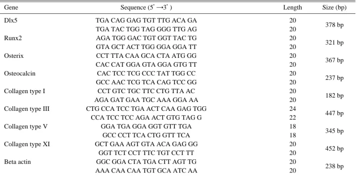
사용하였으며 TINA 2.0 program을 통해 각각의 발현 정도 를 비교 분석하였다.Table 1. Sequences of primers used for reverse transcription-polymerase chain reaction to amplify various cDNA
Gene Sequence (5’→3’) Length Size (bp)
Dlx5 TGA CAG GAG TGT TTG ACA GA 20
378 bp
TGA TAC TGG TAG GGG TTG AG 20
Runx2 AGA TGG GAC TGT GGT TAC TG 20
321 bp
GTA GCT ACT TGG GGA GGA TT 20
Osterix CCT TTA CAA GCA CTA ATG GG 20
367 bp
CAC CAT GGA GTA GGA GTG TT 20
Osteocalcin CAC TCC TCG CCC TAT TGG CC 20
237 bp
GCC AAC TCG TCA CAG TCC GG 20
Collagen type I CCT GTC TGC TTC CTG TTA AC 20
182 bp
AGA GAT GAA TGC AAA GGA AA 20
Collagen type III CTG CCA TCC TGA ACT CAA GAG TGG 24
447 bp
CCA TCC TCC AGA ACT GTG TAG G 22
Collagen type V GGA TGA GGA GGT GTT TGA 18
345 bp
GCC CCT TCA CTG GTT TCA 18
Collagen type XI GCT GAA AGT GTA ACA GAG GG 20
452 bp
GGT TCT CCT TTC TGT CCT TT 20
Beta actin GGC GGA CTA TGA CTT AGT TG 20
238 bp
AAA CAA CAA TGT GCA ATC AA 20
5. Western Blot analysis를 이용한 osteocalcin 발현 검사
Lysis buffer (0.5% Triton X-100, 10 mM HEPES, 150 mM NaCl, 0.02% sodium azide, protease inhibitor mixture) (Sigma)를 이용하여 cell lysate를 만들고 이를 10% tricine- SDS gel을 이용하여 분리하고 전이하였다. Osteocalcin를
분석하기 위해 1×TBST with 5% 탈지유에 block 후 blot-ted membrane을 rabbit anti-osteocalcin antibody (1:10,000 dilution)(Chemicon international, Temecula, CA)에 1시간
노출시켰다. Membrane은 1×TBST으로 세척 후 sec-ondary antibody (1:10,000 dilution of goat anti-rabbit IgG, horseradish perocidase conjugated, Santa Cruz, CA)에 노출
시키고 상온에서 45분간 보관하였다. Immuno-reactivebands는 1×TBST로 3번 세척 후 ECL kit (Amersham Phar- macia, Piscataway, NJ)를 이용하여 분석하였다.
6. Alkaline phosphatase 염색
황색인대 세포를 citrate buffered acetone (Sigma)이 들 어 있는 고정액을 사용하여 30초 동안 고정시킨 후, 증 류수로 45초 동안 씻어 내고 미리 준비해 둔 alkaline-dye
mixture (Sigma)를 각 well에 적용하였다. 이후 상온에서
빛을 차단한 채 30분 동안 반응시켰다. 반응시킨 세포를2분 동안 증류수로 씻어낸 후 Mayer’s Hematoxylin 용액
에서 10분간 반응시켰으며, 반응 후 다시 한 번 증류수 로 1분간 씻어 내어 관찰하였다15).
7. 실험군
황색인대 세포에 0.1% 우태아 혈청, 10 mM β-Glyc-
erophosphate이 함유된 α-MEM, 그리고 활액막 조직 상
층액을 1:1로 혼합한 용액을 각각 처리하여 배양하였다.각 배양군에서 세포 증식 반응 및 골형성 전사인자
(Runx2, Dlx5, Osterix)와 제 1형, 3형, 5형, 11형 교원질,
그리고 osteocalcin에 대한 mRNA 발현을 측정하였으며,Alkaline phosphatase 염색 및 Western Blotting을 이용해
서 골형성 표현형 및 osteocalcin 발현 검사를 하였다. 한 편, 활액막 조직 상층액 대신 우태아 혈청이 함유되지 않은 DMEM/F-12를 동량 처리한 황색인대 세포를 이 실 험의 대조군으로 정하였다.8. 통계
위와 같은 실험을 3회에 걸쳐서 반복하여 결과를 얻었 으며, 모든 자료는 대조군에 대한 백분율로서 평균±표
Fig. 1. Alamarblue assay for cytotoxicity and cellular metabo- lism. Control (white round) denotes ligamentum flavum cell culture with culture media. Supernatant (black square) denotes ligamentum flavum cell culture with supernatant mixture from facet joint synovium culture.
Table 2. Conditions of reverse transcription-polymerase chain reaction
Primer Conditions
Denaturation Annealing Polymerization Cycles
Dlx5 94�C 05 sec 58�C 05 sec 72�C 30 sec 35
Runx2 94�C 05 sec 58�C 05 sec 72�C 30 sec 35
Osterix 94�C 05 sec 55�C 05 sec 72�C 30 sec 30
Osteocalcin 94�C 05 sec 60�C 05 sec 72�C 30 sec 30
Collagen type I 94�C 05 sec 48�C 05 sec 72�C 30 sec 25
Collagen type III 94�C 30 sec 54�C 30 sec 72�C 01 min 30
Collagen type V 94�C 30 sec 60�C 30 sec 72�C 01 min 35
Collagen type XI 94�C 30 sec 54�C 30 sec 72�C 01 min 40
Beta-actin 94�C 05 sec 53�C 05 sec 72�C 30 sec 24
준편차로 표시하였으며, SPSS (SPSS Inc. Chicago IL)를 이용하여 처리하였다. One-way Analysis of variance 및
Fisher’s protected LSD post-hoc test, power analysis로 실
험군간 비교하였다. 통계 방법의 유의 수준은 p<0.05로 정하였다.결 과
1. Alamarblue를 통한 세포 독성 및 세포 증식
Alamarblue solution을 통해 시간에 따른 세포의 독성
및 증식을 알아본 결과, 활액막 조직 상층액을 처리한 황색인대 세포는 대조군보다 약 10% 높은 세포 대사 활 성을 보였다. 또한 세포를 배양한 지 각각 6시간과 12시 간대, 그리고 24시간과 48시간대의 사이에서 세포의 대 사 활성이 급격하게 증가하였다(Fig. 1).Fig. 2. Reverse transcription-polymerase chain reaction prod- ucts of osteogenic transcription factors collagens and osteocalcin. (A) Dlx5, Runx2, Osterix. (B) Collagen type I, III, V, XI and osteocalcin. Early osteogenic (Dlx5, Runx2, Osterix) and late osteogenic marker (osteocalcin) and various collagens (type I, III, V, XI) showed increase in expression with treatment of synovi- um supernatant from degenerated facet joint.
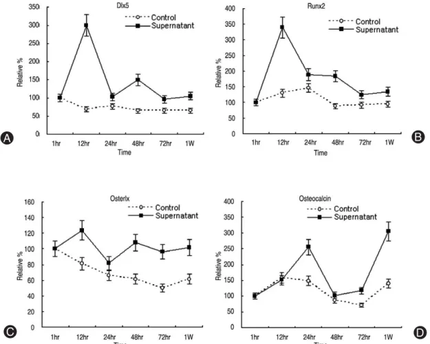
Fig. 3. Densitometry of osteogenic mRNA expression. (A) Dlx5, (B) Runx2, (C) Osterix, (D) Osteocalcin. Dlx5 and Runx2 mRNA showed increase in expression in 6 to 12 hours after synovial supernatant treatment. Late osteogenic marker (osteocalcin mRNA) showed upregulation 1 week after synovium supernatant treatment.
2. 골형성 전사인자인 Dlx5, Runx2, osterix, 그리고 osteocalcin의 mRNA 발현
황색인대 조직의 골화와 관련하여 대표적 골형성 전 사인자인 Dlx5, Runx2, osterix의 초기 mRNA 발현 정도 를 RT-PCR에 대한 전기영동으로 알아보았다. 각각의
mRNA 발현을 densitometry를 통해 분석한 결과, 활액막
조직 상층액을 처리한 황색인대 세포 실험군은 대조군 에 비해 더 높은 발현 증가를 보였다. 특히 배양한 지 6 시간 후의 실험군의 경우, 1시간 후에 비해 Dlx5는300%, Runx2는 340%, 그리고 osterix는 123%의 발현 증
가율을 보였다. 또한 6시간 이후에 시간이 지날수록 대 조군은 골형성 전사인자들의 발현이 줄어드는 반면에, 실험군은 꾸준한 증가율을 보였다. 역시 골형성 표현인 자인 osteocalcin mRNA 발현의 경우도 대조군에 비해 높 은 발현 증가를 하였으며, 1주일째에는 초기 1시간 배양 에 비해 300%의 발현 증가를 보였다(Fig. 2, 3).3. 제 1형, 3형, 5형, 11형 교원질의 mRNA 발현 황색인대 조직의 섬유화와 관련하여 세포의 제 1형, 3 형, 5형, 그리고 11형 교원질 mRNA의 발현 정도를 den-
sitometry를 통해 분석한 결과, 활액막 조직 상층액을 처
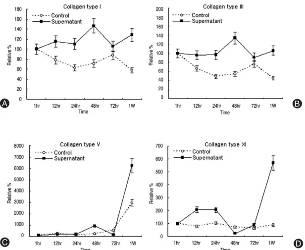
리한 황색인대 세포 실험군은 대조군에 비해 더 높은 발 현 증가를 보였다. 제1형, 3형 교원질의 경우 대조군에Fig. 4. Densitometry of collagen mRNA expression. (A) Collagen type I, (B) Collagen type III, (C) Collagen type V, (D) Collagen type XI. Type I, III, V, XI mRNA expressions were upregulated with course of time after treatment of synovium supernatant.
Fig. 5. Expression of osteocalcin protein in supernatant of liga- mentum flavum cell culture with synovium supernatant (Sup) for 2 weeks. Expression was detected by Western blot analysis. Negative control denotes ligamentum flavum cell with culture media mixture and positive control denotes ligamentum flavum cell with BMP-2 (100 ng/ml).
비해 꾸준한 발현 증가를 하였으며, 48시간 및 1주일째 에서 활발한 증가를 보였다. 특히 제 5형 교원질은 1주 일째에서 6200%의 가장 높은 증가율을 보였으며, 제 11 형 교원질은 배양 초기에 꾸준한 증가율을 보이다가 1 주일째에는 570%의 발현 증가를 하였다(Fig. 2, 4).
4. 활액막액 처리에 의한 인간 황색인대 세포의 Osteocalcin 단백질 발현
Osteocalcin 단백 발현을 검증하기 위해 배양한 지 2주
일 후에 Western Blot analysis를 시행하여 본 결과, 16kDa의 osteocalcin이 검출되었다. 인간 황색인대 세포에
배지의 혼합액만 처리한 배양군을 음성대조군으로, 세 포에 활액막액 상층액과 배지의 혼합액에 BMP-2 (100ng/ml)을 처리한 배양군을 양성대조군으로 설정하였다.
음성대조군에서 osteocalcin 발현이 미미하게 발견되었 으나 양성대조군 및 실험군에서는 많은 양의 osteocalcin 이 검출되었다(Fig. 5).
5. 활액막액 처리에 의한 인간 황색인대 세포의 alkaline phosphatase 염색
황색인대 세포에 활액막액을 처리한 후 1주일이 지나 초기 골형성 표현인자의 생성 여부를 확인하고자 Alka-
line phosphatase 염색을 실시한 결과, 발현됨을 관찰할
수 있었다. 대조군에서는 골형성 인자가 관찰되지 않았 으나 활액막액을 처리한 세포 배양군에서는 명백한 골 형성 표현인자가 생성된 것을 확인할 수 있었다(Fig. 6).고 찰
척추 황색인대의 비후의 기전에 대해서 여러 가지 실
험적 연구가 있었다. 반복적인 기계적 신연 자극에 의한
transforming growth factor beta1 발현에 의한 세포 증식
및 교원질 발현 증가16)노화 및 인대 자체의 퇴행성 변화 에 의한 비후1)등이 제시되고 있다. 본 연구에서는 척추 후관절과 인접한 황색인대가 척추 후관절의 퇴행성 변 화에 의한 염증성 활액의 작용으로 비후 혹은 골화될 수 있다는 가설하에 실험적 연구를 진행하였다.본 연구에서 인간 척추 황색인대에 퇴행된 활액막에 서 얻어진 활액막액을 처리한 결과, 척추 황색인대의 골 화 및 섬유화가 되는 것을 실험적으로 증명하였다. 퇴행 된 척추 활액막으로부터 얻은 활액막액을 처리한 황색 인대 세포는 초기 골형성 전사인자인 Dlx5, Runx2,
Osterix 등의 mRNA 발현이 증가하였고, 후기 골형성 인
자인 osteocalcin mRNA 발현도 증가하였다. 골형성 표현 형인 osteocalcin의 단백 발현이 검출되었으며, 골형성 표 현인자인 alkaline phosphatase 염색에서 양성 반응이 있 었음으로 척추 후관절에서 기인한 활액막액이 척추 황 색인대의 골화에 영향을 끼친다는 것을 확인할 수 있었 다. 그리고 섬유화와 관련된 제 1형, 3형, 5형, 그리고 11 형의 교원질의 mRNA 발현이 시간이 지남에 따라 급격 하게 증가함을 보면 역시 척추 후관절 활액막액이 황색 인대의 섬유화에도 영향을 끼친다는 것을 알 수 있었다.골형성의 완전히 증명하기 위해서는 장기 배양 후 골 형성 결절을 염색하는 von Kossa 염색이 필요하나 본 연 구에서는 장기 배양에서도 의미있는 염색 결과를 보이 지 않았는데 이는 활액막의 골형성능이 bone morpho-
genetic protein-2 같은 강력한 골형성 인자에 비해서는
아주 약한 골형성 능력을 보임으로 실험 기간 중에 의미 있는 골결절은 형성하지 않았다고 추정된다.본 연구의 결과로 기존의 반복적 기계적 신연에 의한
transforming grwth factor beta1 발현
16), 퇴행된 추간판 조
직에서 발현하는 염증성 cytokine류에 의한 자극, 추간 판 간격 감소로 인한 황색인대의 겹침(buckling) 현상 등 에 추가하여 퇴행된 척추 후관절에서 분비되는 염증성 활액막액도 척추 황색인대의 비후와 골화에 중요한 기 전임을 알 수 있다.이상의 연구들과 본 연구의 결과를 종합하면 척추 황 색인대 비후 및 골화는 단순히 인대 자체의 변성 퇴화, 일생에 걸친 반복적 신연에 의한 성장인자(transforming
growth factor-beta1) 발현에 의한 비후, 퇴행된 추간판의
추간판 간격 감소로 인한 후방구조물 황색인대의 겹침 현 상 , 퇴 행 된 추 간 판 조 직 에 서 발 현 되 는 염 증 성cytokine들에 의한 비후 및 골화, 그리고 본 연구에서 증
명된 퇴행된 척추 후관절에서 분비되는 염증성 활액막 에 의한 비후 및 골화로 종합될 수 있다. 기술된 모든 기 전이 황색인대의 교원질 형성을 촉진시켜 섬유화를 이 Fig. 6. Alkaline phosphatase stain. Ligamentum flavum cellculture with synovium supernatant for 1 week showed positive alkaline phosphatase stain.
루는 최종 과정을 거치게 되어 있으므로 치료적 의미로 국소적으로 교원질화의 방해를 유도할 수 있다면 노화 및 퇴행성 변화로 진행되는 황색인대의 비후를 부분적 으로 막을 수 있을 것이며 이는 침습적 수술을 피할 수 있는 방법이 될 수도 있다. 현재 가장 유망한 항 섬유화 인자는 relaxin17)으로 추정되며 이와 관련한 연구가 진행 되어야 할 것이다.
본 연구의 의의는 문헌상 최초로 퇴행된 척추 활액막 으로부터 나온 활액막액이 척추 황색인대의 골화 및 섬 유화에 영향을 끼친다는 사실을 분자생물학적 그리고 조직 면역학적 기법을 동원하여 골성 인자 및 교원질
mRNA의 발현, 골성 표현형의 발현, 그리고 alkaline phosphatase
발현 등을 통해 증명하였다는 것이다. 그럼 에도 불구하고 본연구의 제한점도 있는데 이는 척추 후 관절 활액막액을 활액막의 조직 배양에서 추출하였음 으로 생체 내의 환경을 반영할 수 없다는 점과 해부학적 으로 척추 후관절과 황색인대가 인접한 구조이기는 하 나 적접적으로 척추 후관절 활액막액이 황색인대 조직 에 전체적으로 침투하여 세포 증식 및 섬유화를 야기한 다고 실제 조직에서 증명하기가 어렵다는 점이다.결 론
이상의 결과로 퇴행성 척추협착증 환자로부터 추출한 요추 후관절의 활액막 조직의 상층액은 척추 황색인대 세포의 세포 증식 및 골형성 인자(osteocalcin, Dlx5,
Runx2, osterix mRNA 발현, alkaline phosphatase 염색 반
응, osteocalcin 단백질 발현) 발현과 교원질(제 1형, 3형,5형, 11형 교원질 mRNA) 발현의 증가에 영향을 주었다.
따라서 퇴행성 척추 후관절 활액막액은 황색인대의 비 대 및 골화에 중요한 영향을 발휘한다고 사료된다.
참고문헌
01) Lee HM: Pathophysiology of lumbar spinal stenosis. J Kor Spine Surg 2000; 7: 100-105.
02) Igarashi A, Kikuchi S, Konno S, Olmarker K: Inflam- matory cytokines released from the facet joint tissue in degenerative lumbar spinal disorders. Spine 2004; 29:
2091-2095.
03) Keiseki K, Segami N, Sun W, Sato J, Fujimura K:
Analysis of tumor necrosis factor-alpha, interleukin-6, interleukin-1beta, soluble tumor necrosis factor receptors I and II, interleukin-6 soluble receptor, interleukin-1 solu-
ble receptor type II, interleukin-1 receptor antagonist, and protein in the synovial fluid of patients with temporo- mandibular joint disorders. Oral Surg Oral Med Oral Pathol Oral Radiol Endod 2005; 99: 276-284.
04) Lohmander LS, Atley LM, Pietka TA, Eyre DR: The release of crosslinked peptides from type II collagen into human synovial fluid is increased soon after joint injury and in osteoarthritise. Arthritis Rheum 2003; 48: 3130- 3139.
05) Lettesjo H, Nordstrom E, Strom H, et al: Synovial fluid cytokines in patients with rheumatoid arthritis or other arthritic lesions. Scand J Immunol 1998; 48: 286-292.
06) Haynes MK, Hume EL, Smith JB: Phenotypic charac- terization of inflammatory cells from osteoarthritic synovi- um and synovial fluids. Clin Immunol 2002; 105: 315-325.
07) Specchia N, Pagnotta A, Gigante A, Logroscino G, Toesca A: Characterization of cultured human ligamen- tum flavum cells in lumbar spine stenosis. J Orthop Res 2001; 19: 294-300.
08) Okuda T, Baba I, Fujimoto Y, et al: The pathology of ligamentum flavum in degenerative lumbar disease. Spine 2004; 29: 1689-1697.
09) Li H, Zou Z, Baatrup A, Lind M, Bunger C: Cytokine profiles in conditioned media from culture human inter- vertebral disc tissue. Acta Orthop 2005; 76: 115-121.
10) Nociari MM, Shalev A, Benias P, Russo C: A novel one- step, highly sensitive fluorometric assay to evaluate cell- mediated cytotoxicity. J Immunol Methods 1998; 213: 157- 167.
11) Derfoul A, Carlberg AL, Tuan RS, Hall DJ: Differen- tial regulation of osteogenic marker gene expression by Wnt-3a in embryonic mesenchymal multipotential progen- itor cells. Differentiation 2004; 72: 209-223.
12) Luppen CA, Leclerc N, Noh T, et al: Brief bone mor- phogenetic protein 2 treatment of glucocorticoid-inhibited MC3T3-E1 osteoblasts rescues commitment-associated cell cycle and mineralization without alteration of Runx2.
J Biol Chem 2003; 278: 44995-45003.
13) Ryoo HM, Hoffmann HM, Beumer T, et al: Stage-spe- cific expression of Dlx-5 during osteoblast differentiation:
involvement in regulation of osteocalcin gene expression.
Mol Endocrinol 1997; 11: 1681-1694.
14) Lee MH, Kwon TG, Park HS, John MW, Ryoo HM:
BMP-2-induced osterix expression is mediated by Dlx5 but is independent of Runx2. Biochem Biophys Res Com- mun 2003; 309: 689-694.
15) He J, Jiang J, Safavi KE, Spangberg LS, Zhu Q: Emdo- gain promotes osteoblast proliferation and proliferation and differentiation and stimulates osteoprotegerin expres- sion. Oral Surg Oral Med Oral Pathol Oral Radiol Endod 2004; 97: 239-245.
16) Nakatani T, Marui T, Hitora T, Doita M, Nishida K,
Kurosaka M: Mechanical stretching force promotes col- lagen synthesis by cultured cells from human ligamentum flavum via transforming growth factor-beta1. J Orthop Res 2002; 20: 1380-1386.
17) Sherwood OD: Relaxin’s physiological roles and other diverse actions. Endocrine Rev 2004; 25: 205-234.
※ 통신저자 : 문 성 환
서울특별시 서대문구 신촌동 134 연세대학교 의과대학 정형외과학교실
Tel: 82-2-2228-2188 Fax: 82-2-363-1139 E-mail: shmoon@yumc.yonsei.ac.kr
연구계획: 인간 황색인대 세포에 퇴행성 척추 후관절로부터 나온 활액막액을 처리한 후 배양한다.
연구목적: 시간이 지남에 따라 활액막액이 황색인대 세포의 증식, 교원질 생성 및 골형성에 어떤 영향을 끼치는지에 대하여 알아보았다.
대상 및 방법: 요추협착증으로 수술한 환자에게서 황색인대 조직을 채취하여 황색인대 세포를 배양하였으며 척추 후관절에서 활액막을 제거한 후 조직 배양하였다. 활액막 조직을 96시간 동안 조직 배양하여 모은 상층액을 황색인 대 세포에 처리하여 배양하였으며, 시간에 따른 세포의 대사 활성 및 생존력을 확인하기 위해 alamarblue assay를 실 시하였고, 전사 수준에서의 골형성 관련 유전자 및 osteocalcin, 그리고 교원질 유전자의 변화를 확인하고자 RT-PCR 을 통한 densitometry를 분석하였다. 또한 골형성 표현형 확인을 위해 alkaline phosphatase staining을 실시하였고, osteocalcin 단백질 발현 정도를 알아보고자 western blotting을 실시하였다.
결과: 활액막 조직 상층액을 처리한 황색인대 세포의 실험군에 Alamarblue assay를 한 결과, 대조군보다 활발한 대사 활성을 보임으로써 상층액이 무독성인 것을 확인하였으며, 세포들은 일정 시간에서 급격한 대사 활성의 증가를 보 였다. 또한 대표적인 골형성 전사인자인 Dlx5, Runx2, 그리고 osterix들의 1시간에서 72시간 간의 발현이 대조군에 비 해 유의하게 증가하였으며, osteocalcin과 교원질 유전자의 1시간에서 1주일간의 발현 역시 후반부에서 급격한 증가 를 보였고, alkaline phosphatase staining을 통해 골형성 분화가 진행되는 것을 확인할 수 있었다. 또한 western blotting 을 통해서 osteocalcin 단백질의 발현이 시간이 지남에 따라 대조군에 비해 유의하게 증가하였다.
결론: 이상의 결과로 퇴행성 척추협착증이 있는 환자로부터 추출된 퇴행성 요추 후관절 내의 활액막액은 황색인대 세포의 증식 및 골형성 인자(osteocalcin, Dlx5, Runx2, osterix mRNA expressions, positive alkaline phsphatase stain, osteocalcin protein) 발현을 증가시켰으며, 제 1형, 3형, 5형, 11형의 교원질의 발현도 증가시켰다. 따라서 이전 연구에 서 밝혀진 퇴행된 추간판과 함께 퇴행성 요추 후관절 내의 활액막액은 황색인대의 비후 및 골화에 중요한 영향을 끼 친다고 사료된다.
색인단어: 황색인대 세포, 척추 후관절, 활액막, 활액막액, 척추협착증 국 문 초 록