Received on May 11, 2012. Revised on May 29, 2012. Accepted on May 31, 2012.
CC This is an open access article distributed under the terms of the Creative Commons Attribution Non-Commercial License (http://creativecommons.org/licenses/by-nc/3.0) which permits unrestricted non-commercial use, distribu- tion, and reproduction in any medium, provided the original work is properly cited.
*Corresponding Author. Tel: 82-2-3399-1601; Fax: 82-2-3399-1617; E-mail: [email protected] Keywords: Aloe QDM complex, Type 2 diabetes mellitus, Insulin sensitivity
Dietary Aloe QDM Complex Reduces Obesity-Induced Insulin Resistance and Adipogenesis in Obese Mice Fed a High-Fat Diet
Seulmee Shin1, Seulah Kim1, Hee-Eun Oh1, Hyunseok Kong1, Eunju Shin2, Seon-Gil Do2, Tae Hyung Jo2, Young-In Park3, Chong-Kil Lee4 and Kyungjae Kim1*
1College of Pharmacy, SahmYook University, Seoul 139-742, 2Univera Inc., Seoul 133-120, 3School of Life Sciences and Biotechnology, Korea University, Seoul 136-701, 4College of Pharmacy, Chungbuk National University, Cheongju 361-763, Korea
Obesity-induced disorders contribute to the development of metabolic diseases such as insulin resistance, fatty liver dis- eases, and type 2 diabetes (T2D). In this study, we evaluated whether the Aloe QDM complex could improve metabolic disorders related to blood glucose levels and insulin resistance. Male C57BL/6 obese mice fed a high-fat diet for 54 days received a supplement of Aloe QDM complex or pio- glitazone (PGZ) or metformin (Met) and were compared with unsupplemented controls (high-fat diet; HFD) or mice fed a regular diet (RD). RT-PCR and western blot analysis were used to quantify the expression of obesity-induced inflam- mation. Dietary Aloe QDM complex lowered body weight, fasting blood glucose, plasma insulin, and leptin levels, and markedly reduced the impairment of glucose tolerance in obese mice. Also, Aloe QDM complex significantly enhanced plasma adiponectin levels and insulin sensitivity via AMPK activity in muscles. At the same time, Aloe QDM decreased the mRNA and protein of PPARγ/LXRα and scavenger re- ceptors in white adipose tissue (WAT). Dietary Aloe QDM complex reduces obesity-induced glucose tolerance not only by suppressing PPARγ/LXRα but also by enhancing AMPK activity in the WAT and muscles, both of which are im- portant peripheral tissues affecting insulin resistance. The Aloe QDM complex could be used as a nutritional inter- vention against T2D.
[Immune Network 2012;12(3):96-103]
INTRODUCTION
Obesity, metabolic syndrome, and associated insulin resist- ance are major contributors to cardiovascular disease, the leading cause of mortality in the United States (1-4). Insulin resistance is characterized by hyperglycemia, increased lip- olysis and free fatty acid levels and increased hepatic trigly- ceride secretion (5,6). Excessive expansion of white adipose tissue (WAT), the hallmark of obesity, is a major risk factor for type II diabetes (T2D) and cardiovascular diseases (7).
Adipose tissue acts as a central regulator of energy metabo- lism and vascular homeostasis by secreting a diverse range of adipokines, such as adiponectin, leptin, and proin- flammatory mediators (8). Inflammation in WAT, charac- terized by macrophage infiltration and elevated production of adipokines such as leptin and proinflammatory cytokines, plays a key role in linking obesity with insulin resistance and metabolic dysfunction (9,10).
AMP-activated protein kinase (AMPK) is a phylogenetically conserved intracellular energy sensor that plays a central role in the regulation of glucose and lipid metabolism. AMP-acti- vated protein kinase is a heterotrimeric complex composed of a catalytic subunit and two regulatory subunits, and is acti- vated when cellular energy is depleted (11). Recently, it has been found that AMPK plays an important role in in- flammation (12) and accelerates ATP-generating catabolic pathways, including glucose and fatty acid oxidation (13,14).
In vitro and in vivo studies have demonstrated that the AMPK agonist, 5-aminoimidazole-4-carboxy-amide-1-D-ribofuranoside (AICAR), enhances insulin-mediated glucose transport and in- sulin action in the muscle and liver of insulin-resistant rats fed a high-fat diet (15,16).
One of the most effective of the currently available medi- cations for T2D is the thiazolidione (TZDs) class of in- sulin-sensitizing drugs. The TZDs function by binding to the nuclear receptor peroxisome proliferator-activated receptor γ (PPARγ). In WAT, PPARγ activation promotes adipogenesis and the differentiation of new adipocytes. Despite increasing total adipose tissue mass, TZDs have been suggested to im- prove systemic insulin-sensitive cells and increase the pro- duction of adiponectin, a glucose-sensitizing peptide that has anti-inflammatory properties (17).
Metformin (1,1-dimethylbiguanide hydrochloride) is one of the most widely prescribed drugs for the treatment of type 2 diabetes (18). The main molecular target of metformin is AMPK activation. AMPK is a highly conserved heterotrimeric kinase that functions as a metabolic switch, thereby coordi- nating the cellular enzymes involved in carbohydrate and fat metabolism to enable ATP conservation and synthesis. AMPK is activated by conditions that increase the adenosine mono- phosphate (AMP): adenosine triphosphate (ATP) ratio, such as exercise and metabolic stress. The effects of stress, ex- ercise, hypoxia and ischemia on AMPK activation have been extensively examined. When the adenosine monophosphate (AMP): adenosine triphosphate (ATP) ratio increases, AMPK is activated by AMPK kinase, and a conformational change is induced when it combines with AMP, thereby decreasing the AMP: ATP ratio by switching off ATP-consuming path- ways and switching on ATP-generating pathways (19).
Aloe species have been used for centuries for their laxative, antiinflammatory, immunostimulant, antiseptic (20), burn healing (21), antiulcer (22), and antitumour (23) activities. In the past 15 years, there have also been reports regarding the antidiabetic activity of aloe extracts (24,25).
Our experiments revealed that Aloe QDM complex reduces obesity-induced glucose tolerance and insulin resistance by enhancing AMPK activity in WAT. Hence, Aloe QDM com- plex may be useful as a dietary adjuvant for reducing obe- sity-induced metabolic disorders.
MATERIALS AND METHODS Chemicals and reagents
Aloe QDM complex (26) was provided by Univera, Inc.
(Seoul, Republic of Korea). Pioglitazone (ActosⓇ) was pur- chased from Eli Lilly (Toronto, Canada) and metformin (DiabexⓇ) was purchased from Daewoong Pharm. Co., Ltd.
(Seoul, Republic of Korea); chromium (Cr) was purchased from Lallemand, Inc. (Montreal, Canada); and leupeptin, aprotinin, and phenylmethylsulfonyl fluoride (PMSF) were purchased from Sigma Chemical Co. (St. Louis, MO, USA).
Anti-mouse AMP-activated protein kinase (AMPK) and phos- pho-AMPK (p-AMPK) were purchased from Cell Signaling (Cell Signaling Technology, Beverly, MA, USA), and all other chemicals and reagents used in this study were reagent grade.
Animals and diets
Male C57BL/6NCrjBgi mice were purchased from the Charles River Laboratory of Animal Science (Orient Co., Seoul, Republic of Korea) at four weeks old and fed a normal diet for one week. Animals were housed in individual cages with free access to water and food in a temperature-controlled ani- mal facility under a 12 h light-dark cycle at 22±2oC and 55±5% humidity. Mice were fed either a high-fat diet (HFD) (Open Source diets #D12492; Research Diets Inc., New Brunswick, NJ) to induce obesity, or a regular diet (RD; Open Source diets #D12450B; Research Diets Inc.). The nutritional contents of the HFD were similar to those of the regular diet except for the low carbohydrate content and high level of fat.
At 26 weeks of age, mice exhibiting blood glucose levels
>160 mg/dl were selected as non-insulin-dependent diabetes mellitus (NIDDM) animals and were divided into five groups of 20 animals per group. One group was treated with PBS only and served as diabetic controls; one group received daily 100 mg/kg of PAG (Processed Aloe vera gel) containing 2%
ALS (Aloe QDM) plus 500 mg/kg of Cr-enriched yeast con- taining 0.2% Cr (Aloe QDM complex), the other group was administered pioglitazone (PGZ, 2.5 mg/kg), and the fifth group was administered metformin (Met, 250 mg/kg), an an- ti-diabetic drug currently in clinical use. Mice were weighed and blood samples were collected weekly by tail bleeding in- to heparin-coated tubes after 4-h fasts.
At the end of the experimental period, mice were sacrificed and blood samples were taken from the inferior vena cava to determine plasma insulin. After collecting blood, the liver,
thymus, pancreas, kidney, lung, heart, and spleen were re- moved, rinsed with physiological saline solution, and immedi- ately stored at −70oC. White adipose tissues were immedi- ately removed from periepididymal and perirenal fat for mor- phological examination. Mice were treated in accordance with the guidelines issued by Sahmyook University for the care and use of laboratory animals.
Blood glucose
Blood glucose concentrations were monitored after 4-hour fasts from venous blood from the tail vein using a glucometer (MediSence Optimum, Abbott Laboratories, Bedford, MA, USA) at 7, 14, 24, 34, 44, and 54 days of age, equivalent to days of the supplementation period.
Serum collection and analysis
Blood was collected via cardiac puncture, allowed to clot for 30 minutes, and spun at 7,000 rpm for 10 minutes. Isolated serum was stored at −80oC. Plasma insulin, plasma adipo- nectin, and plasma leptin levels were assayed using en- zyme-linked immunosorbent assay (ELISA) kits (Shibayagi's Insulin Assay Kit, Shibayagi Co., Gunma, Japan, and Millipore, Bedford, MA, USA).
Intraperitoneal glucose tolerance testing (IPGTT) Intraperitoneal glucose tolerance testing was performed dur- ing the last week of the experimental period. Mice that had fasted overnight received an intraperitoneal injection of glu- cose (1.5 g of glucose/kg of body weight), and blood sam- ples in Aloe QDM complex-treated groups were obtained for glucose measurement at 0, 30, 60, 90, 120, and 150 minutes.
Blood glucose was measured using a glucometer (MediSence Optimum, Abbott Laboratories, Bedford, USA) for blood col- lected from the tail vein after glucose administration.
Intraperitoneal insulin tolerance testing (IPITT) Intraperitoneal insulin tolerance testing was performed during the last week of the experimental period. Mice that had fasted overnight received an intraperitoneal injection of insulin (0.75 U of insulin/kg of body weight), and blood samples in Aloe QDM complex-treated groups were obtained for glucose measurement at 0, 30, 60, 90, 120, and 150 minutes. Blood glucose was measured using a glucometer (MediSence Opti- mum, Abbott Laboratories, Bedford, USA) on blood collected from the tail vein after glucose administration.
Isolation of total RNA and reverse transcription poly- merase chain reaction (RT-PCR)
Total RNA was extracted from frozen tissues using the RNeasy Mini kit (QIAGEN, Valencia, USA) in an RNase-free environ- ment. RNA was then quantified based on the absorbance at 260 nm. The reverse transcription of 1μg of RNA was carried out using M-MLV reverse transcriptase (Promega, USA), an oligo (dT) 16 primer, dNTP (0.5μM), and 1 U RNase inhibitor. After incubation at 65oC for 5 min and 37oC for 60 min, M-MLV reverse transcriptase was inactivated by heating at 70oC for 15 min. The polymerase chain reaction (PCR) was performed in 50 mM KCl, 10 mM Tris-HCl (pH 8.3), 1.5 mM MgCl2, and 2.5 mM dNTPs with 5 units of Taq DNA polymer- ase and 10 pM of each primer set for peroxisome pro- liferator-activated receptor gamma (PPARγ), liver X receptor alpha (LXRα), CD36 and scavenger receptor A (SR-A). The cDNA was amplified by 35 cycles of denaturing at 94oC for 45 s, annealing at 62oC for 45 s, and extension at 72oC for 1 min. The final extension was performed at 72oC for 5 min.
The PCR products were then electrophoresed on 1.5% agar- ose gels and stained with ethidium bromide. The primers se- lected were 5' GAG CCT GTG AGA CCA ACA GC 3' (forward) and 5' GAT TCC GAA GTT GGT GGG CC 3' (reverse) for PPARγ, 5' AGG GTT GGA GTC AGC AGA GC 3' (forward) and 5' GGA AGA ATC CCT TGC AGC CC 3' (reverse) for LXRα, 5' AGT AGG CGT GGG TCT GAA GG 3' (forward) and 5' CTT GCT TGC CCA GTC ACA GG 3' (reverse) for CD36, 5' GGA GAC AGA GGG CTT ACT GG 3' (forward) and 5' GTT GAT CCG CCT ACA CTC CC 3' (reverse) for SR-A, and 5' CAA CTT TGG CAT TGT GGA AGG 3' (forward) and 5' ATG GAA ATT GTG AGG GAG ATG C 3' (reverse) for GAPDH, which was used as an internal control.
Western blot analysis
Frozen muscles were homogenized in 3 volumes of ice-cold lysis buffer [20 mM Trizma base, 50 mM NaCl, 250 mM sucrose, 50 mM NaF, 5 mM Na4P2O7ㆍ10H2O, 1%
Triton-X100, 5μg/ml leupeptin, 1 mM phenylmethylsulfonyl fluoride (PMSF), and 5μg/ml aprotinin]. Twenty micrograms of protein from cell lysates were applied to 8∼12% SDS-poly- acrylamide gels and then transferred to nitrocellulose mem- branes. The membranes were blocked with 5% skim milk in TBST solution for 1 hr. They were then incubated with an- ti-AMPK and anti-p-AMPK monoclonal antibodies for 2 hrs and washed 3 times with TBST. After incubation with alkaline phosphatase-labeled secondary antibody for 2 hrs, the bands
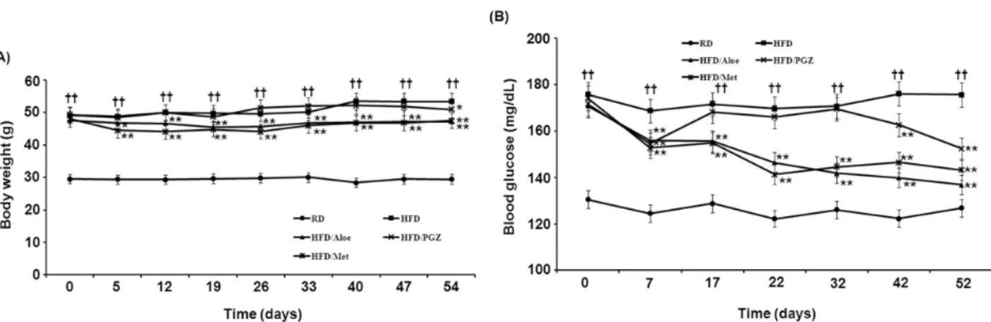
Figure 1. Effects of Aloe QDM complex on body weight and blood glucose change. C57BL/6 mice were fed a high-fat diet supplemented with an aloe formula. (A) Weekly changes in body weight of Aloe QDM complex or anti-diabetic drug-supplemented mice. (B) Blood glucose levels in plasma. Data are means±SEM.values. ††p<0.01 compared with RD-fed mice. *p<0.05, **p<0.01 compared with untreated HFD-fed mice.
were visualized using a Western Blot Kit with an alkaline phosphatase substrate (Vector, Burlingame, USA).
Statistical analysis
All data have been presented as mean±SEM values. Signifi- cant differences (p<0.05) between groups were evaluated using one-way analysis of variance in SPSS (Chicago, IL, USA) for Windows and Duncan s Multiple Range Test where ap- propriate.
RESULTS
Effect of dietary Aloe QDM complex on body weight, blood glucose, and insulin change
The obese mice supplemented with Aloe QDM complex had a lower body weight than that of obese mice on a HFD (Fig.
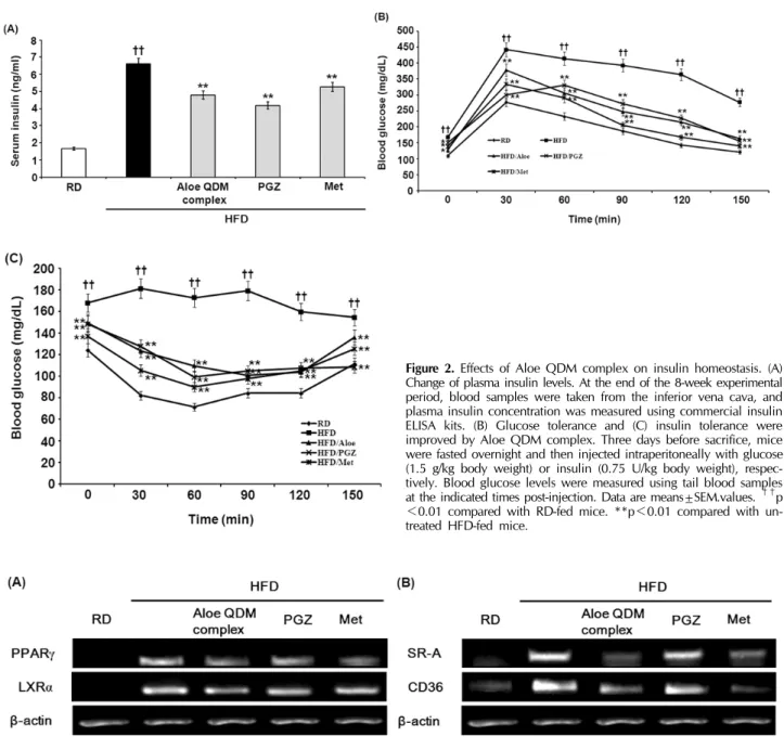
1A). The effects of the Aloe QDM complex, pioglitazone (PGZ), and metformin (Met) supplementation on blood glu- cose levels are shown in Fig. 1B. The HFD-fed mice ex- hibited a significant increase in blood glucose concentrations compared with RD-fed mice. On the other hand, Aloe QDM complex-treated mice significantly decreased fasting blood glucose levels, similar to Met. Further, the fasting plasma in- sulin levels of HFD-fed mice were significantly (3.5-fold) higher than those of regular diet-fed mice. And, treatment of HFD-fed mice with Aloe QDM complex for 8 weeks sig- nificantly reduced plasma insulin levels (Fig. 2A).
Effects of Aloe QDM complex on insulin resistance and glucose tolerance
To test whether Aloe QDM complex reduced obesity-induced insulin resistance, we compared the degree of glucose or in- sulin intolerance in the mice. At the near end of the 8-week experimental period (3 days before sacrifice), glucose toler- ance was examined using the IPGTT method. As shown in Fig. 2B, glucose levels following the i.p. injection were sig- nificantly higher than those in RD-fed mice. Aloe QDM com- plex treatment reduced blood glucose levels at 60 min and 90 min. Insulin tolerance tests revealed that the obese mice were insulin intolerant, and as shown in Fig. 2C, those sup- plemented with dietary Aloe QDM complex had significantly lower levels of plasma glucose than control obese mice at 30 min and 60 min after oral glucose infusion and the levels tended to remain lower. These findings indicated that Aloe QDM complex protected against obesity-induced diabetes.
Inhibition of adipogenic transcription and scavenger receptors in WAT of obese mice
The nuclear receptor, PPARγ, is endogenously activated by some polyunsaturated fatty acids and products of lipid meta- bolism. PPARγ activation has been shown to significantly at- tenuate adipocyte hypertrophy and inhibit WAT inflamma- tion, whereas it increases the total WAT mass. We examined whether the aloe formula could affect mRNA expression of the nuclear receptor PPARγ/LXRα in WAT. PPARγ and LXRα mRNA expression of Aloe QDM complex treatment group was lower in WAT than in the HFD group (Fig. 3A).
CD36 and SR-A are scavenger receptors for oxidized
Figure 2. Effects of Aloe QDM complex on insulin homeostasis. (A) Change of plasma insulin levels. At the end of the 8-week experimental period, blood samples were taken from the inferior vena cava, and plasma insulin concentration was measured using commercial insulin ELISA kits. (B) Glucose tolerance and (C) insulin tolerance were improved by Aloe QDM complex. Three days before sacrifice, mice were fasted overnight and then injected intraperitoneally with glucose (1.5 g/kg body weight) or insulin (0.75 U/kg body weight), respec- tively. Blood glucose levels were measured using tail blood samples at the indicated times post-injection. Data are means±SEM.values. ††p
<0.01 compared with RD-fed mice. **p<0.01 compared with un- treated HFD-fed mice.
Figure 3. Effects of Aloe QDM complex on adipogenesis. WAT were isolated from HFD-fed mice. mRNA expression of (A) PPARγ and LXRα and (B) SR-A and CD36 in the WAT were measured by RT-PCR. Experiments were performed in triplicate with similar results.
low-density lipoprotein (oxLDL) and cellular transporters of long-chain fatty acid (27). In the present study, we examined changes in the level of expression of scavenger receptors and their effect on the amount of adipose tissue in obese mice.
We found that WAT and levels of scavenger receptors such as SR-A and CD36 in the WAT were significantly lower in obese mice supplemented with dietary aloe formula than in the HFD group (Fig. 3B).
This was noteworthy, and it also confirmed that the aloe formula could be adopted as a PPAR antagonist for inhibition of scavenger receptors in ATM (adipose tissue macrophage).
Effect of aloe formula on plasma adiponectin and leptin, and pAMPK Signaling
To further define the possible underlying mechanisms, we measured the expression of adiponectin and leptin and AMPK
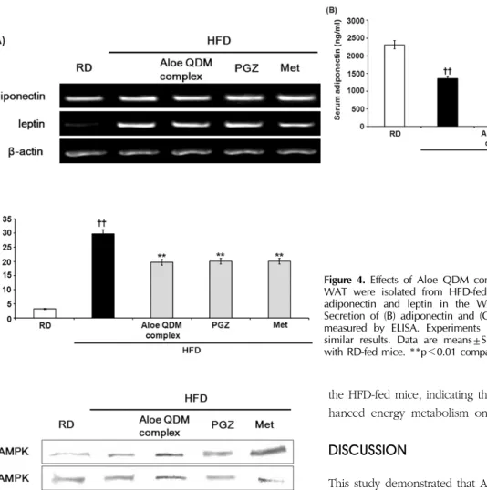
Figure 4. Effects of Aloe QDM complex on adiponectin and leptin.
WAT were isolated from HFD-fed mice. (A) mRNA expression of adiponectin and leptin in the WAT were measured by RT-PCR.
Secretion of (B) adiponectin and (C) leptin protein in the WAT was measured by ELISA. Experiments were repeated in triplicate with similar results. Data are means±SEM.values. ††p<0.01 compared with RD-fed mice. **p<0.01 compared with untreated HFD-fed mice.
Figure 5. Effects of Aloe QDM complex on AMPK phosphorylation.
Muscles were isolated from HFD-fed mice. Protein expression in the WAT was measured by western blot. Experiments were performed in triplicate with similar results.
activity. As shown in Fig. 4, gene expressions of adiponectin and leptin were significantly increased by aloe formula as with PGZ or Met. Circulating adiponectin was significantly re- duced by HFD feeding when compared with normal chow fed, whereas leptin production was increased. Aloe QDM complex increased production of adiponectin while circulat- ing leptin was reduced as with PGZ and Met.
Next, we measured the AMPK expression in the muscles.
Since the AMPK cascade has been shown to activate insulin signaling (16), we hypothesized that AMPK might be involved in the effect cellular Energy Metabolism on tissues. As shown in Fig. 5, the ratio of phospho-AMPKα to AMPKα was sig- nificantly higher in the Aloe QDM complex-fed mice than in
the HFD-fed mice, indicating that the dietary aloe formula en- hanced energy metabolism on WAT.
DISCUSSION
This study demonstrated that Aloe QDM complex can reduce obesity-induced insulin resistance. Herbal prescriptions have been recognized as potentially valid by the scientific medical establishment, and their use has been increasing. Since tradi- tional herbal prescriptions are generally prepared from a combination of crude drugs, on the basis of oriental pre- scriptions and herbology, they may exert combined effects that differ from the sum of the effects of the individual con- stituents (27).
Dietary aloe formula has been demonstrated to affect in- flammation via immunosuppression. Recent studies have sug- gested that inflamed adipocytes in the obese trigger the devel- opment of obesity-related metabolic disorders such as insulin resistance and T2D, indicating that a reduction of tissue in- flammation may be beneficial in obesity-related metabolic diseases. Our previous in vivo study demonstrated a potential effect of PAG, an aloe formula, on hypoglycemia and hypo- lipidemia (26,28). In particular, our results demonstrated that the administration of aloe formulas including PAG, ALS, Aloe
QDM, and Aloe QDM complexes to these mice prevented the development of T2D-related symptoms. However, the pre- vention of or therapeutic effect on obesity-induced metabolic disorders has never been fully established.
The administration of Aloe QDM complex to HFD-fed mice reduced body weight and blood glucose concentration to nor- mal levels despite being continued throughout the 8-week treatment period (Fig. 1). The current understanding of dis- ease progression in T2D is that insulin resistance in peripheral tissues leads to compensatory hyperinsulinemia followed by β-cell failure (29). In the present study, Aloe QDM complex significantly improved insulin resistance (Fig. 2). These results showed that Aloe QDM complex increased insulin sensitivity by decreasing blood glucose and insulin levels. Improved blood glucose homeostasis was also observed in the IPGTT in Aloe QDM complex-supplemented HFD-fed mice (Fig. 2).
High-fat intake and increased levels of free fatty acids in the circulation lead to insulin resistance. Oral administration of aloe formula reduced plasma leptin whereas adiponectin was significantly induced (Fig. 4) by Aloe QDM complex. We also examined the mRNA expression of adipogenic genes in WAT by semiquantitive RT-PCR to determine whether Aloe QDM complex reduced lipogenesis. Aloe QDM complex suppressed the expressions of the adipogenesis genes, PPARγ/LXRα (Fig. 3A), suggesting that the aloe formula may be able to im- prove insulin resistance through a reduction of fat in the WAT.
Also, scavenger receptors, specifically SR-A and CD36, play crucial roles in the pathogenesis of atherosclerotic lesions by identifying and facilitating the uptake of oxLDL (30). In phag- ocytes, CD36 primarily functions as a scavenger receptor, rec- ognizing specific self and nonself molecular patterns and trig- gering internalization and inflammatory signaling pathways to eliminate pathogens and altered self components, such as apoptotic cells (31,32). CD36 cooperates with toll-like receptor (TLR)-4 and -6 to mediate the sterile inflammatory response to altered self components oxLDL (33) and also acts as a cor- eceptor with TLR2 and -6 in the recognition of microbial diac- ylglycerides (34,35).
Despite the importance of adipose tissue-resident macro- phages in the pathogenesis of insulin resistance, this is the first study to our knowledge that measures the expression of scavenger receptor genes in diet-induced obesity (DIO) mice supplemented with Aloe QDM complex. Our data demon- strated that the Aloe QDM complex decreased gene ex- pression of scavenger receptors in WAT of obese mice (Fig.
3B) in WAT, leading to reduced atherosclerotic lesions in
obese mice.
The most important finding in this study was that Aloe QDM complex reduced body fat via activation of AMPK in the muscle (Fig. 5). These results reveal that the Aloe QDM complex increased mitochondrial biogenesis in both WAT and muscles by activating AMPK and further reduced body fat by down-regulating the expression of scavenger receptors in the WAT.
In almost all of the data, Aloe QDM complex significantly improved insulin resistance and suppressed adipogenesis genes in WAT by activating AMPK, suggesting that Aloe QDM complex is a useful dietary phytochemical for improving obe- sity-related metabolic disorders such as insulin resistance.
ACKNOWLEDGMENTS
This work was supported by Univera, Inc. as one of CAP projects and also by the Sahmyook University Research fund in 2011.
CONFLICTS OF INTEREST
The authors have no financial conflict of interest.
REFERENCES
1. Weiser, M., W. H. Frishman, M. D. Michaelson, and M. A.
Abdeen. 1997. The pharmacologic approach to the treatment of obesity. J. Clin. Pharmacol. 37: 453-473.
2. Surwit, R. S., C. M. Kuhn, C. Cochrane, J. A. McCubbin, and M. N. Feinglos. 1988. Diet-induced type II diabetes in C57BL/6J mice. Diabetes 37: 1163-1167.
3. Stunkard, A. J. 1996. Current views on obesity. Am. J. Med.
100: 230-236.
4. Nicolai, A., M. Li, D. H. Kim, S. J. Peterson, L. Vanella, V.
Positano, A. Gastaldelli, R. Rezzani, L. F. Rodella, G.
Drummond, C. Kusmic, A. L'Abbate, A. Kappas, and N. G.
Abraham. 2009. Heme oxygenase-1 induction remodels adi- pose tissue and improves insulin sensitivity in obesity-in- duced diabetic rats. Hypertension 53: 508-515.
5. Peterson, S. J., D. H. Kim, M. Li, V. Positano, L. Vanella, L. F. Rodella, F. Piccolomini, N. Puri, A. Gastaldelli, C.
Kusmic, A. L'Abbate, and N. G. Abraham. 2009. The L-4F mimetic peptide prevents insulin resistance through increased levels of HO-1, pAMPK, and pAKT in obese mice. J. Lipid.
Res. 50: 1293-1304.
6. Porstmann, T., C. R. Santos, B. Griffiths, M. Cully, M. Wu, S. Leevers, J. R. Griffiths, Y. L. Chung, and A. Schulze. 2008.
SREBP activity is regulated by mTORC1 and contributes to Akt-dependent cell growth. Cell Metab. 8: 224-236.
7. Wellen, K. E., and G. S. Hotamisligil. 2005. Inflammation,
stress, and diabetes. J. Clin. Invest. 115: 1111-1119.
8. Rajala, M. W., and P. E. Scherer. 2003. Minireview: The adi- pocyte--at the crossroads of energy homeostasis, in- flammation, and atherosclerosis. Endocrinology. 144: 3765- 3773.
9. Weisberg, S. P., D. McCann, M. Desai, M. Rosenbaum, R.
L. Leibel, and A. W. Jr. Ferrante. 2003. Obesity is associated with macrophage accumulation in adipose tissue. J. Clin.
Invest. 112: 1796-1808.
10. Xu, H., G. T. Barnes, Q. Yang, G. Tan, D. Yang, C. J. Chou, J. Sole, A. Nichols, J. S. Ross, L. A. Tartaglia, and H. Chen.
2003. Chronic inflammation in fat plays a crucial role in the development of obesity-related insulin resistance. J. Clin.
Invest. 112: 1821-1830.
11. Hardie, D. G., and D. Carling. 1997. The AMP-activated pro- tein kinase--fuel gauge of the mammalian cell? Eur. J.
Biochem. 246: 259-273.
12. Sag, D., D. Carling, R. D. Stout, and J. Suttles. 2008.
Adenosine 5'-monophosphate-activated protein kinase pro- motes macrophage polarization to an anti-inflammatory func- tional phenotype. J. Immunol. 181: 8633-8641.
13. Makinde, A. O., J. Gamble., and G. D. Lopaschuk. 1997.
Upregulation of 5'-AMP-activated protein kinase is respon- sible for the increase in myocardial fatty acid oxidation rates following birth in the newborn rabbit. Circ. Res. 80: 482-489.
14. Zong, H., J. M. Ren, L. H. Young, M. Pypaert, J. Mu, M.
J. Birnbaum, and G. I. Shulman. 2002. AMP kinase is re- quired for mitochondrial biogenesis in skeletal muscle in re- sponse to chronic energy deprivation. Proc. Natl. Acad. Sci.
U. S. A. 99: 15983-15987.
15. Ju, J. S., M. A. Gitcho, C. A. Casmaer, P. B. Patil, D. G.
Han, S. A. Spencer, and J. S. Fisher. 2007. Potentiation of insulin-stimulated glucose transport by the AMP-activated pro- tein kinase. Am. J. Physiol. Cell Physiol. 292: C564-572.
16. Iglesias, M. A., J. M. Ye, G. Frangioudakis, A. K. Saha, E.
Tomas, N. B. Ruderman, G. J. Cooney, and E. W. Kraegen.
2002. AICAR administration causes an apparent enhancement of muscle and liver insulin action in insulin-resistant high-fat- fed rats. Diabetes 51: 2886-2894.
17. Josep, B. R., G. Amir, K. Jennifer, and H. Raquel. 2005.
Peroxisome proliferator activated receptors: the nutritionally controlled molecular networks that integrate inflammation, immunity and metabolism. Current Nutrition & Food Science 1: 179-187.
18. Hardie, D. G. 2007. AMP-activated protein kinase as a drug target. Annu. Rev. Pharmacol. Toxicol. 47: 185-210.
19. Hardie, D. G., D. Carling, and M. Carlson. 1998. The AMP-activated/SNF1 protein kinase subfamily: metabolic sen- sors of the eukaryotic cell? Annu. Rev. Biochem. 67: 821-855.
20. Capasso, F., F. Borrelli, R. Capasso, Carlo G. Di, A. Izzo, L. Pinto, N. Mascolo, S. Castaldo, and R. Longo. 1998. Aloe and its therapeutic use. Phytother. Res. 12: S124-127.
21. Heggers, J. P., A. Kucukcelebi, C. J. Stabenau, F. Ko, L. D.
Broemeling, M. C. Robson, and W.D. Winters. 1995. Wound healing effects of Aloe gel and other topical antibacterial agents on rat skin. Phytother. Res. 9: 455-457.
22. Koo, M. W. L. 1994. Aloe vera: Antiulcer and antidiabetic effects. Phytother. Res. 8: 461-464.
23. Winters, W.D., R. Benavides, and W. J. Clouse. 1981. Effects of aloe extracts on human normal and tumor cells in vitro. Econ. Bot. 35: 89-95.
24. Yongchaiyudha, S., V. Rungpitarangsi, N. Bunyapraphatsara, and O. Chokechaijaroenporn. 1996. Antidiabetic activity of Aloe vera L. juice. I. Clinical trial in new cases of diabetes mellitus. Phytomedicine. 3: 241-243.
25. Bunyapraphatsara, N., S. Yongchaiyudha, V. Rungpitarangsi, and O. Chokechaijaroenporn. 1996. Antidiabetic activity of Aloe vera L. juice: II. Clinical trial in diabetes mellitus patients in combination with glibenclamide. Phytomedicine. 3: 245-248.
26. Kong, H., S. Lee, S. Shin, J. Kwon, T. H. Jo, E. Shin, K.
S. Shim, Y. I. Park, C. K. Lee, and K. Kim. 2010. Down-regu- lation of adipogenesis and hyperglycemia in diet-induced obesity mouse model by Aloe QDM. Biomolecules &
Therapeuticss 18: 336-342.
27. Kim, J. O., K. S. Kim, G. D. Lee, and J. H. Kwon. 2009.
Antihyperglycemic and antioxidative effects of new herbal formula in streptozotocin-induced diabetic rats. J. Med. Food.
12: 728-735.
28. Kim, K., H. Kim, J. Kwon, S. Lee, H. Kong, S. A. Im, Y.
H. Lee, Y. R. Lee, S. T. Oh, T. H. Jo, Y. I. Park, C. K. Lee, and K. Kim. 2009. Hypoglycemic and hypolipidemic effects of processed Aloe vera gel in a mouse model of non-in- sulin-dependent diabetes mellitus. Phytomedicine. 16: 856- 863.
29. Martín-Fuentes, P., F. Civeira, D. Recalde, A. L. García-Otín, E. Jarauta, I. Marzo, and A. Cenarro. 2007. Individual varia- tion of scavenger receptor expression in human macrophages with oxidized low-density lipoprotein is associated with a dif- ferential inflammatory response. J. Immunol. 179: 3242-3248.
30. Stewart, C. R., L. M. Stuart, K. Wilkinson, J. M. van Gils, J. Deng, A. Halle, K. J. Rayner, L. Boyer, R. Zhong, W. A.
Frazier, A. Lacy-Hulbert, J. El Khoury, D. T. Golenbock, and K. J. Moore. 2010. CD36 ligands promote sterile inflammation through assembly of a Toll-like receptor 4 and 6 heterodimer.
Nat. Immunol. 11: 155-161.
31. Sahoo, D., and V. Drover. 2006. The role of scavenger re- ceptors in signaling, inflammation and atherosclerosis.
Biochemistry of Atherosclerosis. 1: 70–91.
32. Peiser, L., and S. Gordon. 2001. The function of scavenger receptors expressed by macrophages and their role in the reg- ulation of inflammation. Microbes. Infect. 3:149-159.
33. Stewart, C. R., L. M. Stuart, K. Wilkinson, J. M. van Gils, J. Deng, A. Halle, K. J. Rayner, L. Boyer, R. Zhong, W. A.
Frazier, A. Lacy-Hulbert, J. El Khoury, D. T. Golenbock, and K. J. Moore. 2010. CD36 ligands promote sterile inflammation through assembly of a Toll-like receptor 4 and 6 heterodimer.
Nat. Immunol. 11: 155-161.
34. Hoebe, K., P. Georgel, S. Rutschmann, X. Du, S. Mudd, K.
Crozat, S. L. Sovath, Shamel, T. Hartung, U. Zähringer, and B. Beutler. 2005. CD36 is a sensor of diacylglycerides.
Nature. 433: 523-527.
35. Stuart, L. M., J. Deng, J. M. Silver, K. Takahashi, A. A. Tseng, E. J. Hennessy, R. A. Ezekowitz, and K. J. Moore. 2005.
Response to Staphylococcus aureus requires CD36-mediated phagocytosis triggered by the COOH-terminal cytoplasmic domain. J Cell Biol. 170: 477-485.