Comparison between Conventional Cytogenetics and Interphase Fluorescence in situ Hybridization (FISH) for
Patients with Multiple Myeloma
Sung-Hyun Kim, M.D.1, Jung Hwan Kim, M.D.1, Dong Mee Lee, M.D.1, Suee Lee, M.D.1, Sung Yong Oh, M.D.1, Hyuk-Chan Kwon, M.D.1, Kyung-Eun Kim, M.D.2, Jin-Yeong Han, M.D.2 and Hyo-Jin Kim, M.D.1
1Department of Internal Medicine and 2Laboratory Medicine, Dong-A University College of Medicine, Busan, Korea
Background: For patients with multiple myeloma (MM), different strategies are used to detect chromoso- mal abnormalities (CA). There have been a few studies that have directly compared FISH with conven- tional cytogenetics (CC) for the detection of CA. In this study, we employed a combined approach of metaphase cytogenetics and interphase FISH to investigate the genetic basis for the great heterogeneity observed in the clinical behavior of 28 MM patients.
Methods: Cytogenetic analysis was performed via traditional metaphase karyotype analysis. The FISH studies were done using DNA probes to detect translocations involving the immunoglobulin heavy chain gene (IGH) at 14q32 and deletions of 17p13.1 and 13q14.
Results: CA were detected by CC in 16 patients (57.1%) and by FISH in 14 patients (50.0%) of the 28 patients we studied. 14q32 abnormalities and deletion abnormalities of 13q14 and 17p13.1 were de- tected by CC in five patients (17.9%), three patients (10.7%) and no patients (0%), respectively and these were detected by FISH in 12 (42.8%), four (14.3%) and five (17.8%), respectively, of the 28 patients we studied. The median follow-up timefor the patients was 23.85 months (range: 0.3∼58.13 months).
On the univariate and multivariate analyses, none of the abnormalities detected by cytogenetics and inter- phase FISH affected survival.
Conclusion: On comparing the cytogenetics and interphase FISH results, we can suggest that both studies should be an essential part of the workup for the diagnosis of patients with MM. Also, both studies may complement each other to predict the prognosis. (Korean J Hematol 2009;44:14-21.)
Key Words: Multiple myeloma, Conventional cytogenetics, Fluorescence in situ hybridization
14 접수:2008년 8월 9일, 수정:2009년 1월 12일
승인:2009년 1월 18일
교신저자:김효진, 부산시 서구 동대신동 3가 1번지
602-715, 동아대학교병원 혈액종양내과 Tel: 051-240-2951, Fax: 051-240-2088 E-mail: [email protected]
This paper was supported by the Dong-A University Research Fund in 2005.
Correspondence to:Hyo-Jin Kim, M.D., Ph.D.
Department of Internal Medicine, Dong-A University College of Medicine
3-1, Dongdaeshin-dong, Seo-gu, Busan 602-715, Korea Tel: +82-51-240-2951, Fax: +82-51-240-2088 E-mail: [email protected]
INTRODUCTION
Multiple myeloma (MM) is a B-cell clonal ma- lignancy that is characterized by the accumu- lation of terminally differentiated plasma cells.
MM accounts for 10% of hematological neo- plasm, and it has a median survival of 2∼3 years.1) Among the various prognostic factors for MM, molecular cytogenetic abnormalities (CA) have emerged as a major independent parameter for predicting the clinical outcome.2)
The most common significant structural chro- mosomal changes are rearrangements involving the switch regions of the immunoglobulin heavy chain gene (IGH) at 14q32 with various partner genes, and the incidence of this is 50∼70% for MM patients.3) 13q14 deletions have been de- tected by interphase FISH in 40∼50% of the pa- tients with MM, although it has remained un- clear whether or not del(13q) as detected by FISH would provide only the same prognostic in- formation as that detected by metaphase cyto- genetics.4)
Deletions at the p53 locus can be seen in smaller percentages of cells and this suggests ear- ly clonal evolution.5) In addition, interphase FISH has identified deletions of 17p that involved the p53 gene locus, and these CA in MM were in- dependently correlated with a short survival time after conventional-dose chemotherapy.6)
Cytogenetic analysis is difficult because of the small number and low mitotic index of plasma cells in the bone marrow in those patients with MM. However, metaphase cells can be examined by conventional cytogenetic (CC) methods to de- tect a wide variety of CA in proliferating cells from the patients with MM.7) In addition, FISH can be used to study interphase nuclei (inter- phase FISH) to detect CA in unselected non-pro- liferating nuclei to establish the overall pro- portion of normal and abnormal nuclei. The studies that have directly compared FISH with CC for the detection of CA8) and the clinical effi- cacy of studying metaphase cells versus inter- phase nuclei in those patients with MM4,5,9-11) were reported. We employed a combined ap- proach of CC and interphase FISH to investigate the prognostic relevance in MM.
MATERIALS AND METHODS 1. Patients
During a 6-year period (2000-2005), twenty eight MM patients whose cytogenetic results were available at Dong-A University Medical Center,
Busan, Korea were enrolled in this study. In ad- dition to the CC, all the patients had interphase FISH performed simultaneously. In most cases, the slides were made with the cells collected at presentation, and in some cases, new slides were made from cells that were kept frozen in liquid nitrogen. This study was approved by our institu- tional human ethics committee that oversees re- search involving human subjects. A written in- formed consent form was provided and obtained from all the patients.
2. Conventional cytogenetics (CC)
CC was performed on initial bone marrow samples as previously published.12) Synchroniza- tion culture using methotrexate and thymidine was also performed in some cases. The chromoso- mal aberrations were defined and described ac- cording to the International System for Cytoge- netic Nomenclature 2005.13)
3. Interphase FISH
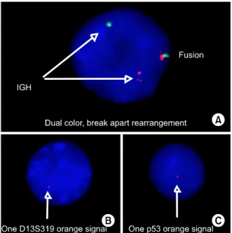
Interphase FISH was performed with p53 (LSI p53 DNA probe), IGH (LSI IGH Dual color, Break Apart Rearrangement probe), and 13q14 (LSI D13S319 DNA probe) probes according to the manufacturer's instructions (Vysis Inc., Dow- ners Grove, IL, USA). We aimed at counting over 500 nuclei in each sample. The background cut-off levels were as follows: rearrangement of IGH: 3.5%, deletion of 13q14: 3.6% and deletion of 17p13.1: 2.5%. Representative cell images were captured using a computer-based imaging system (Fig. 1).
4. Treatment
A total of 26 patients (92.9%) received treat- ment. The conventional chemotherapy consisted of a CP (cyclophosphamide and prednisolone) regimen or a VAD (vincristine, doxorubicin, and dexamethasone) regimen or a MPT (melphalan, prednisolone and thalidomide) regimen. Twenty patients were treated with conventional chemo- therapy (71.4%). Three or four cycles VAD fol-
Fig. 1. Interphase fluorescence in situ hybridization (FISH) findings. (A) A plasma cell shows rearrangement of IGH.
(B) A plasma cell shows deletion 13q14. The locus-specific 13q14 probes are labeled by orange color. (C) A plasma cell shows deletion 17p13.1. The locus-specific p53 probes are labeled by orange color.
lowed by high-dose chemotherapy (melphalan 200mg/m2) with autologous peripheral stem cell transplantation was administered to six patients (21.5%). Two patients (7.1%) did not receive che- motherapy owing to their poor clinical status. If a patient had progressive disease, then we permit- ted the use of second-line chemotherapy.
5. Treatment response
The treatment response criteria we used were described previously.14) Responders included those patients who had achieved a complete response (CR), partial response (PR) or minimal response (MR). In brief, CR was the absence of the origi- nal monoclonal paraprotein in the serum and urine, as determined by immunofixation, and this was maintained for a minimum of six weeks. PR was a ≥50% reduction in the level of the serum monoclonal paraprotein, and this was maintained for a minimum of six weeks. MR was a 25∼49%
reduction in the level of the serum monoclonal paraprotein that was maintained for a minimum
of six weeks. Non-responders included the pa- tients who had no response or they had pro- gressive disease. Overall survival (OS) was de- fined as the time period from the date of diag- nosis to the date of death, regardless of cause.
6. Statistical analysis
Fisher’s exact test was used for the between- group comparisons of the discrete variables. The Mann-Whitney U-test was used to test differences between the patient groups on the basis of their CA, translocation status and clinical character- istics for the continuous variables. Kaplan-Meier survival curves were used for determining the dif- ferences in OS between the groups. Log-rank tests were used to test for differences in OS be- tween the groups. Those factors with statistical significance from the univariate analysis were then tested by multivariate analysis with the Cox proportional hazards regression model with using forward stepwise selection. All directional P val- ues were two-tailed, with a P value of ≤0.05 be- ing considered significant for all tests. All the analyses were performed using SPSS 12.0 soft- ware (SPSS, Inc., Chicago, IL, USA).
RESULTS 1. Patient characteristics
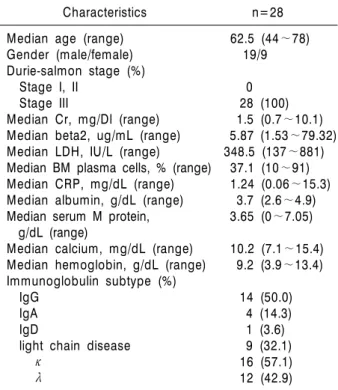
Of the 28 patients with MM, there were 19 males and nine females with a median age of 62.5 years (age range: 44∼78). Their characteristics are given in Table 1. Up to July 2008, the median follow-up time for the patients was 23.85 months (range: 0.3∼58.13). The median OS for all the patients has not yet been determined.
2. Prevalence and details of the chromosomal abnormalities (CA)
Among the 28 patients, CA were detected by CC in 16 patients (57.1%). Table 2 provides a de- scription of the karyotypes.
CA of chromosome 13 were observed in three patients, including monosomy 13 in two patients
Karyotype Patients
n Incidence in
MM patients % FISH Patients
n Incidence in
MM patients %
AA 16 57.1 Detected 14 50
NA only 1 3.6 IGH only 7 25
SA only 3 10.7 del(13q) only 1 3.6
BA 12 42.8 del(17p) only 1 3.6
Any NA Hyperdiploidy Hypodiploidy Tetraploidy
13 10 2 1
46.4 35.7 7.1 3.6
IGH and del(13q)
IGH and del(17p) 1
2 3.6
7.1
Any SA 14q32 -13 or 13q14−
15 5 3
53.6 17.9 10.7
del(13q) and del(17p)
0 0
-17 or 17p− 0 0 IGH, del(13q)
and del(17p) 2 7.1
Any IGH Any del(13q) Any del(17p)
12 4 5
42.8 14.3 17.8
Abbreviations: CC, conventional cytogenetics; FISH, fluorescence in situ hybridization; MM, Multiple myeloma; IGH, immunoglobulin heavy chain gene; AA, any abnormalities; NA, numeric abnormalities; SA, structural abnormalities; BA, both numeric and structural abnormalities.
Table 2. CC and FISH data from 28 MM patients Table 1. Characteristics of the 28 study patients
Characteristics n=28
Median age (range) 62.5 (44∼78)
Gender (male/female) 19/9 Durie-salmon stage (%)
Stage I, II 0
Stage III 28 (100)
Median Cr, mg/Dl (range) 1.5 (0.7∼10.1) Median beta2, ug/mL (range) 5.87 (1.53∼79.32) Median LDH, IU/L (range) 348.5 (137∼881) Median BM plasma cells, % (range) 37.1 (10∼91) Median CRP, mg/dL (range) 1.24 (0.06∼15.3) Median albumin, g/dL (range) 3.7 (2.6∼4.9) Median serum M protein, 3.65 (0∼7.05) g/dL (range)
Median calcium, mg/dL (range) 10.2 (7.1∼15.4) Median hemoglobin, g/dL (range) 9.2 (3.9∼13.4) Immunoglobulin subtype (%)
IgG 14 (50.0)
IgA 4 (14.3)
IgD 1 (3.6)
light chain disease 9 (32.1)
κ 16 (57.1)
λ 12 (42.9)
Abbreviations: Cr, creatinine; beta2, β2 microglobulin;
LDH, lactic dehydrogenase; BM, bone marrow; CRP, C-reactive protein.
and del(13)(q12q22) in one patient. 14q32 re- arrangements were observed in five patients, in-
cluding four with t(11;14)(q13;q32) and three with t(8;14)(q24.1;q32). Among those five pa- tients, two had both t(11;14)(q13;q32) and t(8;14) (q24.1;q32). Another structural abnormality of chromosome 14 was observed in one patient with t(1;14)(p32;q13). Monosomy 14 was observed in four patients.
3. Interphase FISH results
As summarized in Table 2, 14 patients (50.0%) showed evidence of an abnormality with using any of the three FISH probes.
4. Comparison of CC and interphase FISH CC and interphase FISH were both abnormal in 11 of 28 patients (39.3%). Among the patient with all negative three FISH probes (14/28, 50%), five patients (5/14, 35.7%) were abnormal as de- termined by CC. Among the patient with normal karyotype (12/28, 42.9%), three (3/12, 25%) were abnormal by FISH. Both the karyotype and FISH results were normal in nine patients (9/28, 32.1%).
FISH revealed rearrangement of IGH in 12 pa- tients (42.8%); two with t(11;14)(q13;q32), one
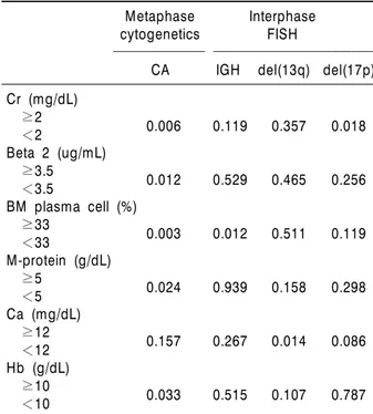
Table 3. P value for the association between the bioclinical features and the abnormalities n=28 Metaphase Interphase
cytogenetics FISH
–––––––––––––––––––––––––––––––––––––––––––
CA IGH del(13q) del(17p) Cr (mg/dL)
≥2 0.006 0.119 0.357 0.018
<2
Beta 2 (ug/mL)
≥3.5 0.012 0.529 0.465 0.256
<3.5
BM plasma cell (%)
≥33 0.003 0.012 0.511 0.119
<33
M-protein (g/dL)
≥5 0.024 0.939 0.158 0.298
<5 Ca (mg/dL)
≥12 0.157 0.267 0.014 0.086
<12 Hb (g/dL)
≥10 0.033 0.515 0.107 0.787
<10
Abbreviations: FISH, fluorescence in situ hybridization; CA, chromosomal abnormalities; IGH, immunoglobulin heavy chain gene; Cr, creatinine; beta2, β2 microglobulin; BM, bone marrow; Ca, calcium; Hb, hemoglobin.
with t(8;14)(q24.1;q32), one t(1;14)(p32;q13), and two patients with both t(11;14)(q13;q32) and t(8;14)(q24.1;q32), as determined by karyotype.
Four patients had monosomy 14 and two patients had normal karyotypes.
Deletion 13q14, as determined by FISH, was observed in four patients (14.3%); two patients with monosomy 13, one t(1;13)(q32;q14), and an- other one patient with del(13)(q12q22).
Deletion 17p13.1 FISH was found in five pa- tients (17.8%). For these patients, there was no monosomy 17 or translocation of chromosome 17 by karyotype study.
5. Association between the bioclinical features and the CA
Table 3 shows associations between the bio- clinical features and the CA. The patients with karyotype abnormalities had significantly higher levels of creatinine and beta2-microglobulin, more plasma cells in their bone marrow, more M-pro- tein amount and a lower level of hemoglobin at the time of diagnosis than those patients without CA (P=0.006, P=0.012, P=0.003, P=0.024 and P=0.033, respectively).
Patients with rearrangement of IGH FISH had more plasma cells in their bone marrow (P=
0.012). Patients with deletion 13q14 by FISH had a higher level of calcium at time of diagnosis (P=0.014). Patients with deletion 17p13.1 had a higher level of creatinine at the time of diagnosis (P=0.003).
6. Response to initial treatment
Twenty patients were treated with conventional chemotherapy and six patients had VAD followed with autologous transplantation. Follow-up data of 21 patients were obtainable among the 26 treated patients. Six patients had CR, 12 patients had PR and three patients had MR, respectively.
There were no significant associations between treatment response and the chromosomal abnor- malities neither by CC nor by FISH (P<0.05).
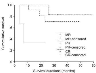
7. Survival
Although the median OS for all the patients has not yet been reached, there were significant differences in survival according to the initial treatment response (P=0.036, Fig. 2). In univa- riate and multivariate analyses, none of the ab- normalities of cytogenetics and interphase FISH affected survival.
DISCUSSION
Recent studies have identified several specific CA as prognostic indicators for patients suffering with MM. Complex karyotypes are seen in 30∼
50% of these cases, and this is combined with a long preclinical clonal evolution. Most newly di- agnosed cases of MM have a normal karyotype, whereas abnormalities are more often seen in ad- vanced disease, and these abnormalities are asso- ciated with increasing proliferative activity of
Fig. 2. The overall survival curves according to the initial treatment response. For patients with a CR or a PR, the median survival has not yet been reached. The median survival was 7.03 months for patients with MR. There were significant differences in survival according to the initial treatment response (P=0.036). (CR; complete response, PR; partial response, MR; minimal response).
malignant cells. The CC techniques have de- tected various rates of CA with according to the culture technique with using bone marrow cells.
Altogether, these techniques have helped identify about 50% of the CA in the patients with newly diagnosed MM.15) In our study, metaphase cyto- genetics showed karyotype abnormalities in 57.1% of the patients and all of them had Durie-Salmon stage III disease.
FISH has enabled the detection of genetic changes in myeloma cases for which CC have proved to be uninformative, and FISH can be performed for the nondividing cells.16,17) FISH al- lows identification of complex rearrangements that cannot be determined by CC and FISH may also reveal cryptic translocations. Previous stud- ies with using FISH indicate that IGH trans- locations are detectable in 43∼70% of MM patients.5,18) Our study found a similar incidence of IGH rearrangements (42.8%). However, this
"break apart" strategy cannot determine the trans- location partner chromosome. Because of the re- cently recognized unbalanced nature of im- munoglobulin (Ig) translocations in MM, it is de- sirable to develop FISH probes that enable a dou- ble fusion strategy.19)
Reciprocal translocations involving chromo- some 8q24 and the Ig genes account for only 25%
of the c-myc rearrangements in MM. The ma- jority of translocations affecting c-myc in myelo- ma involve the non-Ig loci. Apart from 8q24 (c-myc), the common translocation partner chro- mosomes of IGH rearrangements are 11q13 (cy- clin D1; ∼15%), 4p16 (FGFR3 and MMSET;
15%), 16q23 (c-maf; 6%) and 6p21 (cyclin D3;
4%).19) The results of CC in this study showed four cases each of 11q13 rearrangements (4/28;
14.3%) and 8q24 (4/28; 14.3%) rearrangements.
Chromosomal 13 abnormalities are highly pre- valent in MM patients. The frequency of detect- ing monosomy 13 has risen from approximately 15∼20% by CC7) to approximately 50% by FISH.20) Our results showed the presence of mon- osomy 13 or del(13q) in 10.7% of the patients (3/28) by CC and in 14.3% of the patients (4/28) by interphase FISH. Among the four cases with deletion 13q14 by FISH, two patients had monos- omy 13, one had del(13)(q12q22), and the re- maining one patient had t(1;13)(q32;q14), by karyotype. Our results showed the low rate of del(13q) by FISH. These results may reflect the small sample size of this study or biologic differ- ence between Korean and western population.
Further studies with a large number of Korean MM patients are needed to document the rate of del(13q) by FISH.
The previous CC studies have indicated a low frequency of p53 mutations and deletions in mye- loma patients, and this has varied from 3 to 9%.7,15) In most of the previous FISH series, the incidence of p53 deletion among the newly diag- nosed patients was in the range of 5% to 10%.5,21,22) However, functional loss of the gene is present in up to 40% of patients with advanced MM and also in more than 60% of the human myeloma cell lines, which points to this abnor- mality as a marker of tumor progression.23) Dele- tion of the p53 gene locus was identified by FISH is a predictor of shorter survival in several pre- vious studies, and this was independent of the
mode of treatment (conventional chemotherapy or high-dose chemotherapy).5,6,10,24) From the cur- rent FISH results, 17.8% of the patients (5/28) had p53 deletion. There was no chromosome 17p deletion among these patients by CC, while one patient had add(17)(p13) and another had tris- omy 17 abnormalities.
The presence of -13/13q-, as determined by CC, predicts a more unfavorable prognosis than the detection of the same abnormality as de- termined by FISH. Besides, survival differed among the patients depending on whether certain chromosome abnormalities were detected in both the metaphase and interphase cells, or in only the interphase nuclei. This is most likely due to a combination of negative prognostic markers, as reflected by an intrinsic effect of chromosome 13 loss, and the higher rate of proliferating cells.
Thus, interphase FISH is not a substitute for metaphase analysis. Chiecchio et al.25) also showed the unique importance of metaphase analysis in the determination of prognosis in MM. According to the study, when cases with chromosome 13 de- letion detected by interphase FISH only, the poor prognosis of interphase FISH-detectable chromo- some 13 deletion disappeared. They also reported that patients with poor prognostic FISH markers such as t(4;14) or deletion p53 without abnormal metaphase cytogenetics had outcomes comparable to those without these poor prognostic markers and those with normal metaphase cytogenetics.
Gertz et al.10) showed that when both t(4;14) (p16.3;q32) and deletion 13 were present, the OS and progression-free survival times of these pa- tients were significantly worse than those for the patients who had deletion 13, but not t(4;14) (p16.3;q32). They suggested these patients receive only minimal benefit from autologous stem cell transplantation and they may be candidates for novel therapeutic approaches, such as thalido- mide- or bortezomib-based regimen.
Patients with MM display various genetic ab- normalities and detecting all of these genetic ab- normalities is not possible with using FISH pro-
bes. Though this study has a weakness that a small sample size, short follow-up duration, and not uniform treatment approach, our data in- dicate that karyotype analysis may be helpful to detect additional chromosomal abnormalities that may have potential clinical significance. By com- paring the conventional cytogenetics and inter- phase FISH results, both studies should be an es- sential part of the workup for patients with MM.
Further studies with a large number of patients and a longer follow-up period are needed to con- firm the clinical significance of our FISH findings.
요 약
배경: 다발골수종에서의 핵형이상을 발견하기 위 해서는 여러가지 방법들이 사용되는데 이들 중 임 상적 다양성을 나타내는 유전학적 근거를 연구하기 위해 28명의 환자를 고식적 핵형분석과 간기 형광 제자리부합법(이하 FISH)을 시행하여 분석하였다.
방법: 고식적 핵형분석은 분열중기염색체검사법 을 시행하였다. 간기 FISH는 염색체 14q32의 면역 글로불린 중쇄유전자(이하 IGH)와 17p13.1과 13q14 의 결손을 발견하기 위한 DNA 표식자를 사용하였 다.
결과: 고식적 핵형분석 결과 16예(57.1%)에서, 간 기 FISH 결과 14예(50.0%)에서 각각 이상이 있었 다. 14q32 이상, 13번 염색체 결손, 17번 염색체 결 손은 고식적 핵형분석 결과 5예(17.9%), 3예(10.7%), 0 예에서, 간기 FISH 결과 12예(42.8%), 4예(14.3%), 5예(17.8%)에서 각각 검출되었다. 전체 환자의 추적 중앙값은 23.85개월(범위: 0.3∼58.13개월)이었다.
단변량분석과 다변량분석을 시행하였는데, 생존율 에 영향을 미치는 고식적 핵형분석과 간기 FISH의 이상은 없었다.
결론: 고식적 핵형분석과 간기 FISH 비교 결과, 두 검사가 다발골수종 진단에는 필수적으로 함께 시행되어야하며 두 검사가 예후를 예측하는데 있어 상호보완적일 것이라 생각된다.
REFERENCES
1) Bataille R, Harousseau JL. Multiple myeloma. N
Engl J Med 1997;336:1657-64.
2) Seong C, Delasalle K, Hayes K, et al. Prognostic val- ue of cytogenetics in multiple myeloma. Br J Haematol 1998;101:189-94.
3) Bergsagel PL, Kuehl WM. Chromosome trans- locations in multiple myeloma. Oncogene 2001;20:
5611-22.
4) Kaufmann H, Krömer E, Nösslinger T, et al. Both chromosome 13 abnormalities by metaphase cytoge- netics and deletion of 13q by interphase FISH only are prognostically relevant in multiple myeloma. Eur J Haematol 2003;71:179-83.
5) Fonseca R, Blood E, Rue M, et al. Clinical and bio- logic implications of recurrent genomic aberrations in myeloma. Blood 2003;101:4569-75.
6) Drach J, Ackermann J, Fritz E, et al. Presence of a p53 gene deletion in patients with multiple myeloma predicts for short survival after conventional-dose chemotherapy. Blood 1998;92:802-9.
7) Sawyer JR, Waldron JA, Jagannath S, Barlogie B.
Cytogenetic findings in 200 patients with multiple myeloma. Cancer Genet Cytogenet 1995;82:41-9.
8) Chang H, Li D, Zhuang L, et al. Detection of chro- mosome 13q deletions and IgH translocations in pa- tients with multiple myeloma by FISH: comparison with karyotype analysis. Leuk Lymphoma 2004;45:
965-9.
9) Dewald GW, Therneau T, Larson D, et al. Relation- ship of patient survival and chromosome anomalies detected in metaphase and/or interphase cells at di- agnosis of myeloma. Blood 2005;106:3553-8.
10) Gertz MA, Lacy MQ, Dispenzieri A, et al. Clinical im- plications of t(11;14)(q13;q32), t(4;14)(p16.3;q32), and -17p13 in myeloma patients treated with high- dose therapy. Blood 2005;106:2837-40.
11) Shaughnessy J Jr, Tian E, Sawyer J, et al. Prognostic impact of cytogenetic and interphase fluorescence in situ hybridization-defined chromosome 13 deletion in multiple myeloma: early results of total therapy II. Br J Haematol 2003;120:44-52.
12) American College of Medical Genetics. Standards and guidelines for clinical genetics laboratories. 2006 Edition.
13) Shaffer L, Tommerup N. An international system for human cytogenetic nomenclature. Basel, Switzer- land: S. Karger, 2005.
14) Bladé J, Samson D, Reece D, et al. Criteria for evalu- ating disease response and progression in patients with multiple myeloma treated by high-dose therapy
and haemopoietic stem cell transplantation. Myelo- ma Subcommittee of the EBMT. European Group for Blood and Marrow Transplant. Br J Haematol 1998;102:1115-23.
15) Lai JL, Zandecki M, Mary JY, et al. Improved cyto- genetics in multiple myeloma: a study of 151 patients including 117 patients at diagnosis. Blood 1995;85:
2490-7.
16) Tabernero D, San Miguel JF, Garcia-Sanz M, et al.
Incidence of chromosome numerical changes in mul- tiple myeloma: fluorescence in situ hybridization analysis using 15 chromosome-specific probes. Am J Pathol 1996;149:153-61.
17) Drach J, Schuster J, Nowotny H, et al. Multiple mye- loma: high incidence of chromosomal aneuploidy as detected by interphase fluorescence in situ hybridi- zation. Cancer Res 1995;55:3854-9.
18) Nishida K, Tamura A, Nakazawa N, et al. The Ig heavy chain gene is frequently involved in chromo- somal translocations in multiple myeloma and plas- ma cell leukemia as detected by in situ hybridization.
Blood 1997;90:526-34.
19) Fonseca R, Barlogie B, Bataille R, et al. Genetics and cytogenetics of multiple myeloma: a workshop report. Cancer Res 2004;64:1546-58.
20) Zojer N, Königsberg R, Ackermann J, et al. Deletion of 13q14 remains an independent adverse prog- nostic variable in multiple myeloma despite its fre- quent detection by interphase fluorescence in situ hybridization. Blood 2000;95:1925-30.
21) Avet-Loiseau H, Li JY, Godon C, et al. P53 deletion is not a frequent event in multiple myeloma. Br J Haematol 1999;106:717-9.
22) Schultheis B, Krämer A, Willer A, Hegenbart U, Goldschmidt H, Hehlmann R. Analysis of p73 and p53 gene deletions in multiple myeloma. Leukemia 1999;13:2099-103.
23) Mazars GR, Portier M, Zhang XG, et al. Mutations of the p53 gene in human myeloma cell lines.
Oncogene 1992;7:1015-8.
24) Chang H, Qi C, Yi QL, Reece D, Stewart AK. p53 gene deletion detected by fluorescence in situ hy- bridization is an adverse prognostic factor for pa- tients with multiple myeloma following autologous stem cell transplantation. Blood 2005;105:358-60.
25) Chiecchio L, Protheroe RK, Ibrahim AH, et al.
Deletion of chromosome 13 detected by conventional cytogenetics is a critical prognostic factor in mye- loma. Leukemia 2006;20:1610-7.