www.i-mri.org 110
MR Findings of Breast Implant Rupture Presenting with Unusual Breast
Enlargement
INTRODUCTION
Silicone-gel-filled breast implants are widely used for breast augmentation and breast reconstruction following mastectomy (1). Postoperative conditions such as hematoma, infection, capsular contracture, silicone granuloma formation, and implant rupture have been reported, and rupture is one of the most common complications of silicone implant augmentation (2). The gold standard for diagnosing and evaluating implant rupture is magnetic resonance imaging (MRI) (3). Numerous publications have outlined MRI features of implant rupture (2-6).
We present the case of a patient who experienced silicone breast implant rupture 10 years after implantation. MRI of the breast showed intra- and extracapsular ruptures of the implant and mass suggesting silicone granuloma. Unlike most cases of silicone implant ruptures, which exhibit collapse of the implant shell and decreased breast size, MRI showed swelling of the breast and maintenance of implant volume rather than collapse.
CASE REPORT
A 50-year-old woman presented with a history of gradual enlargement of her right breast that had persisted for about one month. She reported mild pain but no skin changes or fever. The results of all laboratory tests were within normal ranges. She
This is an Open Access article distributed under the terms of the Creative Commons Attribution Non-Commercial License (http://creativecommons.org/licenses/
by-nc/3.0/) which permits unrestricted non-commercial use, distribution, and reproduction in any medium, provided the original work is properly cited.
Received: March 14, 2018 Revised: March 28, 2018 Accepted: April 17, 2018 Correspondence to:
Ok Hee Woo, M.D., Ph.D.
Department of Radiology, Korea University Guro Hospital, Korea University College of Medicine, 148 Gurodong-ro, Guro-gu, Seoul 08308, Korea.
Tel. +82-2-2626-1338 Fax. +82-2-863-9282 E-mail: wokhee@korea.ac.kr
Copyright © 2018 Korean Society of Magnetic Resonance in Medicine (KSMRM)
iMRI 2018;22:110-112 https://doi.org/10.13104/imri.2018.22.2.110
Case Report
We report the case of a patient who presented with rupture of a silicone breast implant showing acute and chronic inflammation. Magnetic resonance imaging (MRI) showed silicone foci outside the implant shell and inside the pectoralis muscles that represented intra- and extracapsular ruptures of the implant and silicone granuloma.There were distinct fluid-fluid levels of various signal intensities and no signs of implant collapse such as ‘linguine sign.’ Rather, we detected enlargement of both the implant shell and the breast.
Keywords: Breast implants; Silicone elastomers; Magnetic resonance imaging
pISSN 2384-1095 eISSN 2384-1109
So Yeon Park1, Ok Hee Woo1, Eun Sang Dhong2
1Department of Radiology, Korea University Guro Hospital, Korea University College of Medicine, Seoul, Korea
2Department of Plastic Surgery, Korea University Guro Hospital, Korea University College of Medicine, Seoul, Korea
Magnetic resonance imaging
www.i-mri.org 111
https://doi.org/10.13104/imri.2018.22.2.110
had undergone augmentation mammoplasty with silicone implants 10 years prior to presentation. A physician examined her breasts and decided to perform breast MRI to evaluate possible implant complications.
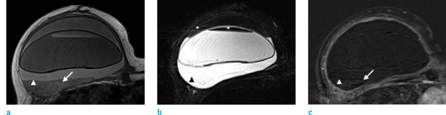
On breast MRI, bilateral implants were seen in subpectoral locations, and the right breast showed asymmetric enlargement. On T2-weighted images (T2WI), silicone gel with high signal intensity was seen both inside and outside the implant shell, representing intracapsular rupture and gel leakage (Fig. 1b). Although it was difficult to evaluate the fibrous capsule, silicone gel with high signal intensity was seen within the pectoralis minor and major muscles, superior and inferior to the enlarged breast, respectively, and possibly represented extracapsular rupture. In addition, a 5.0 × 1.6 cm lesion with an irregular shape was seen in the posterior portion of the right breast outside the implant
shell. The lesion showed iso-signal intensity compared with the silicone inside the implant shell along with small foci of subtle high signal intensity on a T1-weighted image (T1WI) (Fig. 1a).
The small foci showed subtle high signal intensity on T1WI with mild enhancement on gadolinium-enhanced subtraction images and was considered indicative of silicone granuloma (Fig. 1c). Different fluid-fluid levels were noted inside and outside the implant shell, and nondependent portions of low signal intensity on T2WI were thought to reflect recent or old hemorrhages. There was no evidence of implant shell collapse; rather, the volume of the implant was maintained and even slightly increased compared with the intact implant on the contralateral side (Figs. 1, 2).
During surgery, the surgeons observed a rupture in the silicone implant shell and a silicone granuloma. The implant,
Fig. 1. T1- (a), fat-suppressed T2- (b), and 2 minutes delayed gadolinium-enhanced subtraction T1-weighted (c) MR images.
A mass-like lesion of irregular shape and iso-signal intensity as compared with the silicone inside the implant shell in the posterior of the right breast represents silicone granuloma (arrowheads in a-c). It has small foci of subtle high signal intensity on T1WI with mild enhancement (arrows in a, c). Inside and outside the implant shell, a different fluid-fluid level and nondependent portion of low signal intensity on T2WI (asterisks in b) are noticed. Unlike other cases of silicone-implant rupture, this shows enlargement of breast and implant instead of the collapse usually expected.
a b c
Fig. 2. Fat-suppressed T2-weighted MR images. Silicone gel of high signal intensity inside and outside the implant shell, and within pectoralis muscles superior and inferior to breast (arrows in a, b) represents intra- and extracapsular rupture.
a b
www.i-mri.org 112
Implant Rupture with Breast Enlargement | So Yeon Park, et al.
fibrous capsule, and granuloma were removed via the inframammary folds. Upon histopathologic examination, chronic granulomatous inflammation was discovered in the removed mass lesion and diagnosed as granuloma. A fluid analysis was also performed to detect infection, but there was no bacterial growth in culture.
DISCUSSION
Silicone-implant ruptures can be classified as intra- capsular or extracapsular, depending on the location of the rupture. Intracapsular rupture is defined as rupture of the implant shell elastomer with silicone leakage that does not macroscopically extend beyond the fibrous capsule that normally forms around silicone implants. Most implant ruptures (80-90%) are intracapsular. On MRI, the well- known ‘linguine sign,’ which represents multiple curvilinear low-signal-intensity lines within the high-signal-intensity silicone gel, is considered the hallmark of intracapsular rupture. These curvilinear lines represent the collapsed implant shell floating within the silicone gel. Minor ruptures of recently manufactured silicone-gel implants with multilayer shells, a barrier layer, and thick silicone gel show as the ‘keyhole sign’ or ‘noose sign’ on MRI, which represents free silicone within a radial fold without collapse of the implant shell. Extracapsular silicone- implant rupture is less common and is defined as rupture of both the implant shell and the fibrous capsule, with silicone leakage that extends beyond the fibrous capsule into surrounding tissues. On MRI, focal areas of iso- to low signal intensity on T1 fat-suppressed images and high signal intensity on water-suppressed T2WI represent free silicone (4). When free silicone gel incites intense fibrotic reactions in the surrounding tissue, silicone granulomas (also called siliconomas) are formed. Silicone granuloma shows as nearly iso- or slightly hypointense to fat on water- suppressed fast spin-echo T2WI and may increase much as breast carcinomas do (2, 5). On ultrasonography, silicone granuloma may show complex cystic lesions or a ‘snowstorm appearance’, depending on the amount of extravagated silicone gel and the extent of the fibrous and foreign-body reactions (1). Siliconomas in axillary, supraclavicular, or internal mammary lymph nodes may mimic the metastatic lymphadenopathy of breast cancer (7).
There are many studies reporting intra- or extracapsular ruptures of the silicone bag and silicone granuloma (2-6).
Our case was unusual, and this is the first report of breast enlargement after silicone-implant rupture. We detected fluid-fluid levels of various signal intensities both inside and outside the implant elastomer. The implant volume was maintained instead of collapsing, as is usually observed.
As noted above, collapse of the implant shell is a natural result of silicone-implant rupture. We hypothesized that a small rupture and chronic leakage of silicone gel can result in silicone granuloma formation and acute inflammation or hemorrhage. Consequently, it can cause swelling of the breast and implant. Additionally, the high osmotic gradient across the hydrogel implant may cause swelling of the implant and resistance against implant collapse (8).
In conclusion, we report a rare case of intra- and extracapsular silicone breast implant rupture that resulted in fluid-fluid levels of various signal intensities, silicone granulomas, and unexpected enlargement of the breast.
REFERENCES
1. Grubstein A, Cohen M, Steinmetz A, Cohen D. Siliconomas mimicking cancer. Clin Imaging 2011;35:228-231
2. Yang N, Muradali D. The augmented breast: a pictorial review of the abnormal and unusual. AJR Am J Roentgenol 2011;196:W451-460
3. Colombo G, Ruvolo V, Stifanese R, Perillo M, Garlaschi A.
Prosthetic breast implant rupture: imaging--pictorial essay.
Aesthetic Plast Surg 2011;35:891-900
4. Gorczyca DP, Gorczyca SM, Gorczyca KL. The diagnosis of silicone breast implant rupture. Plast Reconstr Surg 2007;120:49S-61S
5. Berg WA, Nguyen TK, Middleton MS, Soo MS, Pennello G, Brown SL. MR imaging of extracapsular silicone from breast implants: diagnostic pitfalls. AJR Am J Roentgenol 2002;178:465-472
6. Orel SG. MR imaging of the breast. Radiol Clin North Am 2000;38:899-913
7. Steinke K, Brook P, Ramuz O. Radiological pitfall:
Siliconoma in internal mammary lymph node mimics breast cancer recurrence. Radiol Case Rep 2011;6:601
8. Choi JJ, Lee JH, Kang BJ, et al. Clinical and imaging characteristics of Polyimplant Prosthesis hydrogel breast implants. J Comput Assist Tomogr 2010;34:449-455