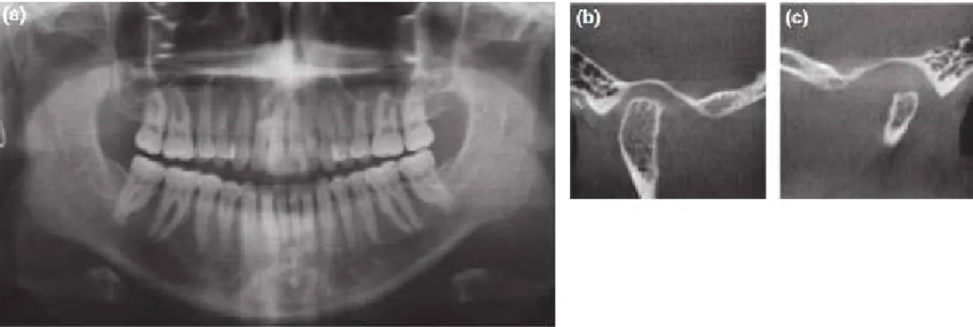
턱관절 장애의 영상검사
전체 글
수치


관련 문서
The ability to visualize simulated construction operations in 3D augmented reality can be of significant help in alleviating model engineering problems that
– Cross section of a beam in Cross section of a beam in pure bending remain plane – (There can be deformation in (There can be deformation in.
Postoperative Postoperative Postoperative Postoperative Magnetic Magnetic Magnetic Magnetic resonance resonance resonance image resonance image image image shows
Also, they can be regarded as the most optimal tools which can be used to manufacture the environmental sculpture which reflects various demands of the public society, which
This study aimed to evaluate the site and extent of injury, injury mechanism, player position, and the reinjury incidence in the hamstring by using magnetic
CT (Computed Tomography) – TDLAS (Tunable Diode Laser Absorption Spectroscopy) is a non-intrusive diagnostic technique that allows for spatially resolved measurements
Moreover, a battery imaging technique to visualize the current distribution pattern using a magnetic field induced at batteries under external current load has
The magnetostatic energy of the domain wall in thin films can be minimized by forming Neel wall, and thus Neel walls are observed stable in various magnetic films for thickness up