아토피피부염 환아에서 interleukin-31 및
25-hydroxyvitamin D의 혈청 수치와 아토피피부염의 중증도와의 상관관계
양윤석, 이정원, 심재원, 김덕수, 정혜림, 박문수, 심정연 성균관대학교 의과대학 강북삼성병원 소아청소년과
Relationship between serum interleukin-31/25-hydroxyvitamin D levels and the severity of atopic dermatitis in children
Yun Seok Yang, Jung Won Lee, Jae Won Shim, Deok Soo Kim, Hye Lim Jung, Moon Soo Park, Jung Yeon Shim Department of Pediatrics, Kangbuk Samsung Hospital, Sungkyunkwan University School of Medicine, Seoul, Korea
Purpose: Atopic dermatitis (AD) is a chronic relapsing inflammatory skin disease. Vitamin D and interleukin-31 (IL-31) are known to be related to the pathogenesis of AD with pruritus. The purpose of this study was to investigate the relationship between serum lev- els of 25-hydroxyvitamin D (25(OH)D) and IL-31 and the disease severity of AD in children with AD.
Methods: We recruited 160 children with AD and 42 controls. We used the SCORing Atopic Dermatitis (SCORAD) index to measure the severity of AD. Serum IL-31 and 25(OH)D levels were assayed using enzyme-linked immunosorbent assay and high-performance liquid chromatography, respectively. Serum levels of total IgE, specific IgE to common allergens and peripheral blood total eosino- phil count were carried out in children with AD.
Results: Serum IL-31 level was significantly higher in AD group compared to control group and 25(OH)D level was significantly lower in AD group than control group. Serum IL-31 level showed the highest level in severe AD group followed by moderate and mild AD group, whilst serum 25(OH)D level was the lowest in severe AD group compared to moderate and mild AD group. There was no difference in serum IL-31 level between AD group and nonatopic dermatitis group. IL-31 level was positively correlated with subjective SCORAD index indicating pruritus in children with AD, and 25(OH)D was inversely correlated with SCORAD index.
Conclusion: IL-31 and vitamin D may be related to the pathogenesis of AD, especially with regard to the pruritus. (Allergy Asthma Respir Dis 2015;3:396-401)
Keywords: Atopic dermatitis, Interleukin-31, 25-hydroxyvitamin D, Pediatrics
서 론
아토피피부염은 소아에서 가장 흔한 만성 재발성 피부 질환으 로, 건조한 피부 및 심한 소양증을 특징으로 하며 그 유병률도 과거 에 비해 현저히 증가하고 있다.1 피부소양증은 아토피피부염의 가 장 특징적인 증상으로 이를 완화시켜주는 것이 삶의 질 향상과 피 부병변의 악화를 막을 수 있는 효과적인 방법이다. 하지만 피부소 양증을 유발하는 인자와 기전은 아직 명확히 밝혀지지 않았다.2
체내에 존재하는 비타민 D는 태양광에 포함된 자외선 B (290–
315 nm 파장)에 피부의 상피 세포가 노출되어 피부의 각질 세포와 섬유 모 세포 형질막에 있는 7-dehydrocholesterol이 비타민 D3 인 cholecalciferol로 전환되어 합성되며 고등어, 정어리, 연어, 참치 같 은 생선의 기름과 계란노른자를 섭취함으로써 얻어진다. 이렇게 피 부에서 합성되거나 음식물을 통해 섭취된 비타민 D는 간을 거치면 서 25-hydroxylase를 통해 대사되어 25-hydroxyvitamin D (25(OH)D)로 전환되며, 이후 신장을 거치면서 1α-hydroxylase에 Allergy Asthma Respir Dis 3(6):396-401, November 2015 http://dx.doi.org/10.4168/aard.2015.3.6.396 ORIGINAL ARTICLE
Correspondence to: Jung Yeon Shim http://orcid.org/0000-0001-9367-2233
Division of Pediatric Allergy & Pulmonology, Department of Pediatrics, Kangbuk Samsung Hospital, Sungkyunkwan University School of Medicine, 29 Saemunan-ro, Jongno-gu, Seoul 04516, Korea Tel: +82-2-2001-2484, Fax: +82-2-2001-2199, E-mail: [email protected]
Received: June 8, 2015 Revised: July 28, 2015 Accepted: August 13, 2015
© 2015 The Korean Academy of Pediatric Allergy and Respiratory Disease The Korean Academy of Asthma, Allergy and Clinical Immunology This is an Open Access article distributed under the terms of the Creative Commons Attribution Non-Commercial License
의해 hydroxyl기가 더해져 활성형비타민 D인 1,25-dihydroxyvita- min D로 전환된다.3 비타민 D가 체내 칼슘 농도 조절 및 뼈의 대사 에 관여하는 기존의 역할을 넘어 악성 종양, 심혈관계 질환, 면역 질 환 등 다양한 조직 및 장기들에도 영향을 미치고 있다는 것이 많은 연구들을 통해 밝혀지고 있다.3,4 특히 최근 들어 비타민 D와 알레 르기 질환과의 연관성에 대한 연구들이 활발히 이루어지고 있는데 아토피피부염, 알레르기비염 및 천식과 같은 알레르기 질환을 가지 고 있는 환자에서 비타민 D가 감소되어 있다는 연구도 있으나,5-7 알 레르기 질환과 비타민 D의 농도와는 연관성이 없다는 연구도 발표 된 바 있어8,9 알레르기 질환에 있어 비타민 D의 역할은 아직 논란 의 여지가 있다고 생각된다.
Interleukin (IL)-31은 비교적 최근에 발견된 사이토카인으로 네 개의 나선다발구조를 가지며 T helper (Th)-2 세포에 의해 생성이 증가되고, IL-31 receptor alpha (IL-31RA)로 구성되어 있는 het- erodimeric 수용체와, 상피 세포와 각질 세포에 발현되어 있는 on- costatin M 수용체를 통해 신호를 보내게 된다.10 이러한 세포들은 IL-31의 자극에 반응하여 아토피피부염에서 나타나는 피부병변을 유발하고 가려움증과 같은 증상들이 나타나게 된다.11 뿐만 아니라 기도과민반응을 유발시킨 동물실험을 통해 IL-31 수용체는 폐의 상피 세포 및 기관지 폐포세척액에서 상향조절된 소견을 보인다는 연구도 있으며,11 알레르기비염 환자의 비강점막에서 IL-31 및 IL- 31RA가 상승된 소견이 관찰된다는 연구도 보고된 바 있다.7 아직 논란의 여지가 있지만 이를 통해 IL-31은 아토피피부염, 천식, 알레 르기비염 등과 같은 알레르기 질환에서 중요한 역할을 하고 있는 것으로 생각된다.
지금까지 아토피피부염 환자들에서 비타민 D와 IL-31을 따로 측 정한 연구 결과는 있었으나 이들을 함께 본 연구는 거의 없다. 저자 들은 이 연구를 통해 IL-31과 비타민 D (25(OH)D)의 상관관계 및 아토피피부염군과 정상군 사이에 차이가 있는지 알아보고자 하였 고, 특히 아토피피부염의 중증도 및 항원감작 여부에 따른 차이가 있는지 알아보고자 하였다.
대상 및 방법
1. 연구 대상
2012년 3월부터 2014년 2월까지 강북삼성병원 소아청소년과 알 레르기클리닉을 방문한 아토피피부염 환아 160명과 같은 기간 내 원한 알레르기 질환의 증상, 징후 및 과거력이 없고 아토피피부염 환아와 나이와 성별이 맞는 42명의 대조군을 대상으로 하였다. 아 토피피부염 진단은 Hanifin and Rajka Criteria에 근거 하였다.12 아 토피피부염의 중증도는 SCORing Atopic Dermatitis (SCORAD) Index를 이용하여 평가하였으며,13 SCORAD 점수 25점 미만을 경 증 아토피피부염, 25점에서 50점 사이를 중등증 아토피피부염, 그
리고 50점 초과를 중증 아토피피부염으로 분류하여 세 군 사이의 IL-31, 25(OH)D, 혈청 총 IgE 농도와 혈액 총 호산구 수(total eosin- ophil count, TEC)를 비교하였다. 가려움의 정도를 평가하기 위해 서 SCORAD index 중 subjective score를 이용하였으며 IL-31, 25(OH)D, SCORAD index, 그리고 SCORAD index 중 subjective score의 관계를 분석하였다. 본 연구는 강북삼성병원 연구윤리위 원회의 심사(2012-01-030)를 받고 시행하였다.
2. 혈청 IL-31, 25(OH)D 및 항원 특이 IgE 측정
아토피피부염군과 대조군에서 정맥혈을 채취하여 원심분리 후 (3,000 rpm, 15분) 혈청을 영하 20°C에서 보관하였다가 일시에 IL-31과 25(OH)D를 측정하였다. 25(OH)D의 농도는 high-perfor- mance liquid chromatography (Neodin, Seoul, Korea), IL-31 농도 는 enzyme-linked immunosorbent assay kit (R&D systems, Min- neapolis, MN, USA)를 이용하여 분석하였다. 혈청 총 IgE와 Der- matophagoides farinae, cat, dog, alternaria, weed mixture, tree mixture, egg white, soy bean, milk에 대한 특이항체는 immuno- CAP (Pharmacia Diagnostics, Uppsala, Sweden)을 이용하여 분석 하였다. 항원감작은 한 개 이상 특이항원에 대한 IgE가 0.35 IU/mL 이상일 때로 정의하였으며, 항원감작 여부에 따라 아토피군, 비아 토피군으로 나누어 각 군에서 IL-31, 25(OH)D, 혈청 총 IgE 농도, 혈액 총 호산구 수가 어떠한 차이를 보이는지 알아보았다.
3. 통계 분석
통계적인 분석은 IBM SPSS Statistics ver. 19.0 (IBM Co., Ar- monk, NY, USA)을 이용해 분석이 이루어졌으며 두 그룹 사이의 차이는 t-test 혹은 chi-square test를 이용하였고, 세 그룹 사이의 평 균값 비교는 one-way analysis of variance를 이용하였다. 연속변수 들은 평균±표준편차로 표현하거나, 정규분포를 따르지 않을 때는 중앙값과 사분범위(interquartile range)로 나타내거나 로그치환하 여 분석하였다. 상관관계의 분석은 Pearson correlation test를 이용 하였으며, P-value가 0.05 미만일 때 통계적으로 의미 있다고 해석 하였다.
결 과
1. 아토피피부염군과 대조군의 인구학적 비교
아토피피부염군과 정상 대조군을 비교하여 보았을 때, 두 그룹 사이에 나이 및 성별은 통계학적으로 차이가 없었으며 알레르기 질 환의 가족력은 아토피피부염군에서 34%로 정상 대조군의 9.4%보 다 더 높았다. 항원감작이 되어있는 아토피피부염군은 58.1%였다.
혈청 총 IgE 농도와 혈액 총 호산구 수는 아토피피부염군에서 정상 대조군보다 통계적으로 유의하게 높았다(Table 1).
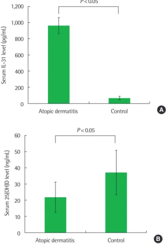
2. 아토피피부염군과 대조군의 혈청 IL-31, 25(OH)D 농도 비교 IL-31의 농도는 아토피피부염군에서 평균 961.5±105.3 pg/mL 로 정상 대조군의 63.1±23.2 pg/mL보다 유의하게 높았고, 25(OH) D의 경우 아토피피부염군에서 평균 21.9±9.5 ng/mL로 정상 대조 군의 36.9±13.7 ng/mL보다 유의하게 감소되었다(Fig. 1).
3. 아토피피부염의 중증도에 따른 비교
아토피피부염의 중증도가 증가할수록 IL-31 농도는 의미 있게 증 가하였으며(경증, 127.3±24.7 pg/mL; 중등증, 882.7±108.7 pg/
mL; 중증, 1,810.4±256.9 pg/mL; P= 0.00), 25(OH)D의 농도는 아 토피피부염의 중증도가 증가할수록 감소하였다(경증, 29.9±9.9
ng/mL; 중등증, 21.7±8.4 ng/mL; 중증, 15.7±4.5 ng/mL;
P= 0.00). 혈액 총 호산구 수는 경증의 아토피피부염군과 중등증의 아토피피부염군 사이에는 차이가 없었으나 중증의 아토피피부염 군의 경우 다른 두 군에 비해 유의하게 증가되었다(P= 0.00). 하지 만 혈청 총 IgE 농도의 경우 중증도에 따른 의미 있는 차이는 보이 지 않았다(Table 2).
4. 항원감작에 따른 비교
혈청 IL-31 농도는 아토피군과 비아토피군에서 통계적으로 유의 한 차이를 보이지 않았다(아토피군, 818.9±104.4 pg/mL; 비아토피 군, 1,169.8±207.9 pg/mL; P= 0.10). 혈청 25(OH)D 농도는 아토피 군에서 23.7±9.8 ng/mL로 비아토피군의 19.2±8.4 ng/mL보다 유 의하게 증가하였으며(P= 0.00), 혈청 총 IgE의 로그값(logIgE)은 아 토피군에서 5.6±1.1 IU/mL로 비아토피군의 3.7±1.4 IU/mL에 비 해 유의하게 상승되었다(P = 0.00). 혈액 총 호산구 수의 로그값 (logTEC)도 아토피군에서 6.0±0.6/mm3로 비아토피군의 5.7±
Table 1. Clinical and laboratory characteristics of study subject
Characteristic AD (n= 160) Control (n= 42) P-value
Age (yr) 6.2± 3.2 5.4± 2.4 0.14
Male sex (%) 47.5 54.8 0.40
FHx of AD (%) 34.0 9.4 0.01
LogIgE (IU/mL) 4.9± 1.5 4.0± 1.2 0.01
LogTEC (/mm3) 5.9± 0.7 5.1± 1.1 0.00
Atopic sensitization (%) 58.1 0 0.00
Values are presented as mean± standard deviation unless otherwise indicated.
AD, atopic dermatitis; Fhx of AD, family history of AD; logIgE, logarithmic transfor- mation of serum total IgE; logTEC, logarithmic transformation of blood total eosino- phil count.
1,200
1,000
800
600
400
200
0
Serum IL-31 level (pg/mL)
Atopic dermatitis Control P < 0.05
A
60
50
40
30
20
10
0
Serum 25(OH)D level (ng/mL)
Atopic dermatitis Control P < 0.05
B Fig. 1. Serum levels of 25(OH)D and IL-31 in children with atopic dermatitis and control group. (A) Serum IL-31 level was significantly higher in AD group com- pared to control group and (B) 25(OH)D level was significantly lower in AD group compared to control group. 25(OH)D, 25-hydroxyvitamin D; IL, interleukin; AD, atopic dermatitis.
Table 2. Comparison of IL-31, 25(OH)D, total IgE, and blood eosinophil count by severity of atopic dermatitis
Variable Atopic dermatitis severity
P-value
Mild Moderate Severe
IL-31 (pg/mL) 127.3± 24.7 882.7± 108.7 1,810.4± 256.9 0.00 25(OH)D (ng/mL) 29.9± 9.9 21.7± 8.4 15.7± 4.5 0.00 LogIgE (IU/mL) 4.6± 1.5 4.7± 1.4 4.9± 1.7 0.34 LogTEC (/mm3) 5.7± 0.3 5.7± 0.7 6.2± 0.7 0.00 Values are presented as mean± standard deviation.
IL, interleukin; 25(OH)D, 25-hydroxyvitamin D; logIgE, logarithmic transformation of serum total IgE; logTEC, logarithmic transformation of blood total eosinophil count.
Table 3. Comparison of IL-31, 25(OH)D, total IgE, and blood eosinophil count in children with atopic dermatitis by atopic sensitization
Variable Atopic dermatitis
P-value
Atopic Nonatopic
IL-31 (pg/mL) 818.9± 104.4 1,169.8± 207.9 0.10
25(OH)D (ng/mL) 23.7± 9.8 19.2± 8.4 0.00
LogIgE (IU/mL) 5.6± 1.1 3.7± 1.4 0.00
LogTEC (/mm3) 6.0± 0.6 5.7± 0.8 0.00
Values are presented as mean ± standard deviation.
IL, interleukin; 25(OH)D, 25-hydroxyvitamin D; logIgE, logarithmic transformation of serum total IgE; logTEC, logarithmic transformation of blood total eosinophil count.
0.8/mm3에 비해 유의하게 상승되었다(P= 0.00) (Table 3).
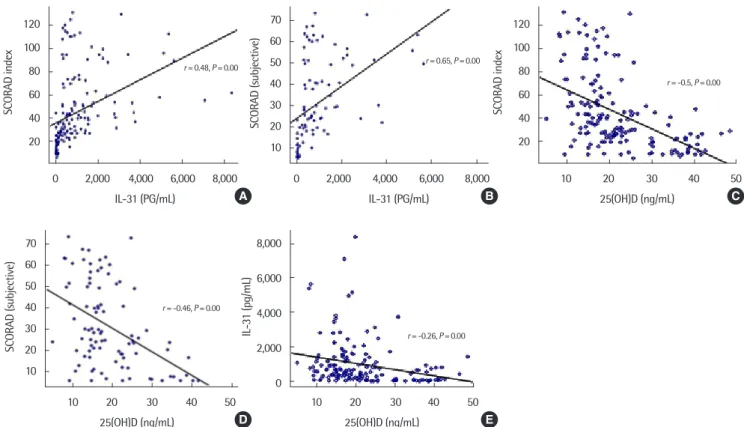
5. IL-31, 25(OH)D, 아토피피부염 중증도와의 상관관계 아토피피부염 환아의 혈청 IL-31의 농도는 SCORAD index와 양 의 상관관계를 보였고 SCORAD index의 subjective score와도 양 의 상관관계를 보였다. 아토피피부염 환아의 혈청 25(OH)D의 농도 는 SCORAD Index 및 SCORAD index의 subjective score와 음의 상관관계를 보였다. 또한 아토피피부염 환아에서 25(OH)D와 IL-31 의 농도는 서로 음의 상관관계를 보였다(Fig. 2).
고 찰
아토피피부염의 특징적 증상인 소양증이 나타나는 기전은 아직 까지 완전히 밝혀지지는 않았지만 IL-31이 소양증과 관련이 있다 는 연구 결과들이 있다. IL-31과 소양증에 관한 동물실험에 의하면, IL-31을 과발현시킨 형질변환 쥐(transgenic mice)에서 아토피피부 염과 유사한 심한 소양증 및 피부병변이 발생하였고11 아토피피부 염 유전자 형질변환 쥐(NC/Nga mice) 연구에서는 긁는 행동을 보 이는 쥐에서 그렇지 않은 쥐보다 IL-31 mRNA의 발현이 더 증가되 었다.14 또한 사람을 대상으로 한 연구에서도 정상 대조군보다 아토
피피부염 환자의 피부병변에서 IL-31 mRNA가 과발현되었으며15 아 토피피부염 환아의 질병활성도가 IL-31의 수치와 연관되어 있다고
했다.16-18 이렇듯 최근 많은 연구들을 통해 T 세포에서 분비되는 향
염증성 사이토카인인 IL-31은 아토피피부염에 있어 소양증과 밀접 한 연관이 있다고 알려져 있고, 본 연구에서도 아토피피부염의 중 증도 및 소양증을 나타내는 주관적 증상과 상관관계가 있음이 확 인되었다. 본 연구에서는 아토피피부염군에서 항원감작 여부에 따 른 혈청 IL-31의 농도가 의미 있는 차이를 보이지 않았는데, 이는 IL-31이 아토피피부염의 면역병태생리에 중요한 역할을 하지만 Th2 세포매개 면역반응과는 관련이 없다는 것을 시사한다. 쥐를 대상으로 한 동물연구에 따르면 IL-31을 과발현시킨 형질변환 쥐 와 정제된 IL-31 단백을 주입한 BALB/c 혹은 C57BL/6 쥐의 피부병 변 및 소양증을 비교한 결과 소양증은 두 그룹 모두에서 나타났지 만, 피부병변은 형질변환 쥐에서만 나타났다.11 또한 두 쥐의 림프 절, 골수, 흉선, 혈청 IgE 농도를 비교해보았는데, IL-31 단백을 주입 한 쥐에 비해 형질변환 쥐의 말단림프절이 커져있고 그 림프절 내에 서 T 세포 수치가 증가한 것 외에 혈청 IgE 농도는 두 그룹 사이에 차이가 없었다는 결과로 미루어보아 IL-31로 인해 유발된 가려움 은 비아토피성 아토피피부염처럼 IgE와는 무관하게 나타나는 증 상임을 시사한다. 하지만 어떤 기전에 의해 IL-31이 가려움을 유발 120
100 80 60 40 20
SCORAD index
0 2,000 4,000 6,000 8,000 IL-31 (PG/mL)
r = 0.48, P = 0.00
120 100 80 60 40 20
SCORAD index
10 20 30 40 50 25(OH)D (ng/mL)
r = -0.5, P = 0.00
70 60 50 40 30 20 10
SCORAD (subjective)
0 2,000 4,000 6,000 8,000 IL-31 (PG/mL)
r = 0.65, P = 0.00
A B C
70 60 50 40 30 20 10
SCORAD (subjective)
10 20 30 40 50 25(OH)D (ng/mL)
r = -0.46, P = 0.00
8,000
6,000
4,000
2,000
0
IL-31 (pg/mL)
10 20 30 40 50 25(OH)D (ng/mL)
r = -0.26, P = 0.00
D E
Fig. 2. Correlation between the SCORAD index, subjective SCORAD index, serum levels of 25(OH)D, and IL-31. The levels of IL-31 showed a positive correlation with the SCORAD index (A), and subjective SCORAD index (B). The levels of 25(OH)D were inversely correlated with the SCORAD index (C), and subjective SCORAD index (D).
(E) The levels of 25(OH)D were inversely correlated with the IL-31 concentration. SCORAD, SCORing Atopic Dermatitis; 25(OH)D, 25-hydroxyvitamin D; IL, interleukin.
하는지에 대해서는 더 구체적인 연구 및 논의가 필요하겠다.
비타민 D 역시 아토피피부염에 있어 중요한 역할을 할 것이라 알 려져 있다. 비타민 D는 filaggrin과 같은 피부장벽 형성에 필요한 단 백질의 합성을 촉진하는데, 비타민 D의 부족은 피부장벽에 결핍을 유발하고 이로 인해 균이나 알레르기항원 등이 피부로 쉽게 침투 하여 증상이 악화될 수 있다.19 또한 항균 효과를 갖는 antimicro- bacterial peptide의 발현에 비타민 D가 관여하여 황색포도상구균 의 집락화를 막아 아토피피부염의 증상 악화를 감소시킬 수 있다.20 또한 아토피피부염 환자에게 비타민 D를 보충해 주었을 때 그 임상 증상이 호전되었다는 연구들도 있다.21-23 이런 연구 결과는 비타민 D가 아토피피부염의 병태생리에 중요한 역할을 함을 시사한다. 본 연구 결과에서도 아토피피부염 환아의 혈청 비타민 D 수치가 대조 군에 비해 감소되어 있었으며, 아토피피부염의 중증도가 증가할수 록 비타민 D의 수치가 감소되어 있는 것을 확인할 수 있었다. 또한 비타민 D의 수치와 아토피피부염의 중증도 및 소양감은 음의 상관 관계를 보였으며 비타민 D와 IL-31도 음의 상관관계를 보였다. 또 한 혈청 비타민 D의 농도는 항원감작에 따른 차이를 보였다. 비타 민 D와 아토피피부염의 면역학적 기전에 대한 연구는 아직 논란의 여지가 많은데 비타민 D가 Th1 세포면역을 억제할 뿐만 아니라, Th2 면역반응도 억제한다는 연구 결과가 있는 반면,24 비타민 D가 Th1 면역반응은 억제하지만 Th2 면역반응은 자극한다는 연구 결
과도 있다.25,26 또 다른 연구에서는 calcitriol이 interferon-gamma
분비 세포는 억제하지만 IL-4 분비 세포에는 영향을 미치지 않아 비타민 D가 Th2 면역반응에는 관여하지 않는다고 얘기하고 있
다.27,28 영국에서 시행된 대규모의 코호트 연구에서는 비타민 D와
혈청 IgE는 선형적인 관계를 보이지 않고 비타민 D가 너무 낮거나 (<25 nmol/L) 너무 높으면(>135 nmol/L) 혈청 IgE의 수치가 증가 되어 있다는 연구가 발표된 바도 있다.29 한편 비타민 D 수용체를 없 앤 형질변환 쥐 연구에서 혈청 IL-5, IL-13 및 IgE가 상승해 있었음 에도 불구하고 외부항원에 노출시켰을 때 천식이 유발되지 않아 비 타민 D 수용체 발현이 알레르기반응을 일으키는 데 필요한 요소라 고 했다.8 이로 미루어 보건대 비타민 D가 구체적으로 어떠한 면역 기전을 통해 아토피피부염에 영향을 미치는지에 대해서는 아직 논 란이 되고 있으며 이는 추후 연구를 통해 밝혀야 될 과제라고 생각 된다.
한편 알레르기 질환에서 비타민 D와 IL-31의 상관관계에 대한 연구는 거의 없다. 알레르기비염과 알레르기비염 및 천식을 동시에 가지고 있는 환자의 연구에서6 IL-31과 비타민 D의 농도는 서로 상 관관계를 보이지 않았다고 하여 비타민 D와 IL-31은 서로 독립적 으로 알레르기 질환에 영향을 미친다고 제시하였으며 연구자들의 이전 연구에서도 역시 비타민 D와 IL-31은 연관성이 없었다.30 본 연구에서는 비타민 D와 IL-31이 음의 상관관계를 보여 비타민 D 결 핍이 Th2 면역반응에 관여한다고 생각할 수도 있겠으나 본 연구의
결과에서 보인 아토피군과 비아토피군 사이에서의 비타민 D 및 IL-31 수치의 차이를 함께 고려하여 보았을 때, 비타민 D는 다른 복 합적인 기전으로 아토피피부염에 영향을 미친다고 생각할 수 있다.
추후 비타민 D 결핍과 IL-31의 발현에 관한 기전에 대해서 자세한 연구가 이루어져야 하겠다.
본 연구의 강점으로는 비타민 D, IL-31과 아토피피부염의 중증 도와의 관계뿐 아니라 비타민 D와 IL-31의 상관관계도 알아보았다 는 데 있다고 생각되며 단점으로는 비타민 D 결핍 환자에서 비타민 D 투여에 따른 아토피피부염의 증상 변화 여부를 조사하지 못하였 고 각 개인의 신체활동이나 음식 섭취에 대해 조사하지 못한 것에 있다. 또한 9종의 항원 특이 항체의 존재 유무만으로 아토피 여부 를 판단한 제한점이 있다.
결론적으로, IL-31과 비타민 D는 아토피피부염의 병태생리, 특 히 가려움과 관련된 면역학적 기전에 중요한 역할을 한다고 생각되 며, Th2 매개 면역반응과의 관계에 대해서는 추가적인 연구가 필요 할 것으로 생각한다.
REFERENCES
1. Asher MI, Montefort S, Bjorksten B, Lai CK, Strachan DP, Weiland SK, et al. Worldwide time trends in the prevalence of symptoms of asthma, al- lergic rhinoconjunctivitis, and eczema in childhood: ISAAC Phases One and Three repeat multicountry cross-sectional surveys. Lancet 2006;368:
733-43.
2. Stander S, Steinhoff M. Pathophysiology of pruritus in atopic dermatitis:
an overview. Exp Dermatol 2002;11:12-24.
3. Holick MF. Vitamin D deficiency. N Engl J Med 2007;357:266-81.
4. Bikle D. Nonclassic actions of vitamin D. J Clin Endocrinol Metab 2009;
94:26-34.
5. Grober U, Spitz J, Reichrath J, Kisters K, Holick MF. Vitamin D: Update 2013: From rickets prophylaxis to general preventive healthcare. Derma- toendocrinol 2013;5:331-47.
6. Bonanno A, Gangemi S, La Grutta S, Malizia V, Riccobono L, Colombo P, et al. 25-Hydroxyvitamin D, IL-31, and IL-33 in children with allergic disease of the airways. Mediators Inflamm 2014;2014:520241.
7. Baumann R, Rabaszowski M, Stenin I, Gaertner-Akerboom M, Scheck- enbach K, Wiltfang J, et al. The release of IL-31 and IL-13 after nasal aller- gen challenge and their relation to nasal symptoms. Clin Transl Allergy 2012;2:13.
8. Wittke A, Weaver V, Mahon BD, August A, Cantorna MT. Vitamin D re- ceptor-deficient mice fail to develop experimental allergic asthma. J Im- munol 2004;173:3432-6.
9. Chiu YE, Havens PL, Siegel DH, Ali O, Wang T, Holland KE, et al. Serum 25-hydroxyvitamin D concentration does not correlate with atopic der- matitis severity. J Am Acad Dermatol 2013;69:40-6.
10. Zhang Q, Putheti P, Zhou Q, Liu Q, Gao W. Structures and biological functions of IL-31 and IL-31 receptors. Cytokine Growth Factor Rev 2008;19:347-56.
11. Dillon SR, Sprecher C, Hammond A, Bilsborough J, Rosenfeld-Franklin M, Presnell SR, et al. Interleukin 31, a cytokine produced by activated T cells, induces dermatitis in mice. Nat Immunol 2004;5:752-60.
12. Hanifin JM, Rajka G. Diagnostic features of atopic dermatitis. Acta Derm
Venereol Suppl (Stockh) 1980;92:44-7.
13. Severity scoring of atopic dermatitis: the SCORAD index. Consensus Re- port of the European Task Force on Atopic Dermatitis. Dermatology 1993;
186:23-31.
14. Takaoka A, Arai I, Sugimoto M, Honma Y, Futaki N, Nakamura A, et al.
Involvement of IL-31 on scratching behavior in NC/Nga mice with atop- ic-like dermatitis. Exp Dermatol 2006;15:161-7.
15. Sonkoly E, Muller A, Lauerma AI, Pivarcsi A, Soto H, Kemeny L, et al.
IL-31: a new link between T cells and pruritus in atopic skin inflamma- tion. J Allergy Clin Immunol 2006;117:411-7.
16. Kim S, Kim HJ, Yang HS, Kim E, Huh IS, Yang JM. IL-31 serum protein and tissue mRNA levels in patients with atopic dermatitis. Ann Dermatol 2011;23:468-73.
17. Raap U, Weibmantel S, Gehring M, Eisenberg AM, Kapp A, Folster-Holst R. IL-31 significantly correlates with disease activity and Th2 cytokine lev- els in children with atopic dermatitis. Pediatr Allergy Immunol 2012;23:
285-8.
18. Ezzat MH, Hasan ZE, Shaheen KY. Serum measurement of interleukin-31 (IL-31) in paediatric atopic dermatitis: elevated levels correlate with sever- ity scoring. J Eur Acad Dermatol Venereol 2011;25:334-9.
19. Palmer CN, Irvine AD, Terron-Kwiatkowski A, Zhao Y, Liao H, Lee SP, et al. Common loss-of-function variants of the epidermal barrier protein filaggrin are a major predisposing factor for atopic dermatitis. Nat Genet 2006;38:441-6.
20. Hata TR, Kotol P, Jackson M, Nguyen M, Paik A, Udall D, et al. Adminis- tration of oral vitamin D induces cathelicidin production in atopic indi- viduals. J Allergy Clin Immunol 2008;122:829-31.
21. Samochocki Z, Bogaczewicz J, Jeziorkowska R, Sysa-Jedrzejowska A, Glinska O, Karczmarewicz E, et al. Vitamin D effects in atopic dermatitis.
J Am Acad Dermatol 2013;69:238-44.
22. Camargo CA Jr, Ganmaa D, Sidbury R, Erdenedelger Kh, Radnaakhand N, Khandsuren B. Randomized trial of vitamin D supplementation for win- ter-related atopic dermatitis in children. J Allergy Clin Immunol 2014;134:
831-5.e1.
23. Amestejani M, Salehi BS, Vasigh M, Sobhkhiz A, Karami M, Alinia H, et al. Vitamin D supplementation in the treatment of atopic dermatitis: a clinical trial study. J Drugs Dermatol 2012;11:327-30.
24. Pichler J, Gerstmayr M, Szepfalusi Z, Urbanek R, Peterlik M, Willheim M.
1 alpha,25(OH)2D3 inhibits not only Th1 but also Th2 differentiation in human cord blood T cells. Pediatr Res 2002;52:12-8.
25. Boonstra A, Barrat FJ, Crain C, Heath VL, Savelkoul HF, O'Garra A.
1alpha,25-Dihydroxyvitamin d3 has a direct effect on naive CD4(+) T cells to enhance the development of Th2 cells. J Immunol 2001;167:4974-80.
26. Jirapongsananuruk O, Melamed I, Leung DY. Additive immunosuppres- sive effects of 1,25-dihydroxyvitamin D3 and corticosteroids on TH1, but not TH2, responses. J Allergy Clin Immunol 2000;106:981-5.
27. Staeva-Vieira TP, Freedman LP. 1,25-dihydroxyvitamin D3 inhibits IFN- gamma and IL-4 levels during in vitro polarization of primary murine CD4+ T cells. J Immunol 2002;168:1181-9.
28. Lemire JM, Archer DC, Beck L, Spiegelberg HL. Immunosuppressive ac- tions of 1,25-dihydroxyvitamin D3: preferential inhibition of Th1 func- tions. J Nutr 1995;125(6 Suppl):1704S-1708S.
29. Hypponen E, Berry DJ, Wjst M, Power C. Serum 25-hydroxyvitamin D and IgE: a significant but nonlinear relationship. Allergy 2009;64:613-20.
30. Cheon BR, Shin JE, Kim YJ, Shim JW, Kim DS, Jung HL, et al. Relationship between serum 25-hydroxyvitamin D and interleukin-31 levels, and the severity of atopic dermatitis in children. Korean J Pediatr 2015;58:96-101.