Received August 30, 2013 /Revised September 23, 2013 /Accepted September 25, 2013
Trichinella spiralis (T. spiralis) has been reported to induce angiogenesis and a supply of nutrients and to act as a reliable waste disposal system by induction of the expression of the angiogenic molecule vascular endothelial cell growth factor (VEGF) during nurse cell formation. However, the mechanism underlying the induction of VEGF in nurse cells by T. spiralis has not yet been defined. Some research has pointed to the possibility of hypoxia in nurse cells, but whether hypoxia occurs in infected muscle or nurse cells has not been studied. It is also a matter of debate whether hypoxia induces the ex- pression of VEGF and subsequent angiogenesis in infected muscle. Recent studies showed that thymo- sin β4, a potent VEGF-inducing protein, was expressed at a very early stage of muscle infection by T. spiralis, suggesting that VEGF is induced at an early stage in nurse cells. Furthermore, hypoxia was not detected in any nurse cell stage but was detected in inflammatory cells. The findings suggest that induction of angiogenesis by VEGF in T. spiralis-infected nurse cells is mediated by thymosin β4 and unrelated to hypoxia.
Key words : Thymosin β4, Trichinella spiralis, nurse cell, hypoxia, vascular endothelial cell growth factor (VEGF)
*Corresponding author
*Tel : +82-51-990-6428, Fax : +82-51-990-3081
*E-mail : [email protected]
This is an Open-Access article distributed under the terms of the Creative Commons Attribution Non-Commercial License (http://creativecommons.org/licenses/by-nc/3.0) which permits unrestricted non-commercial use, distribution, and reproduction in any medium, provided the original work is properly cited.
서 론
기생충은 그 자체로는 생존이 불가능하므로 숙주에 침입하 여 숙주의 여러 가지 영양분을 섭취하고 숙주의 세포 및 기관 을 자신의 생존에 적합하게 변형, 이용하여 성장하고 번식한 다. 따라서 숙주가 병에 걸리거나 사망하면 기생충도 더 이상 숙주에서 살아남지 못하게 되므로 오랜 세월 동안 기생충은 숙주에 가능한 영향을 미치지 않으면서 숙주의 시스템 및 영 양분을 효율적으로 이용하여 살아남는 방식으로 진화해 왔다.
숙주의 시스템을 효율적으로 이용하는 기생충중의 하나인 선 모충(Trichinella spiralis) 감염은 감염된 육고기를 익히지 않은 상태로 섭취함으로서 일어나며 유충은 위에서 탈낭되고 소장 에서 성숙하여 성충이 된다. 이후 선모충은 소장의 점막에 박 힌 채 기생하며, 암컷은 교미 후 유충을 낳는다. 유충은 장벽을 뚫고 혈류를 따라 뇌와 심장을 포함한 전신을 순환하다 작은 모세혈관에 이르면 혈관 밖으로 나와 횡문근으로 향한다. 근 육섬유를 따라 들어간 선모충 유충은 근육을 구성하는 긴 방 추형 세포에 침입한다. 호발 근육은 목, 턱, 혀, 후두, 횡경막 등이며, 유충이 횡문근에 도달한 뒤 5일째 정도부터 콜라겐 막으로 감염된 세포를 둘러싸기 시작하여 capsule 또는 nurse
cell 복합체를 형성한다(Fig. 1)[5, 22]. 이후 선모충은 근육세포 에서 살아남고 지속적인 영양분을 공급받기 위해 nurse cell 주위로 혈관 형성을 유도하는데 실제로 초기의 nurse cell 주 변에 혈관내피세포증식인자(Vascular Endothelial Cell Growth Factor, VEGF) 형성을 유도하여 주변의 혈관 네트워크 형성을 촉진한다[2, 3]. Baruch and Despommier (1991)은 선모충 감 염 근육세포(nurse cell) 주위에 혈관그물망을 관찰하고, 이러 한 혈관 조직이 정상 근육세포 주변 혈관과는 형태학적 성상 이 뚜렷이 구분된 기생충 감염에 따른 혈관신생(angiogensis) 임을 밝혔다[2]. 이러한 혈관신생 유도는 혈관신생을 유도하는 물질을 기생충이 분비하거나 혹은 숙주의 유전체 조작을 통한 숙주유래 혈관신생물질에 의해 일어날 수 있다고 보고하였다.
또 Capo 등(1998)은 nurse cell 내에서 VEGF mRNA 및 pep-
tide가 증가됨을 확인하고 VEGF 증가 이전에 nurse cell에 hy-
poxia 상태가 올 것이라 가정하였으며 hypoxia는 감염된 근육
세포의 미토콘드리아 손상에 의한 것으로 추정하였다. 또한
VEGF가 nurse cell에서 16개월까지 존재하는 것을 근거로 hy-
poxia 상태가 nurse cell 내에서 오랫동안 지속될 것이라고 예
측하였다[3]. 하지만, 이러한 연구의 한계점은 VEGF mRNA
및 단백질 분비가 유도되어 혈관 신생을 촉진한다는 현상에
그치고 있으며VEGF가 유도되는 기작에 대한 구체적인 연구
가 이루어지지 못했다. 또Nurse cell 형성과정에 hypoxia가
올 것으로 가정하였지만 실제 nurse cell 형성과정에 hypoxia
가 발생하는지에 대한 구체적인 실험도 이루어지지 않아
nurse cell 형성 과정에 따른 hypoxia 발생 여부에 대해서 증명
하지 못하였다. 따라서 선모충 감염에 의한 nurse cell 형성과
정에 일어나는 VEGF 발현 증가 현상이 hypoxia에 의해 일어
Fig. 1. Life cycle of
Trichinella spiralis
("Parasitic Diseasers" 4th Ed. Apple Trees Productions, LLC., Pub. P.O.Box280, New York, NY 100332).나는지 다른 작용 기작으로 일어나는지를 먼저 규명할 필요가 있다.
Hypoxia가 VEGF의 발현을 유도하는 과정에 관해서는 많 은 연구가 이루어져 있다. VEGF의 발현 유도 기작 중에 가장 많이 알려진 전사 조절 인자로 hypoxia-inducible factor (HIF) 를 들 수 있다. HIF-1α 단백질은 HIF-1β 단백질과 결합한 후 VEGF의 전사조절 영역에 있는HIF-1 responsive elements (HRE)에 결합하여 VEGF의 유전자의 발현을 유도한다[20]. 이 때 HIF-1β은 항시 존재하나 HIF-1α의 경우 정상 산소 농도에 서는 von Hippel–Lindau (VHL) 단백질과 결합하여 ubiq-
uitination 과정을 거처 분해된다[1, 16]. 하지만 hypoxia 상태
에서는 HIF-1α의 분해가 일어나지 않고 HIF-1α는 핵으로 들어
가 HIF-1β와 결합하여 VEGF 전사 조절 부위의 HRE 상에 결
합하여 VEGF의 발현을 유도한다[1, 16]. 그러나 HIF-1α 단백
질의 안정화는 hypoxia에 의해서만 조절 받는 것이 아님이
규명되었다. Hypoxia와 무관한 HIF-1α 단백질의 안정화가 re-
ceptor of activated protein kinase C (RACK1)과 heat-shock
protein 90 (HSP90)의 HIF-1α 단백질 PAS domain에 경쟁적
으로 결합하면서 조절 되는 것이 규명되었다(Fig. 2)[11, 12,
16].
Fig. 2. Oxygen-dependent and oxygen-independent regulation of HIF-1α[16]. HIF-1α contains a basic helix-loop-helix PAS DNA-bind- ing domain (bHLH) and two transactivation domains (NTAD and CTAD). In the presence of oxygen, PHDs hydroxylate proline residues (Pro) within the HIF-1α oxygen-dependent degradation domain (ODD) near the NTAD. pVHL binds to hydroxylated HIF-1α and elongin-C. Consequently, HIF-1α is ubiquitinated and degraded by the 26S proteasome. Oxygen also influences the transcriptional activity of HIF-1α/HIF-2α through factor inhibiting HIF (FIH), which hydroxylates an aspar- aginyl residue (ASN) within the HIF-1α CTAD. During hypoxia, both HIF-1α and HIF-2α are stabilized and enter the nucleus, where they heterodimerize with HIF-1β and initiate downstream transcription. In the oxygen-independent pathway, heat shock protein 90 (HSP90) and receptor for activated C-kinase 1 (RACK1) compete to bind to the PAS domain of HIF-1α.
HSP90 binding stabilizes HIF-1α by excluding RACK1, which recruits elongin-C to HIF-1α. This interaction promotes the poly-ubiquitination and degradation of HIF-1α, while HIF-2α can still activate downstream transcription. Thus, both oxy- gen-dependent and oxygen-independent pathways bring HIF-1α to elongin-C and other subunits of the E3 ubiquitin ligase complex for degradation by the 26S proteasome.
본 연구 그룹에서 최근 보고한 내용에 의하면 선모충 감염 후 nurse cell 형성과정에 있어서 VEGF의 발현은 선모충이 근육에 감염된 초기에 일어나는 것으로 확인되었다. 이러한 현상에 의해 혈관형성이 nurse cell 형성의 초기 단계에서 이 루어질 가능성이 높고 따라서 nurse cell 형성과정에서 예상된 hypoxia 현상은 일어나지 않을 것으로 추정하였다. 실제로 선 모충 감염 단계별로 nurse cell 주변의 hypoxia를 hypoxic probe를 사용하여 관찰한 결과 nurse cell이 완성되는 기간 동안 hypoxia는 nurse cell에서 관찰되지 않았으며 면역현상
에 의해 파괴되는 nurse cell에 다량의 임파구들이 밀집되고 이러한 임파구의 밀집에 의해 hypoxia 현상이 일어나며 이에 따른 HIF-1α 및 임파구가 분비하는 VEGF가 감염 말기의 nurse cell에서 발현이 증가함이 관찰되었다[9]. 이러한 결과는 nurse cell의 VEGF 증가와 이에 따른 혈관신생 작용이 hypo- xia에 의해 일어나는 것이 아님을 시사해 준다[9].
선모충 감염에 의한 nurse cell 형성 과정에 대한 본 연구
그룹의 보고에 의하면 nurse cell 감염 초기부터 티모신베타4
(thymosin β4)의 발현이 증가하였는데[9] 이러한 결과는 티모
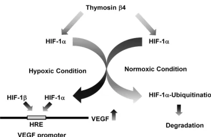
Fig. 3. Proposed mechanism of Thymosin β4 (T β4)-mediated VEGF induction. Tβ4 stim- ulates the induction, stability or activity of HIF-1α. HIF-1α binds to HIF-1β on the hypoxia response element (HRE) in the VEGF promoter region, triggering the ex- pression of VEGF mRNA.
신베타4와 VEGF 및 혈관신생 과정을 관련지어 생각해 볼 수 있게 한다. 티모신베타4는 생체 내에 존재하는 아주 작은 크기 의 단백질로 43개의 아미노산으로 구성된 4.9 kDa의 단백질이 다[7, 13]. 티모신베타4는 actin과 결합하는 기능이 보고되면서 활발히 연구되기 시작했다. 사이모신 베타 4는 낱개의 분리된 형태의 G-actin과 결합하여 G-actin이 결합된 형태인 F-actin 의 형성을 막아주는 기능을 담당하며 G-actin과 F-actin의 전 환을 조절하여 actin cytoskeleton의 구조를 변경하고 세포의 이동을 조절하는 것으로 보고되었다[21]. 티모신베타4의 생체 내 기능에 관해서도 활발히 연구가 진행되어 최근에는 티모신 베타4가 혈관신생을 증가시키고[6, 14] 상처치유[15, 17] 및 모 발 성장[18, 19]을 촉진하며 암의 성장과 전이도 증가시킨다고 보고되었다[4, 10]. 이와 더불어 티모신베타4는 본 연구 그룹에 의해 최초로 VEGF의 발현을 유도하여 혈관 신생을 촉진시키 는 것으로 보고되었다[4]. 즉, 티모신베타4를 과다발현 하는 아데노바이러스에 감염된 암세포에서 VEGF의 발현이 증가하 였으며 티모신베타4 펩티드를 처리한 세포군에서 VEGF가 mRNA 및 단백질 수준에서 발현이 증가되는 것이 증명되었다 [4, 8]. 이후 티모신 베타4에 의한 VEGF의 발현 증가는 티모신 베타4가 VEGF의 발현을 증가시키는 HIF-1α 단백질의 안정화 를 유도함으로써 이루진 다는 것이 규명되었다[8]. 따라서 티 모신베타 4는 hypoxia 유무에 관계 없이 VEGF의 발현을 유도 하는 능력이 있음이 입증되었다(Fig. 3)[16]. 따라서 선모충 감 염 초기부터 티모신베타4의 발현이 증가하는 것은 티모신베 타 4에 의해 VEGF의 발현이 증가되어 nurse cell의 혈관 신생 을 촉진할 가능성을 시사해 준다. 선모충 감염 후 nurse cell이 완성되는 시기까지 nurse cell에 hypoxia가 발생하지 않는 다 는 결과[9]는 티모신베타4가 nurse cell에서 VEGF를 유도하여 nurse cell의 혈관신생을 매개할 가능성을 한층 더 높여 준다.
그러나 이러한 결과에도 불구하고 티모신베타4가 nurse cell의 혈관신생을 직접적으로 매개한다고 단정 짓기에는 많 은 부족함이 있다. 우선 티모신베타 4가 nurse cell의 VEGF
발현을 촉진시키거나 혈관신생을 매개한다는 직접적인 증거 가 부족하다. 이를 위해서는 티모신베타4를 과다발현 하거나 억제하였을 때 선모충 감염 nurse cell에서의 VEGF 발현이 어떻게 변하는지 또 혈관신생에는 어떠한 영향을 주는지에 대한 연구가 필요하다. 이러한 연구를 통해 티모신베타4의 변 화가 VEGF 발현 및 혈관신생에 영향을 준다면 nurse cell의 혈관 신생이 티모신베타4에 의해 매개됨이 증명될 수 있을 것이다. 이와 더불어 선모충 감염이 티모신베타4의 발현을 어 떻게 유도시키는지에 관한 연구도 함께 수행되어 기생충 감염 에 의한 티모신베타4의 발현 유도기작을 규명할 수 있다면 선모충 감염 기작 및 혈관신생 연구에 보다 많은 진전을 기대 할 수 있을 것이다.
지금까지의 기생충 연구는 기생충의 감염경로, 숙주 내에서 의 생활사 및 감염 예방에 초점을 맞추어 기생충 감염을 막고 이에 대한 치료제를 개발하는 것이 그 주된 목적이었다. 하지 만 최근의 연구경향은 기생충의 감염, 감염 생리학 및 분자생 물학적 기전 연구를 통해 인간 질병의 치료제로 사용하는 등 기생충을 질병 치료 및 예방 분야에 이용하고자 하는 시도들 이 이루어지고 있다. 선모충의 혈관신생 작용에 관한 연구도 이러한 맥락에서 혈관신생 촉진제 및 억제제등의 개발에 새로 운 지평을 제공하는 연구과제가 될 수 있을 것이다.
감사의 글
본 연구는 2013년도 고신대학교 의과대학 의과학연구소 학 술연구비의 지원에 의해 수행되었음.
References
1. Baldewijns, M. M., van Vlodrop, I. J., Vermeulen, P. B., Soetekouw, P. M., van Engeland, M. and de Bruine, A. P.
2010. VHL and HIF signalling in renal cell carcinogenesis.
J Pathol
221, 125-138.4. Cha, H. J., Jeong, M. J. and Kleinman, H. K. 2003. Role of thymosin beta4 in tumor metastasis and angiogenesis.
J Natl Cancer Inst
95, 1674-1680.5. Despommier, D., Aron, L. and Turgeon, L. 1975. Trichinella spiralis: growth of the intracellular (muscle) larva.
Exp Parasitol
37, 108-116.6. Grant, D. S., Rose, W., Yaen, C., Goldstein, A., Martinez, J. and Kleinman, H. 1999. Thymosin beta4 enhances endo- thelial cell differentiation and angiogenesis.
Angiogenesis
3, 125-135.7. Hannappel, E. and Huff, T. 2003. The thymosins. Prothymosin alpha, parathymosin, and beta-thymosins: structure and function.
Vitam Horm
66, 257-296.8. Jo, J. O., Kim, S. R., Bae, M. K., Kang, Y. J., Ock, M. S., Kleinman, H. K. and Cha, H. J. 2010. Thymosin beta4 in- duces the expression of vascular endothelial growth factor (VEGF) in a hypoxia-inducible factor (HIF)-1alpha-depend- ent manner.
Biochim Biophys Acta
1803, 1244-1251.9. Kang, Y. J., Jo, J. O., Cho, M. K., Yu, H. S., Ock, M. S. and Cha, H. J. 2011. Trichinella spiralis infection induces angio- genic factor thymosin beta4 expression.
Vet Parasitol
181, 222-228.10. Kobayashi, T., Okada, F., Fujii, N., Tomita, N., Ito, S., Tazawa, H., Aoyama, T., Choi, S. K., Shibata, T., Fujita, H.
and Hosokawa, M. 2002. Thymosin-beta4 regulates motility and metastasis of malignant mouse fibrosarcoma cells.
Am J Pathol
160, 869-882.11. Liu, Y. V., Baek, J. H., Zhang, H., Diez, R., Cole, R. N. and Semenza, G. L. 2007. RACK1 competes with HSP90 for bind- ing to HIF-1alpha and is required for O(2)-independent and HSP90 inhibitor-induced degradation of HIF-1alpha.
Mol Cell
25, 207-217.umbilical vein endothelial cells.
FASEB J
11, 474-481.15. Malinda, K. M., Sidhu, G. S., Mani, H., Banaudha, K., Maheshwari, R. K., Goldstein, A. L. and Kleinman, H. K.
1999. Thymosin beta4 accelerates wound healing.
J Invest Dermatol
113, 364-368.16. Ock, M. S., Song, K. S., Kleinman, H. and Cha, H. J. 2012.
Thymosin beta4 stabilizes hypoxia-inducible factor-1alpha protein in an oxygen-independent manner.
Ann N Y Acad Sci
1269, 79-83.17. Philp, D., Goldstein, A. L. and Kleinman, H. K. 2004.
Thymosin beta4 promotes angiogenesis, wound healing, and hair follicle development.
Mech Ageing Dev
125, 113-115.18. Philp, D., Nguyen, M., Scheremeta, B., St-Surin, S., Villa, A. M., Orgel, A., Kleinman, H. K. and Elkin, M. 2004.
Thymosin beta4 increases hair growth by activation of hair follicle stem cells.
FASEB J
18, 385-387.19. Philp, D., St-Surin, S., Cha, H. J., Moon, H. S., Kleinman, H. K. and Elkin, M. 2007. Thymosin beta 4 induces hair growth via stem cell migration and differentiation.
Ann N Y Acad Sci
1112, 95-103.20. Sang, N., Fang, J., Srinivas, V., Leshchinsky, I. and Caro, J. 2002. Carboxyl-terminal transactivation activity of hypo- xia-inducible factor 1 alpha is governed by a von Hippel- Lindau protein-independent, hydroxylation-regulated asso- ciation with p300/CBP.
Mol Cell Biol
22, 2984-2992.21. Smart, N., Rossdeutsch, A. and Riley, P. R. 2007. Thymosin beta4 and angiogenesis: modes of action and therapeutic potential.
Angiogenesis
10, 229-241.22. Teppema, J. S., Robinson, J. E. and Ruitenberg, E. J. 1973.
Ultrastructural aspects of capsule formation in Trichinella spiralis infection in the rat.

![Fig. 2. Oxygen-dependent and oxygen-independent regulation of HIF-1α[16]. HIF-1α contains a basic helix-loop-helix PAS DNA-bind- DNA-bind-ing domain (bHLH) and two transactivation domains (NTAD and CTAD)](https://thumb-ap.123doks.com/thumbv2/123dokinfo/5015858.550037/3.892.159.739.189.702/oxygen-dependent-oxygen-independent-regulation-contains-transactivation-domains.webp)