Plant Cell Contact-Dependent Virulence Regulation of hrp Genes in Pseudomonas syringae pv. tabaci 11528
Jun Seung Lee, Ji Young Cha and Hyung Suk Baik*
Department of Microbiology, College of Natural Science, Pusan National University, Busan 609-735, Republic of Korea
Received February 7, 2011 /Accepted February 23, 2011The hrp gene cluster in the plant pathogen Pseudomonas syringae is a key determinant of pathogenicity.
Recent studies have demonstrated that specific host cell induction of the Ralstonia solanacearum hrp gene cluster is controlled by the PrhA (plant regulator of hrp) receptor. To characterize the role that P. syringae PrhA plays in the virulence of plant cells, a prhA homolog was isolated from P. syringae pv. tabaci and a ΔprhA mutant was constructed by allelic exchange. The ΔprhA mutant had reduced virulence in the host plant, and co-culture of P. syringae pv. tabaci and plant cell suspensions induced a much higher level of hrpA gene transcription than culture in hrp-inducing minimal medium. These results indicate that PrhA of P. syringae is a putative pathogen-plant cell contact sensor, therefore, we used a hrpA-gfp reporter fusion to monitor the in situ expression of PrhA. The results of this study demonstrated that PrhA induces hrp gene expression in P. syringae pv. tabaci in the presence of plant cells.
Key words : Plant regulator of hrp (PrhA), plant-pathogen interaction, Pseudomonas syringae pv. Tabaci, TonB-dependent siderophore receptor
*Corresponding author
*Tel:+82-51-510-2271, Fax:+82-51-514-1778
*E-mail : [email protected]
Introduction
Pseudomonas syringae is an important plant pathogenic bacterium commonly used to study plant–microbe interactions. P. syringae causes leaf spots and necrosis in host plants and a hypersensitive response (HR) in non-host plants [13]. In host plants, disease symptoms usually develop after several days of bacterial growth in the leaf apoplast.
However, on non-host plants, the defense-associated pro- grammed cell death that characterizes the HR occurs within 24 hr of the plant cells coming in contact with the bacteria [25]. More than 50 P. syringae pathovars have been identified based on their virulence and host specificity [9]. For exam- ple, P. syringae pv. tabaci causes disease on tobacco plants, but induces a HR on many other non-host plant species. The ability of P. syringae to cause diseases on their hosts and to elicit the HR in non-host plants is controlled by the hrp (hypersensitive response and pathogenicity) / hrc (hypersensitive response and conserved) genes, which reside in a pathogenicity island also known as the Hrp PAI [2,5,18].
The hrp/hrc genes (hrp gene cluster) are conserved among many gram-negative plant pathogenic bacteria, including P.
syringae, Ralstonia solanacearum, Xanthomonas campestris,
Erwinia amylovora, Pantoea stewartii sp. stewartii and Erwinia chrysanthemi, as well as bacteria associated with animals [3,4,6,10,18]. The hrp/hrc genes are expressed at a very low level in nutrient-rich media, but are induced in plants or in artificial hrp-inducing minimal media that mimics apo- plastic conditions [14,24,30]. In addition, recent studies have shown that specific host factors induce expression of the Ralstonia solanacearum hrp gene cluster, and that this specific host cell induction of the hrp gene cluster is controlled by PrhA (plant regulatory of hrp), a protein that is homologous to outer-membrane siderophore receptors [1,20].
In this study, we amplified a prhA gene of P. syringae pv.
tabaci (ATCC 11528) by PCR and then constructed a ΔprhA
mutant by allelic exchange. We evaluated prhA to determine
if it regulates the transcription of hrpA by comparing the
levels of β-galactosidase activity after growth in hrp-inducing
minimal medium. Despite no differences being found in any
of the tested strains, the ΔprhA mutant had reduced viru-
lence in the host plant. However, co-culture of P. syringae
and plant cell materials induced a much higher level of hrpA
gene transcription than culture in hrp-inducing minimal
medium. Taken together, these results indicate that PrhA of
P. syringae is a putative pathogen-plant cell contact sensor.
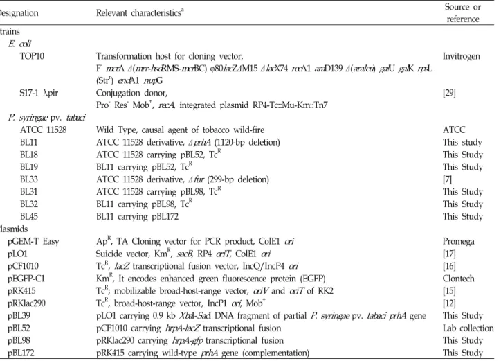
Table 1. Bacterial strains and plasmids used in this study
Designation Relevant characteristicsa Source or
reference Strains
E. coli
TOP10 Transformation host for cloning vector,
F-
mcr
A Δ(mrr-hsd
RMS-mcr
BC) φ80lac
ZΔM15 Δlac
X74rec
A1ara
D139 Δ(araleu
)gal
Ugal
Krps
L (Strr)end
A1nup
GInvitrogen
S17-1 λpir Conjugation donor,
Pro-Res-Mob+,
recA
, integrated plasmid RP4-Tc::Mu-Km::Tn7 [29]P. syringae
pv.tabaci
ATCC 11528 Wild Type, causal agent of tobacco wild-fire ATCC
BL11 ATCC 11528 derivative, Δ
prhA
(1120-bp deletion) This studyBL18 ATCC 11528 carrying pBL52, TcR This Study
BL19 BL11 carrying pBL52, TcR This Study
BL33 ATCC 11528 derivative, Δ
fur
(299-bp deletion) [7]BL31 ATCC 11528 carrying pBL98, TcR This Study
BL32 BL11 carrying pBL98, TcR This Study
BL45 BL11 carrying pBL172 This Study
Plasmids
pGEM-T Easy ApR, TA Cloning vector for PCR product, ColE1
ori
PromegapLO1 Suicide vector, KmR,
sacB
, RP4oriT
, ColE1ori
[17]pCF1010 TcR,
lacZ
transcriptional fusion vector, IncQ/IncP4ori
[16]pEGFP-C1 KmR, It encodes enhanced green fluorescence protein (EGFP) Clontech pRK415 TcR; mobilizable broad-host-range vector,
oriV
andoriT
of RK2 [15]pRKlac290 TcR, broad-host-range vector, IncP1
ori
, Mob+ [12]pBL39 pLO1 carrying 0.9 kb
Xba
I-Sac
I DNA fragment of partialP. syringae
pv.tabaci prhA
gene This StudypBL52 pCF1010 carrying
hrpA-lacZ
transcriptional fusion Lab collectionpBL98 pRKlac290 carrying
hrpA-gfp
transcriptional fusion This StudypBL172 pRK415 carrying wild-type
prhA
gene (complementation) This StudyaApR, Ampicillin resistance; KmR, Kanamycin resistance; TcR, Tetracycline resistance;
ori
(oriV
), origin of replication;oriT
, transfer origin.Materials and Methods Bacterial strains, plasmids, and culture conditions
The bacterial strains and plasmids used in this study are listed in Table 1. P. syringae strains were grown in King’s B (hrp-repressing medium) or M9 minimal medium supple- mented with 5 mM mannitol (hrp-inducing medium, pH 5.5) at 30℃. When necessary, antibiotics were added to the me- dia at the following concentrations (μg/ml): ampicillin, 50;
kanamycin, 20; tetracycline 10. LB agar containing 10% su- crose was employed for sacB gene based counter-selection using the allelic exchange method [11]. Siderophore pro- duction was examined by the addition of either FeCl
3(100 μM) or 2,2′-dipyridyl (200 μM) to chrome azure S (CAS) agar plates to induce iron-depleted or iron-replete conditions, re- spectively [7,28]. Preparation of cell wall material from Arabidopsis cell suspensions was performed using the meth- od described by Aldon et al [1].
General DNA manipulations
DNA manipulations were conducted as described by Sambrook et al. [27] or using the protocols described by the manufacturers of the reagents. The recombinant suicide plas- mid was transferred conjugally into P. syringae pv. tabaci us- ing E. coli S17-1 (pro
-) as a plasmid donor. Standard PCR amplifications were performed with EF-Taq DNA polymer- ase (Solgent, Korea) using GeneAmp 9700 thermal cycler (Applied Biosystems). The oligonucleotides used for cloning are shown in Table 2. Sequencing was performed using a BigDye Terminator v1.1 Ready Reaction cycle sequencing kit on an ABI Prism 377 genetic analyzer (Applied Biosystems).
Similarity searches were performed using the BLAST family of programs in National Center for Biotechnology Information (NCBI), http://www.ncbi.nlm.nih.gov/.
Construction of a prhA deletion mutant by in vivo allelic exchange and complementation
The 2.1-kb fragment of the partial prhA gene was ampli-
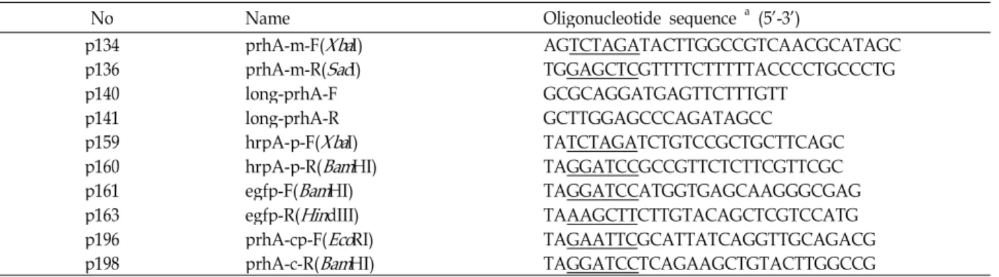
Table 2. Primers used in this study
No Name Oligonucleotide sequence a (5'-3')
p134 prhA-m-F(
Xba
I) AGTCTAGATACTTGGCCGTCAACGCATAGCp136 prhA-m-R(
Sac
I) TGGAGCTCGTTTTCTTTTTACCCCTGCCCTGp140 long-prhA-F GCGCAGGATGAGTTCTTTGTT
p141 long-prhA-R GCTTGGAGCCCAGATAGCC
p159 hrpA-p-F(
Xba
I) TATCTAGATCTGTCCGCTGCTTCAGCp160 hrpA-p-R(
Bam
HI) TAGGATCCGCCGTTCTCTTCGTTCGCp161 egfp-F(
Bam
HI) TAGGATCCATGGTGAGCAAGGGCGAGp163 egfp-R(
Hin
dIII) TAAAGCTTCTTGTACAGCTCGTCCATGp196 prhA-cp-F(
Eco
RI) TAGAATTCGCATTATCAGGTTGCAGACGp198 prhA-c-R(
Bam
HI) TAGGATCCTCAGAAGCTGTACTTGGCCGaUnderlines are the restriction enzyme sites for the enzyme indicated in the primer names
fied by PCR from P. syringae pv. tabaci 11528 genomic DNA using the primer pairs p134 and p136. The PCR product was then subcloned into pGEM-T Easy vector and digested with HindIII/SmaI enzymes. Next, the resultant 3.9-kb DNA frag- ment was blunted with Mung Bean nuclease and self-ligated, which gave pBL26. A 0.9-kb XbaI-SacI fragment of pBL26 was then cloned into pLO1 vector to generate the recombi- nant suicide plasmid, pBL39. Next, the pBL39 plasmid was transferred from E. coli S17-1 to P. syringae pv. tabaci 11528 by a spot mating technique [29] and then introduced into P. syringae pv. tabaci 11528 by allelic exchange [17] using the suicide plasmid pBL39, yielding P. syringae pv. tabaci BL11.
The presence of the 1.2-kb deletion was confirmed by Southern blotting (data not shown).
To complement the ΔprhA mutant (BL11), the complete prhA gene was PCR-amplified from genomic DNA using pri- mer pairs p196 and 198, and then cloned into pRK415 vector.
The construct was then introduced into the strain BL11 by mating, yielding P. syringae pv. tabaci BL45. Complementa- tion tests were then conducted as phenotypic experiments.
Construction of a hrpA::gfp transcriptional fusion plasmid
In order to determine the hrpA expression at the transcrip- tional levels, an hrpA::gfp fusion plasmid was constructed in the pRKlac290 vector. A 0.6-kb hrpA promoter region en- compassing the putative hrpA promoter was then synthe- sized by PCR amplification using the P. syringae pv. tabaci 11528 genomic DNA as a template and primers p159 and p160. Next, a 0.7-kb egfp (enhanced green fluorescent pro- tein) gene was amplified from the pEGFP-C1 plasmid using primers p161 and p163. The resulting PCR products were then restricted using the BamHI enzyme and ligated together
within the pGEM-T Easy vector to give pBL96. Next, the 1.3-kb XbaI-HindIII fragment of pBL96 was cloned into the pRKlac290 vector to generate pBL98. The resultant plasmid, pBL98, was then confirmed by PCR and subsequent DNA sequencing using primers p159 and p163. The pBL98 plas- mid was then transferred from E. coli S17-1 to P. syringae pv. tabaci by a spot mating technique [29].
β -Galactosidase assays
β
-galactosidase activity in bacterial cells was estimated us- ing the procedures described by Miller [21]. Briefly, P. sy- ringae cells grown under various conditions were per- meabilized with chloroform and 1% SDS in Z buffer. The enzyme activity was then determined by measuring the ab- sorbance at 420 nm to determine the rate at which the chro- mogenic substrate, ONPG (ο-nitrophenyl-β-
D-galactopyrano- side), was cleaved. The activity was expressed in Miller units.
Phenotypic and virulence tests
Phenotypic (growth curve, CAS universal siderophore de- tection assay, swarming motility) [7] and virulence tests [26]
were conducted as described previously. For these tests, bac- teria were grown overnight at 30℃, after which, the cultures were adjusted to approximately 2×10
8CFU/ml in distilled water. Swarming motility was observed on King’s B agar (0.4% agar) plates using the method described by Quinones et al [23]. The inoculated plates were incubated for 48 hr at 30℃ and photographs were taken with an Olympus C3020 zoom digital camera. The image contrast and brightness were adjusted using Photoshop CS2 (Adobe, San Jose, CA).
All assays were repeated in three separate experiments.
Confocal laser scanning microscopy
Fluorescence images were recorded on a Zeiss LSM510 confocal laser scanning microscope (Zeiss, Germany). An ar- gon laser was then used to generate an excitation source at 488 nm, and enhanced GFP fluorescence was recorded using a BP 505-515 nm filter set. A HeNe laser was used to generate an excitation source at 543 nm, and the chlor- ophyll fluorescence was recorded using an LP 550 nm filter set.
Statistics
Data are reported as the means±S.D., with the overall stat- istical significance of differences within the groups being de- termined using a student’s t-test. All analyses were per- formed using SPSS for Windows (version 12.0K, SPSS Inc., Chicago, IL). For all statistical analyses, a p value <0.05 was considered significant.
Nucleotide sequence accession number
The sequence of the 2,169-nucleotide, which encodes the prhA gene from P. syringae pv. tabaci 11528, has been de- posited in the GenBank database under accession no.
DQ672633.
Results and Discussion
Nucleotide sequence and characterization of the prhA gene in Pseudomonas syringae pv. tabaci 11528
In our attempt to identify a Ralstonia solanacearum prhA homolog [20] in P. syringae pv. tabaci 11528, degenerate pri- mers (Table 2) were used to PCR-amplify a 17-kb DNA fragment. These primers were designed using the sequences of P. syringae pv. phaseolicola 1448A (GenBank database un- der accession no. NC_005773), P. syringae pv. syringae B728a (GenBank database under accession no. CP_000075), and P.
syringae pv. tomato DC3000 (GenBank database under ac- cession no. NC_004578). Sequence analysis of the prhA gene was performed using the p196 and p198 primers (Table 2).
The putative protein encoded by the prhA gene (2,169 bp) shares homology with outer membrane siderophore receptor proteins and encodes a protein of 722 amino acids, with a calculated molecular mass of 79.5 kDa. PrhA showed sig- nificant similarities to numerous TonB-dependent side- rophore receptor proteins (data not shown) that are believed to bind specific iron-siderophore complexes to form a chan-
nel that allows the transport of these complexes into the peri- plasm [20].
Characterization of a ΔprhA mutant strain of P.
syringae pv. tabaci 11528
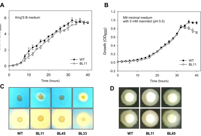
Allelic exchange mutagenesis was used to construct a prhA deletion mutant strain that harbored a 1,172-bp dele- tion of the internal fragment of the prhA gene. When com- pared to the parent strain, growth of the ΔprhA mutant strain was slower, however, the stationary phase was reached at the same cell density as of the parent strain when grown in King’s B media (Fig. 1a). Conversely, the growth rate of the ΔprhA mutant strain was equal to that of the wild-type strain, however, the stationary phase of the mutant strain was reached at a lower cell density than that of the wild-type strain when grown in hrp-inducing media (Fig. 1b). These results indicate that the phenotypic differences between the mutant and wild-type strains were most likely not due to a serious defect in the growth of the mutant. Swarming mo- tility in the plant-pathogen interaction is critical as a viru- lence factor, most likely because it facilitates entry of the pathogen into plant tissues [7]. As shown in Fig. 1c, the swarming motility of the wild-type strain and the mutant were equal, which indicates that PrhA does not regulate swarming motility. In addition, the ΔprhA mutant produced slightly more siderophores than the wild-type strain under iron-depleted and iron-replete conditions (Fig. 1d), which in- dicates that prhA is not a key determinant in iron homeostasis.
The ΔprhA mutant had reduced virulence in the host plant
The ability of the ΔprhA mutant strain to cause wild-fire
disease was assessed in tobacco leaves (host plant). P. sy-
ringae pv. tabaci 11528, BL11, and BL45 overnight cultures
were adjusted to an OD
600of 1.0 (approximately 2.0×10
8CFU/ml) and then diluted serially to a concentration of
2×10
5CFU/ml. After 3 days, the wild-type strain appeared
to elicit disease symptoms at all concentrations, however,
the ΔprhA mutant strain only appeared to elicit weak disease
symptoms at a concentration of 2×10
8CFU/ml (4×10
7CFU/20 µl inoculum). In addition, the wild-type strain eli-
cited small brown necrotic lesions that were surrounded by
chlorosis after 24 hr of incubation, but no such symptoms
were observed when the plants were infected with less than
2×10
7CFU/ml (4×10
6CFU/20 ul) of strain BL11. As shown
Time (hours)
0 10 20 30 40
Growth (OD600)
-0.2 0.0 0.2 0.4 0.6 0.8 1.0 1.2
WT BL11 M9 minimal medium
with 5 mM mannitiol (pH 5.5)
WT BL11 BL45 BL33 WT BL11 BL45
C D
Time (hours)
0 10 20 30 40
0 2 4 6
WT BL11 Growth (OD600)
King'S B medium
A B
Fig. 1. Phenotypic characterization of the Δ
prhA
mutant. Growth curves of the wild-type (WT) and ΔprhA
mutant (BL11) strains ofP. syringae
pv.tabaci
11528 cultured inhrp
-repressing (A) andhrp
-inducing medium (B), respectively. Values represent independent means±standard deviations of OD600readings obtained for triplicate cultures. Siderophore biosynthesis was com- pared using a CAS universal siderophore detection assay under iron-depleted or iron-replete conditions (C). BL33 (Δfur
mutant) was used as a CAS assay control strain. BL45 is BL11 carrying pBL172 (prhA
complementation). Swarming motility test in 0.4% King’s B agar plate (D).Fig. 2. Pathogenicity test of the Δ
prhA
mutant on tobacco leaves. (A) Disease symptoms caused by theP. syringae
pv.tabaci
wild-type (11528; WT), ΔprhA
mutant (BL11), and its complementation strain (BL45). (B) Size of lesions on tobacco leaves produced by infection with the wild-type, BL11, and BL45 strains at a concentration of 4×10⁵ CFU (20 ul). The lesion diameter was measured in millimeters. The vertical lines above each bar indicate the standard errors of the mean of three individual tests.A B
C D
WT on its non-host ΔprhA mutant on its non-host
WT on its host ΔprhA mutant on its host
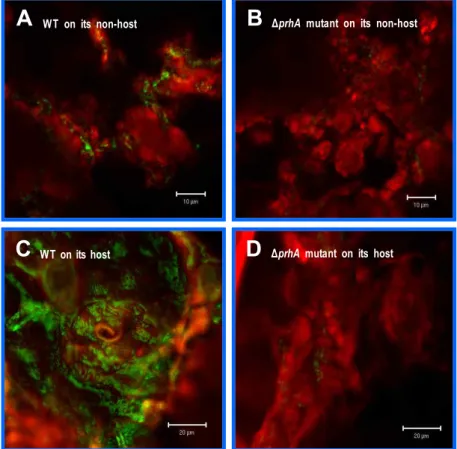
Fig. 4. The plant signal recognized by PrhA, which induces
hrpA
gene expression, is present in plant leaves. Observations by confocal laser scanning microscopy after 4 hr of co-cultivation ofArabidopsis thaliana
(non-host) (A, B) andNicotiana tabacum
(host) (C, D) cell wall materials with theP. syringae
pv.tabaci
BL31 (11528 carrying pBL98) (A, C) or BL32 (BL11 carrying pBL98) (B, D). The bar represents 10 or 20 um. The red color is plant cell wall material and the green color isP. syringae
carrying pBL98 (hrpA-gfp
reporter fusion).in Fig. 2a, the ability of the ΔprhA mutant strain BL11 to produce symptoms in tobacco leaves was significantly weak- er than that of the wild-type. In addition, strain BL11 is less virulent than the wild-type, therefore, PrhA may play an important role in the interaction between tobacco leaves and P. syringae pv. tabaci 11528 (Fig. 2a).
PrhA does not regulate transcription of the hrpA gene in hrp-inducing or nutrient rich media
The hrp gene cluster can be classified into three categories according to their functions, a regulatory system, a type III secretion system (TTSS), and the substrates of TTSS (harpin, Hop and pilus proteins, etc) [8,19]. Three intracellular regu- latory proteins, HrpR, HrpS, and HrpL, are known to acti- vate the hrp gene cluster expression. In addition, HrpL is an alternative sigma factor in the ECF (extracytoplasmic fac- tor) family [30], and HrpR, HrpS, and HrpL appear to func- tion in a regulatory cascade in which HrpS and HrpR syn- ergistically activate the expression of hrpL in response to sig-
King's B hrp-inducing Arabidopsis cell M9MM medium medium wall co-cultures
b-galactosidase activity (Miller units)
0 500 1000 1500 2000 2500 3000 3500
BL18 BL19
Fig. 3. Effect of a
prhA
mutation on expression of thehrpA
gene during co-culture ofP. syringae
withA. thaliana
cell wall material. β-galactosidase activity was measured in BL18 (11528 carrying pBL52) and BL19 (BL11 carrying pBL52) grown inhrp
-repressing medium andhrp
-inducing me- dium, respectively. The co-culture medium used was M9 minimal medium (M9MM).nals from plants or in hrp-inducing minimal medium. HrpL is also presumed to induce expression of the hrp and avr genes by recognizing a consensus sequence motif (hrp box) in the upstream regions [31]. Finally, the HrpL-dependent hrpA gene is a member of TTSS, therefore expression of the hrpA gene represents an hrp gene cluster expression. The transcription of the P. syringae pv. tabaci hrp gene cluster, which was identified by measuring the expression level of the hrpA-lacZ promoter fusion (pBL52), is induced in hrp-in- ducing medium (including M9 minimal medium with 5 mM mannitol, pH 5.5) and repressed in rich medium (including King’s B medium) [20,22]. As shown in Fig. 3, PrhA does not regulate transcription of the hrpA gene in either hrp-in- ducing or nutrient rich media. Taken together, these results indicate that PrhA may regulate expression of the hrp gene cluster in response to an unknown plant cell factor.
Transcription of the hrpA gene is induced upon co-culture with its host or non-host plant cells, and this induction is dependent on the presence of PrhA
Co-culture of P. syringae pv. tabaci 11528 and A. thaliana cell wall material induced a much higher level of hrp gene transcription than culture in hrp-inducing medium (Fig. 3).
In addition, an enhanced green fluorescent protein (eGFP) reporter gene system was used to monitor the in situ ex- pression of the hrpA gene upon cultivation of P. syringae pv.
tabaci 11528 with plant cell material. This hrpA-gfp reporter fusion was also strongly induced upon contact of the bac- teria with plant (both host and non-host) cell wall material, but was not as strongly expressed in an ΔprhA mutant strain (Fig. 4). Based on these observations, we conclude that P.
syringae pv. tabaci PrhA is required for full virulence in plant.
Acknowledgement
This work was supported for two years by Pusan National University Research Grant.
References
1. Aldon, D., B. Brito, C. Boucher, and S. Genin. 2000. A bacte- rial sensor of plant cell contact controls the transcriptional induction of
Ralstonia solanacearum
pathogenicity genes.EMBO
J.
19, 2304-2314.2. Alfano, J. R., A. O. Charkowski, W. L. Deng, J. L. Badel, T. Petnicki-Ocwieja, K. van Dijk, and A. Collmer. 2000. The Pseudomonas syringae Hrp pathogenicity island has a tri-
partite mosaic structure composed of a cluster of type III secretion genes bounded by exchangeable effector and con- served effector loci that contribute to parasitic fitness and pathogenicity in plants.
Proc. Natl. Acad. Sci. USA
97, 4856-4861.3. Arlat, M., C. L. Gough, C. E. Barber, C. Boucher, and M.
J. Daniels. 1991. Xanthomonas campestris contains a cluster of
hrp
genes related to the largerhrp
cluster ofPseudomonas solanacearum
.Mol. Plant-Microbe Interact.
4, 593-601.4. Bauer, D. W., A. J. Bogdanove, S. V. Beer, and A. Collmer.
1994.
Erwinia chrysanthemi hrp
genes and their involvement in soft rot pathogenesis and elicitation of the hypersensitive response.Mol. Plant-Microbe Interact.
7, 573-581.5. Bogdanove, A. J., S. V. Beer, U. Bonas, C. A. Boucher, A.
Collmer, D. L. Coplin, G. R. Cornelis, H. C. Huang, S. W.
Hutcheson, N. J. Panopoulos, and F. Van Gijsegem. 1996.
Unified nomenclature for broadly conserved
hrp
genes of phytopathogenic bacteria.Mol. Microbiol.
20, 681-683.6. Boucher, C. A., F. Van Gijsegem, P. A. Barberis, M. Arlat, and C. Zischek. 1987.
Pseudomonas solanacearum
genes con- trolling both pathogenicity on tomato and hypersensitivity on tobacco are clustered.J. Bacteriol.
169, 5626-5632.7. Cha, J. Y., J. S. Lee, J. I. Oh, J. W. Choi, and H. S. Baik.
2008. Functional analysis of the role of Fur in the virulence of
Pseudomonas syringae
pv.tabaci
11528: Fur controls ex- pression of genes involved in quorum-sensing.Biochem.
Biophys. Res. Commun.
366, 281-287.8. Collmer, A., J. L. Badel, A. O. Charkowski, W. L. Deng, D.
E. Fouts, A. R. Ramos, A. H. Rehm, D. M. Anderson, O.
Schneewind, K. van Dijk, and J. R. Alfano. 2000.
Pseudomonas syringae
Hrp type III secretion system and ef- fector proteins.Proc. Natl. Acad. Sci. USA
97, 8770-8777.9. Deng, W. L., A. H. Rehm, A. O. Charkowski, C. M. Rojas, and A. Collmer. 2003.
Pseudomonas syringae
exchangeable ef- fector loci: sequence diversity in representative pathovars and virulence function inP. syringae
pv.syringae
B728a.J.
Bacteriol.
185, 2592-2602.10. Frederick, R. D., M. Ahmad, D. R. Majerczak, A. S.
Arroyo-Rodriguez, S. Manulis, and D. L. Coplin. 2001.
Genetic organization of the
Pantoea stewartii
subsp.stewartii
hrp gene cluster and sequence analysis of thehrpA
,hrpC
,hrpN
, andwtsE
operons.Mol. Plant-Microbe Interact.
14, 1213-1222.11. Gay, P., D. Le Coq, M. Steinmetz, T. Berkelman, and C. I.
Kado. 1985. Positive selection procedure for entrapment of insertion sequence elements in gram-negative bacteria.
J.
Bacteriol.
164, 918-921.12. Gober, J. W. and L. Shapiro. 1992. A delvelopmentally regu- lated
Caulobacter
flagellar promoter is activated by 3' en- hancer and IHF binding elements.Mol. Biol. Cell
3, 913-926.13. Hirano, S. S. and C. D. Upper. 2000. Bacteria in the leaf ecosystem with emphasis on
Pseudomonas syringae
-a patho- gen, ice nucleus, and epiphyte.Microbiol. Mol. Biol. Rev.
64, 624-653.14. Huynh, T. V., D. Dahlbeck, and B. J. Staskawicz. 1989.
Bacterial blight of soybean: regulation of a pathogen gene
초록:Pseudomonas syringae pv. tabaci 에서 식물세포접촉에 의한 병원성 유전자의 조절 이준승․차지영․백형석*
(부산대학교 미생물학과)
Pseudomonas syringae pv. tabaci는 숙주인 담배에 감염하여 들불병(wild fire)을 일으키는 식물 병원성 세균이다.
이 세균의 pathogenicity island (PAI)는 Type III secretion system 및 병원성 유전자들을 암호화하고 있으며, 병 원성 조절에 있어 핵심적인 역할을 한다. 최근 식물 병원성 세균인 Ralstonia solanacearum에서 식물 세포 접촉을 매개로 하여 hrp gene cluster를 양성조절하는 PrhA (plant regulator of hrp) receptor가 발견되었다. 본 연구에서 는 P. syringae 에서 식물세포에 의해 hrp 유전자가 유도되는지 확인하기 위해, prhA 유사체를 동정하고 PrhA 결 실돌연변이주(BL11)를 구축하였다. BL11은 숙주 감염 실험에서 병원성이 현저히 감소하였고, 식물 세포현탁액에
서 hrpA 유전자의 발현수준이 hrp 유도배지에서 보다 3배 더 높게 나타났다. 이러한 결과들을 근거로 PrhA가
식물세포접촉에 의한 조절에 중요한 역할을 한다는 것을 확인하였으며, hrpA-gfp reporter fusion을 사용하여 이 를 다시 검증하였다.
determining host cultivar specificity.
Science
245, 1374-1377.15. Keen, N. T., S. Tamaki, D. Kobayashi, and D. Trollinger.
1988. Improved broad-host-range plasmids for DNA cloning in gram-negative bacteria.
Gene
70, 191-197.16. Lee, J. K. and S. Kaplan. 1995. Transcriptional regulation of puc operon expression in
Rhodobacter sphaeroides
. Analysis of thecis-
acting downstream regulatory sequence.J. Biol.
Chem.
270, 20453-20458.17. Lenz, O., E. Schwartz, J. Dernedde, M. Eitinger, and B.
Friedrich. 1994. The
Alcaligenes eutrophus
H16hoxX
gene participates in hydrogenase regulation.J. Bacteriol.
176, 4385-4393.18. Lindgren, P. B., R. C. Peet, and N. J. Panopoulos. 1986. Gene cluster of
Pseudomonas syringae
pv. "phaseolicola
" controls pathogenicity of bean plants and hypersensitivity of non- host plants.J. Bacteriol.
168, 512-522.19. Lindgren, P. B. 1997. The role of
hrp
genes during plant-bac- terial interactions.Annu. Rev. Phytopathol.
35, 129-152.20. Marenda, M., B. Brito, D. Callard, S. Genin, P. Barberis, C.
Boucher, and M. Arlat. 1998. PrhA controls a novel regu- latory pathway required for the specific induction of
Ralstonia solanacearum hrp
genes in the presence of plant cells.Mol. Microbiol.
27, 437-453.21. Miller, J. H. 1972. Experiments in molecular genetics. 1972.
New York: Cold Spring Harbor.
22. Noel, L., F. Thieme, D. Nennstiel, and U. Bonas. 2002. Two novel type III-secreted proteins of
Xanthomonas campestris
pv.vesicatoria
are encoded within thehrp
pathogenicity island.J. Bacteriol.
184, 1340-1348.23. Quinones, B., G. Dulla, and S. E. Lindow. 2005. Quorum sensing regulates exopolysaccharide production, motility,
and virulence in
Pseudomonas syringae
.Mol. Plant-Microbe Interact.
18, 682-693.24. Rahme, L. G., M. N. Mindrinos, and N. J. Panopoulos. 1992.
Plant and environmental sensory signals control the ex- pression of hrp genes in
Pseudomonas syringae
pv.phaseolicola
.J. Bacteriol.
174, 3499-3507.25. Richael, C. and D. Gilchrist. 1999. The hypersensitive re- sponse: A case of hold or fold?
Physiol. Mol. Plant Pathol.
55, 5-12.
26. Salch, Y. P. and P. D. Shaw. 1988. Isolation and character- ization of pathogenicity genes of
Pseudomonas syringae
pv.tabaci
.J. Bacteriol.
170, 2584-2591.27. Sambrook, J. and D. W. Russell. 2001. Molecular Cloning:
a laboratory manual, third eds. 2001. New York: Cold Spring Harbor.
28. Schwyn, B. and J. B. Neilands. 1987. Universal chemical as- say for the detection and determination of siderophores.
Anal. Biochem.
160, 47-56.29. Simon, R., U. Priefer, and A. Pühler. 1983. A broad host range mobilization system for
In vivo
genetic engineering:Transposon mutagenesis in Gram negative bacteria.
Nat.
Biotechnol.
1, 784-791.30. Xiao, Y., Y. Lu, S. Heu, and S. W. Hutcheson. 1992.
Organization and environmental regulation of the
Pseudomonas syringae
pv. syringae 61hrp
cluster.J. Bacteriol.
174, 1734-1741.
31. Xiao, Y., S. Heu, J. Yi, Y. Lu, and S. W. Hutcheson. 1994.
Identification of a putative alternate sigma factor and char- acterization of a multicomponent regulatory cascade con- trolling the expression of