Cadms/SynCAMs/Necls/TSLCs Interact with Multi-PDZ Domain Protein MUPP1
Won Hee Jang1, Young Joo Jeong1, Sun Hee Choi1, Sang-Jin Kim2, Sang-Hwa Urm3, Il Soo Moon4 and Dae-Hyun Seog1*
1Department of Biochemistry and u-HARC, Inje University College of Medicine, Busan 614-735, Korea
2Department of Neurology, Inje University College of Medicine, Busan 614-735, Korea
3Department of Preventive Medicine, Inje University College of Medicine, Busan 614-735, Korea
4Department of Anatomy & Dongguk Medical Institute, College of Medicine, Dongguk University, Gyeongju 780-714, Korea Received September 15, 2014 /Revised October 13, 2014 /Accepted October 14, 2014
Cell adhesion molecules determine the cell-cell binding and the interactions between cells and ex- tracellular signals. Cell-cell junctional complexes, which maintain the structural integrity of tissues, consist of more than 50 proteins including multi-PDZ domain protein 1 (MUPP1). MUPP1 contains 13 postsynaptic density-95/disks large/zonula occludens-1 (PDZ) domains and serves a scaffolding function for transmembrane proteins and cytoskeletal proteins or signaling proteins, but the mecha- nism how MUPP1 links and stabilizes the juxtamembrane proteins has not yet been elucidated. We used the yeast two-hybrid system to identify proteins that interact with PDZ domains of MUPP1. We found an interaction between MUPP1 and cell adhesion molecule 1 (Cadm1, also known as SynCAM1, Necl-2, or TSLC1). Cadm1 bound to the second PDZ domain of MUPP1. The carboxyl (C)-terminal end of Cadm1 has a type II PDZ-association motif (-Y-F-I) which was essential for the interaction with MUPP1 in the yeast two-hybrid assay. MUPP1 also bound to the C-terminal cytoplasmic tail region of other Cadm family members (Cadm2, Cadm3, and Cadm4). In addition, these protein-protein inter- actions were observed in the glutathione S-transferase (GST) pull-down assay and by co- immunoprecipitation. Anti-MUPP1 antibody co-immunoprecipitated Cadm1 and Cadm4 with MUPP1 from mouse brain extracts. These results suggest that MUPP1 could mediate interaction between Cadms and cytoskeletal proteins.
Key words : Cadm1 (cell adhesion molecule 1), cell junction, MUPP1 (multi-PDZ domain protein 1), PDZ (postsynaptic density-95/disks large/zonula occludens-1) domain, scaffold protein
*Corresponding author
*Tel : +82-51-890-6974, Fax : +82-51-894-5801
*E-mail : [email protected]
This is an Open-Access article distributed under the terms of the Creative Commons Attribution Non-Commercial License (http://creativecommons.org/licenses/by-nc/3.0) which permits unrestricted non-commercial use, distribution, and reproduction in any medium, provided the original work is properly cited.
Journal of Life Science 2014 Vol. 24. No. 12. 1276~1283 DOI : http://dx.doi.org/10.5352/JLS.2014.24.12.1276
Introduction
The protein-protein interactions play an important role in biological function. Cell adhesion molecules (CAMs) and neuronal adhesion molecules are important for recognition of the information in the extracellular environment. The cy- toplasmic regions of CAMs and neuronal adhesion mole- cules are essential for cell signaling, cell spreading, pro- liferation, or migration [17]. CAMs and neuronal adhesion molecules interact, through postsynaptic density-95/disks large/zonula occludens-1 (PDZ)-binding domain in their cytoplasmic regions, with many different scaffolding pro- teins such as multi-PDZ domain protein 1 (MUPP1), mem-
brane-associated guanylate kinases (MAGUKs), calcium/
calmodulin-dependent serine kinase (CASK), and protein associated with Lin seven (Pals-2), which are anchored to both transmembrane proteins and proteins in juxtamem- brane region [3, 26]
PDZ domains are ~80 amino acid residues long and fold- ed into a compact globular structure. PDZ domains are of- ten found in multidomain scaffolding proteins [13]. They are small protein-protein interaction modules and typically bind to specific PDZ-association motif in the carboxyl (C)-terminal end of partner proteins, which are most often transmembrane receptors and channel proteins [24, 29].
PDZ-domain containing proteins play an important role in the targeting of partner proteins to specific subcellular com- partments and in the assembly of partner proteins into large molecular complexes [26]. They also can regulate the function of partner proteins [1, 24]. Defects of PDZ do- main-containing protein can cause the human diseases.
Mutations in a gene encoding harmonin, a PDZ-containing protein, cause Usher syndrome type 1C, an autosomal re-
cessive disorder [34]. Mutations in the periaxin gene, which also encodes a PDZ-containing protein, have been identified to cause Dejerine-Sottas neuropathy, a severe demyelinating form of peripheral neuropathy [9].
MUPP1 was originally identified as an endogenous bind- ing partner that interacts with the C-terminus of the seroto- nin 5-hydroxytryptamine type 2C (5-HT2C) receptor [33]. It is a ubiquitously expressed PDZ domain-containing protein, is found in tight junctions, post-synaptic density (PSD), and Schwann cell incisures, and associates with the plasma membrane [6, 28]. MUPP1, which possesses 13 PDZ do- mains, acts as a scaffold for attaching different proteins at the proper location in the membrane [12, 33]. MUPP1 has been reported to interact with GABAB neurotransmitter re- ceptors, olfactory sensory receptors, synaptic Ras GTPase- activating protein SynGAP, and Ca2+/calmodulin-depend- ent kinase (CaMKII) to regulate neuronal signaling and dendritic spine morphology [5, 12, 18, 21]. MUPP1 also in- teracts several additional proteins in non-neuronal cells, in- cluding claudin-1, junctional adhesion molecule-1 (JAM1), and Kalirin-7, a Rho-GEF, which indicates that MUPP1 po- tentially integrates multiple signal transduction pathways and can regulate cell junction integrity for epithelial func- tion [2, 14, 22].
To help define the role of MUPP1 in cells, it is necessary to identify the binding proteins of MUPP1. We screened for proteins that interact with the PDZ domains of MUPP1 through the yeast two-hybrid assay and identified cell ad- hesion molecule 1 (Cadm1, also known as SynCAM1, Necl-2, or TSLC1), a cell adhesion molecule involved in cell-cell interaction and the formation and maintenance of epithelial structure [15]. The MUPP1 and Cadm1 interaction suggests that MUPP1 may contribute as an adaptor or scaf- fold protein between cell adhesion molecules and the sub- cellular proteins.
Materials and Methods Plasmid constructs
Full-length rat MUPP1 cDNA in the pCMV vector (a gift from Dr. H. Lubbert, Ruhr-Universitat, Denmark) was tag- ged with a FLAG-epitope at the amino (N)-terminus.
Truncations of MUPP1 corresponding to different PDZ do- mains were prepared by PCR amplification using the ap- propriate primers. The amplified fragment was subcloned into T-vector. The fragment was then EcoRI-restricted and
subcloned into the EcoRI site of pLexA vector (Clontech, Palo Alto, CA, USA). The correct orientation and in-frame cloning of cDNA inserts were verified by restriction en- zyme analysis and DNA sequencing. The cDNA fragments corresponding to the cytoplasmic regions of Cadm2 (accession NM_178721), Cadm3 (accession NM_053199), and Cadm4 (accession NM_153112) were amplified by PCR from mouse Marathon-ReadyTM cDNA library (Clontech) and cloned into pGEM-T Easy vector (Promega Corp, Madison, WI, USA). The resulting recombinant plasmid was then cut with EcoRI and XhoI and the insert was subcl- oned into pLexA vector. General recombinant DNA techni- ques were performed according to standard protocol [25].
Screening of MUPP1-binding proteins by yeast two-hybrid assay
The Matchmaker LexA two-hybrid system was used for screening according to the manufacturer’s manual (Clon- tech). In brief, a part of the rat MUPP1 cDNA was fused to the DNA-BD region of the pLexA vector and the result- ing plasmid DNA was transformed into yeast strain EGY48 carrying the p8op-lacZ gene. Transformed EGY48 yeast cells containing the MUPP1 bait plasmid were transformed with the mouse brain cDNA library and grown on synthetic dextrose (SD) plates supplemented with glucose but with no histidine, tryptophan, or uracil (SD/-His/-Trp/-Ura).
The selection of positive clones was performed on an SD/-His/-Trp/-Ura/-Leu plate containing galactose, raffi- nose, X-gal, and BU salts. Plasmids from positive clones were analyzed by restriction digestion. Unique inserts were sequenced and protein sequence analysis was performed with the BLAST algorithm at the National Center for Biotechnology Information (NCBI). Sequence-verified clones were tested again for interaction with the bait in yeast by retransformation.
β-Galactosidase activity in liquid cultures of yeast The β-galactosidase activity of yeast was assayed as de- scribed previously [31]. Mid-log phase yeast cells were col- lected and permeabilized with 0.1% sodium dodecyl sul- phate (SDS) and chloroform. An excess amount of o-nitro- phenyl-β-D-galactoside (ONPG) was added to yeast lysate, and the mixture was incubated at 30°C, and then the re- action was stopped by increasing pH to 11 by the addition of 1 M Na2CO3. The formation of the reaction product, o-ni- trophenol, was determined by measuring absorbance at 420
nm on a spectrophotometer and normalizing for the re- action time. The units of enzyme activity were calculated by the following equation: units = 1000 × [(OD420–1.75 × OD550)] / (reaction time (min) × culture volume (ml) × OD600) [4]. All experiments were independently performed at least three times.
Glutathione S-transferase (GST) pull-down assays cDNA encoding the cytoplasmic region of each Cadm protein was cloned in pET41a. The recombinant GST-Cadm fusion proteins were expressed in bacterial strain BL21 GOLD (Stratagene, La Jolla CA, USA) after induction with 0.5 mM isopropyl thio-β-D-galactopyranoside (IPTG) for 3 hr. The fusion proteins were purified using glutathione- agarose beads (Sigma-Aldrich, St. Louis, MO, USA) accord- ing to the manufacturer’s protocol. The mouse brain S2 frac- tion was incubated overnight at 4°C with the GST fusion protein-coupled glutathione beads. The beads were pelleted by centrifugation, washed three times with the extraction buffer (1% Triton X-100 in PBS containing 10 μg/ml each aprotinin, leupeptin, and pepstatin, and 1 μM phenyl- methanesulfonyl fluoride), and once with PBS. The bound proteins were eluted from the glutathione beads with 100 μl of Laemmli’s loading buffer. The pulled-down proteins were analyzed by immunoblotting with anti-MUPP1 anti- body (BD science, San Jose, CA, USA).
Co-immunoprecipitation and Immunoblot analysis Mouse brains were homogenized in ice-cold homoge- nization buffer (0.32 M sucrose, 4 mM HEPES, pH 7.3) sup- plemented with protease inhibitor cocktail (Sigma-Aldrich).
For immunoprecipitation, mouse brain lysate was diluted in the same volume of 2X binding buffer (50 mM HEPES, 200 mM KCl, 0.2% Triton X-100, pH 7.0) and incubated with an- ti-MUPP1 antibody (BD science) or preimmune serum over- night at 4°C, followed by precipitation with protein-A Sepharose (Amersham Pharmacia, Piscataway, NJ, USA).
The beads were collected by brief centrifugation and wash- ed three times with TBS-T (20 mM Tris-HCl, pH 7.5, 0.2 M NaCl, 0.1% Tween 20). The washed beads were re- suspended with Laemmli’s loading buffer and the proteins were eluted and denatured by boiling for 2 min and then separated by SDS-PAGE. The proteins were transferred from the gel to a nitrocellulose membrane and incubated with anti-Cadm1 and anti-Cadm4 antibodies (Sigma-Aldrich).
Results
Identification of Cadm1 as a MUPP1 interacting protein by yeast two-hybrid screening
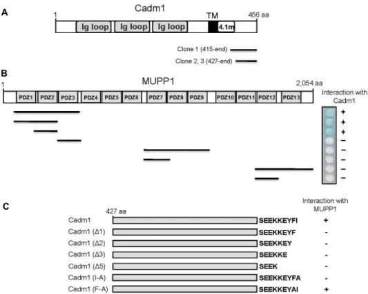
To identify MUPP1-interacting proteins, we screened a mouse brain cDNA library through the yeast two-hybrid as- says using the N-terminal region of MUPP1 containing 1st-3rd PDZ domains as bait. From 1×10⁷ colonies screened, we obtained three positive clones, each of which possesses a cDNA fragment of Cadm1 (Fig. 1A). MUPP1 is composed of an L27 domain and 13 PDZ domains [33]. To determine the binding domain of MUPP1 that is required for the inter- action with Cadm1, we constructed various fragments of MUPP1. Yeast two-hybrid assays with Cadm1 showed that the binding to Cadm1 was critically dependent on the sec- ond PDZ domain of MUPP1 (Fig. 1B). The cytoplasmic re- gion of Cadm1 contains a class II PDZ-association motif (φX φ), where φ is a hydrophobic residue, at its C-terminus (Y-F-I) [13, 26]. Next we investigated whether the C-termi- nal PDZ-association motif of Cadm1 mediates the inter- action with MUPP1. For this purpose, a series of C-terminal deletion and substitution mutants of Cadm1 were con- structed (Fig. 1C) and co-transfected into yeast cells with pLexA-MUPP1. As shown in Fig. 1C, the deletion as well as the substitution of the last C-terminal residue of Cadm1 disrupted the interaction with MUPP1. These result in- dicated that Cadm1 associates with MUPP1 through its C-terminal PDZ-association motif.
MUPP1 interacts with Cadm2, Cadm3, and Cadm4, as well as Cadm1.
The Cadms are a family of type I transmembrane pro- teins consisting of four members: Cadm1, Cadm2, Cadm3, and Cadm4 [7]. They contain a highly conserved short cyto- plasmic tail containing protein-protein interaction domains [7, 23]. Therefore, we tested whether MUPP1 also interacts with the other Cadms (Cadm2, Cadm3, and Cadm4) using yeast two-hybrid assay. As shown in Fig. 2A, MUPP1 also interacted with the cytoplasmic regions of Cadm2, Cadm3, and Cadm4 in yeast system. A quantitative β-galactosidase assay showed that MUPP1 has similar binding affinity to four Cadms (Fig. 2B). These results are not surprising in view of the fact that all four Cadms have a class II PDZ-as- sociation motif at C-terminal tail [7, 23]. As shown in Fig.
2B, Cadm1 did not interact with other PDZ domain-contain- ing protein, PSD-95 (Fig. 2B). Next we determined the inter-
A
B
C
Fig. 1. Identification of the proteins interacting with MUPP1 by yeast two-hybrid screening. (A) Schematic diagram of Cadm1.
Cadm1 contains three extracellular Ig-like loops (Ig loop), a single transmembrane domain (TM), a protein 4.1-binding site (4.1m), and a short C-terminal tail. Clones 1, 2, and 3 were isolated from the yeast two-hybrid screen and were overlapped at the C-terminal region of Cadm1. aa, the amino acid residue number. (B) Minimal Cadm1-binding region in MUPP1.
Different truncations of MUPP1 were constructed by PCR. Several truncated forms of MUPP1 were tested in the yeast two-hybrid assay for interaction with Cadm1. +, interaction with Cadm1; -, no interaction with Cadm1. aa, the amino acid residue number. (C) Specific interaction of MUPP1 with the C-terminus of Cadm1. Several deletion and substitution mutants of Cadm1 were tested in the yeast two-hybrid assay for interaction with MUPP1. +, interaction with MUPP1; -, no interaction with MUPP1.
A B
Fig. 2. Interaction between MUPP1 and Cadms. The cytoplasmic region of each Cadm was fused to the pLexA DNA binding domain. (A) Cadms interacted with MUPP1. +, interaction with MUPP1; -, no interaction with MUPP1.
(B) The strength of interactions between Cadms and MUPP1 were examined quantita- tively using β-galactosidase activity in yeast two-hybrid reporter assay.
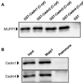
action of MUPP1 with Cadms at the protein level using GST pull-down experiments. Each recombinant GST-Cadm fusion protein was expressed in E. coli. The purified GST
fusion protein was allowed to interact with mouse brain lysates. Immunoblotting analyses revealed that MUPP1 in- teracted with each GST-Cadm, but not with GST (Fig. 3A).
A
B
Fig. 3. Association of MUPP1 with Cadms in the GST pull- down assay and co-immunoprecipitation. (A) Proteins in the mouse brain lysate were allowed to bind to GST-Cadm fusion proteins or GST alone. The elution fractions were resolved by SDS-PAGE and analyzed by immunoblotting with anti-MUPP1 antibody. (B) Mouse brain lysates were immunoprecipitated with anti- MUPP1 antibody or preimmune serum and then the precipitates were immunoblotted with anti-Cadm1 or Cadm4 antibodies.
Together, these data indicated a specific interaction between MUPP1 and Cadms through the second PDZ domain of MUPP1 and a class II PDZ-binding motif of Cadms.
Endogenous interaction of MUPP1 with Cadm1 and Cadm4 in neurons
Cadm1 is expressed in most epithelia and neuroepithelia, such as lung, liver, dorsal root ganglia, various regions of the central nervous system [23]. In contrast to the wide- spread expression of Cadm1, the expression of Cadm3 and Cadm4 seems to be restricted to neurons and glial cells [19, 30]. Cadm1 and Cadm4 were major components of the Cadm family in neurons [23, 32]. To address the question whether MUPP1 interacts with Cadm1 and Cadm4 at the endogenous level of expression in neurons, we per- formed co-immunoprecipitation analyses. Lysates from mouse brain were incubated with anti-MUPP1 antibody or preimmune serum. Protein A-agarose beads selectively pre- cipitated the immuno-complexes, which were then sub- sequently separated by SDS-PAGE and immunoblotted with anti-Cadm1 and anti-Cadm4 antibodies. As shown in Fig. 3B, anti-MUPP1 antibody efficiently precipitated Cadm1 and Cadm4. This result suggests that MUPP1 interacts en-
dogenously with Cadm1 and Cadm4 in neurons.
Discussion
In this study, we have shown that MUPP1 associates with Cadm1. Using the N-terminal PDZ domain-containing region of MUPP1 as bait, we identified Cadm1 in a yeast two-hybrid assay of a mouse brain cDNA library. We have further shown that MUPP1 also interacts with the other Cadms (Cadm2, Cadm3, and Cadm4) and the interactions between MUPP1 and Cadms are mediated by the second PDZ domain of MUPP1 and the C-terminal PDZ-association motif of Cadms. Furthermore, using GST pull-down assay, we confirmed that Cadms interacts with MUPP1 at the pro- tein level. Finally, we have shown that the major neuronal Cadms, Cadm1 and Cadm4, were co-immunoprecipitated with MUPP1 at the endogenous level of expression in neurons. Although we did not investigate the other Cadm1 binding partner, actin cytoskeleton [7] in this study, these results suggest that MUPP1 may act as an adaptor or a scaf- fold protein between Cadms and subcellular proteins.
Cadm1, a multifunctional immunoglobulin-like cell adhe- sion molecule, is involved in cell-cell interaction and the formation and maintenance of epithelial structure [7, 23].
Cadm1 is associated with the actin cytoskeleton through di- rect interaction with the protein 4.1-binding site in its cyto- plasmic domain [7, 23]. Cell adhesion molecules interact with membrane proteins/receptors and adaptor molecules in juxtamembrane regions [3, 17, 23]. For instance, N-cad- herin interacts with fibroblast growth factor receptor (FGFR) in neuronal cells [11]. Cadm1 has potential to form heterophilic trans-interaction with other Cadms [10, 20, 27].
In this study, we demonstrated that Cadms, interacted with an adaptor/scaffold protein MUPP1.
What would the association between MUPP1 and Cadm1 mean? First, MUPP1 may have a role in subcellular local- ization and maintenance of Cadm1. Several PDZ proteins, such as Zonal occludens (ZO)-1 and postsynaptic density protein (PSD)-95 act as scaffolding/targeting proteins that have potential to bring their interacting proteins to tight junction or PSD [2, 12, 26]. Likewise, the association of Cadm1 with MUPP1 could localize Cadm1 to plasma mem- brane and stabilize Cadm1 at juxtamembrane regions in ep- ithelial cells. Second, the association may transmit the in- formation to the cytoplasmic region through intracellular signaling pathway. Cell adhesion proteins recognize the
change in extracellular environment and transmit the in- formation into a cell [16, 20]. For instance, E-cadherin inter- acts with β-catenin in its cytoplasmic region to organize cell adhesion machinery. β-catenin acts as an effector in the Wnt signaling pathway [8]. MUPP1 also interacts with sev- eral signaling proteins such as CaMKII [1]. CaMKII exhibits broad substrate specificity and regulates diverse responses to physiological changes of intracellular Ca2+ concentrations [1]. The protein complex of Cadm1-MUPP1 may possibly recruit CaMKII in the juxtamembrane region to induce cy- toskeletal remodeling and the formation and maintenance of epithelial structure, or induce cell motility. Further func- tional studies on this possibility may help to shed light on the role of MUPP1 in the formation and maintenance of cell junction.
Acknowledgments
The research was supported by the Basic Science Research Program through the National Research Foundation of Korea (NRF) by the Ministry of Science, ICT and Future Planning (NRF-2012R1A1A2020689).
References
1. Ackermann, F., Zitranski, N., Borth, H., Buech, T., Guder- mann, T. and Boekhoff, I. 2009. CaMKIIalpha interacts with multi-PDZ domain protein MUPP1 in spermatozoa and prevents spontaneous acrosomal exocytosis. J Cell Sci 122, 4547-4557.
2. Adachi, M., Hamazaki, Y., Kobayashi, Y., Itoh, M., Tsukita, S., Furuse, M. and Tsukita, S. 2009. Similar and distinct properties of MUPP1 and Patj, two homologous PDZ do- main-containing tight-junction proteins. Mol Cell Biol 29, 2372-2389.
3. Assémat, E., Crost, E., Ponserre, M., Wijnholds, J., Le Bivic, A. and Massey-Harroche, D. 2013. The multi-PDZ domain protein-1 (MUPP-1) expression regulates cellular levels of the PALS-1/PATJ polarity complex. Exp Cell Res 319, 2514- 2525.
4. Ausubel, F. M., Brent, R., Kingston, R. E., Moore, D. D., Seidman, J. G., Smith, J. A. and Struhl, K. 1998. Current Protocols in Molecular Biology. John Wiley & Sons.
5. Balasubramanian, S., Fam, S. R. and Hall, R. A. 2007.
GABAB receptor association with the PDZ scaffold Mupp1 alters receptor stability and function. J Biol Chem 282, 4162- 4171.
6. Becamel, C., Figge, A., Poliak, S., Dumuis, A., Peles, E., Bockaert, J., Lubbert, H. and Ullmer, C. 2001. Interaction of serotonin 5-hydroxytryptamine type 2C receptors with
PDZ10 of the multi-PDZ domain protein MUPP1. J Biol Chem 276, 12974-12982.
7. Biederer, T. 2006. Bioinformatic characterization of the SynCAM family of immunoglobulin-like domain contain- ing adhesion molecules. Genomics 87, 139-150.
8. Bienz, M. and Clevers, H. 2000. Linking colorectal cancer to Wnt signaling. Cell 103, 311-320.
9. Boerkoel, C. F., Takashima, H., Stankiewicz, P., Garcia, C.
A., Leber, S. M., Rhee-Morris, L. and Lupski, J. R. 2001.
Periaxin mutations cause recessive Dejerine-Sottas neuro- pathy. Am J Hum Genet 68, 325-333.
10. Boles, K. S., Barchet, W., Diacovo, T., Cella, M. and Colonna, M. 2005. The tumor suppressor TSLC1/NECL-2 triggers NK-cell and CD8+ T-cell responses through the cell-surface receptor CRTAM. Blood 106, 779-786.
11. Doherty, P. and Walsh, F. S. 1996. CAM-FGF receptor inter- actions: a model for axonal growth. Mol Cell Neurosci 8, 99-111.
12. Dooley, R., Baumgart, S., Rasche, S., Hatt, H. and Neuhaus, E. M. 2009. Olfactory receptor signaling is regulated by the post-synaptic density 95, Drosophila discs large, zona-oc- cludens 1 (PDZ) scaffold multi-PDZ domain protein 1.
FEBS J 276, 7279-7290.
13. Doyle, D. A., Lee, A., Lewis, J., Kim, E., Sheng, M. and MacKinnon, R. 1996. Crystal structures of a complexed and peptide-free membrane protein-binding domain: molecular basis of peptide recognition by PDZ. Cell 85, 1067-1076.
14. Hamazaki, Y., Itoh, M., Sasaki, H., Furuse, M. and Tsukita, S. 2002. Multi-PDZ domain protein 1 (MUPP1) is con- centrated at tight junctions through its possible interaction with claudin-1 and junctional adhesion molecule. J Biol Chem 277, 455-461.
15. Ito, A., Hagiyama, M. and Oonuma, J. 2008. Nerve-mast cell and smooth muscle-mast cell interaction mediated by cell adhesion molecule-1, CADM1. J Smooth Muscle Res 44, 83- 93.
16. Jones, K. A., Srivastava, D. P., Allen, J. A., Strachan, R. T., Roth, B. L. and Penzes, P. 2009. Rapid modulation of spine morphology by the 5-HT2A serotonin receptor through ka- lirin-7 signaling. Proc Natl Acad Sci USA 106, 19575-19580.
17. Juliano, R. L. 2002. Signal transduction by cell adhesion re- ceptors and the cytoskeleton: functions of integrins, cadher- ins, selectins, and immunoglobulin-superfamily members.
Annu Rev Pharmacol Toxicol 42, 283-323.
18. Krapivinsky, G., Medina, I., Krapivinsky, L., Gapon, S. and Clapham, D. E. 2004. SynGAP-MUPP1-CaMKII synaptic complexes regulate p38 MAP kinase activity and NMDA receptor-dependent synaptic AMPA receptor potentiation.
Neuron 43, 563-574.
19. Maurel, P., Einheber, S., Galinska, J., Thaker, P., Lam, I., Rubin, M. B., Scherer, S. S., Murakami, Y., Gutmann, D. H.
and Salzer, J. L. 2007. Nectin-like proteins mediate axon Schwann cell interactions along the internode and are es- sential for myelination. J Cell Biol 178, 861-874.
20. Murakami, S., Sakurai-Yageta, M., Maruyama, T. and Mur- akami, Y. 2014. Trans-Homophilic interaction of CADM1
activates PI3Kby forming a complex with MAGuK-family proteins MPP3 and Dlg. PLoS One 9, e82894.
21. Pei, L., Teves, R. L., Wallace, M. C. and Gurd, J. W. 2001.
Transient cerebral ischemia increases tyrosine phosphor- ylation of the synaptic RAS-GTPase activating protein, SynGAP. J Cereb Blood Flow Metab 21, 955-963.
22. Penzes, P., Johnson, R. C., Sattler, R., Zhang, X., Huganir, R. L., Kambampati, V., Mains, R. E. and Eipper, B. A. 2001.
The neuronal Rho-GEF Kalirin-7 interacts with PDZ do- main-containing proteins and regulates dendritic morpho- genesis. Neuron 29, 229-242.
23. Pietri, T., Easley-Neal, C., Wilson, C. and Washbourne, P.
2008. Six cadm/SynCAM Genes Are Expressed in the Ner- vous System of Developing Zebrafish. Dev Dyn 237, 233- 246.
24. Ponting, C. P., Phillips, C., Davies, K. E. and Blake, D. J.
1997. PDZ domains: targeting signalling molecules to sub- membranous sites. Bioessays 19, 469-479.
25. Sambrook, J., Fritsch, E. F. and Maniatis, T. 1989. Molecular cloning: a laboratory manual. Cold Spring Habor Laboratory, Cold Spring Habor, New York.
26. Sheng, M. and Sala, C. 2001. PDZ domains and the organ- ization of supramolecular complexes. Annu Rev Neurosci 24, 1-29.
27. Shingai, T., Ikeda, W., Kakunaga, S., Morimoto, K., Takeku- ni, K., Itoh, S., Satoh, K., Takeuchi, M., Imai, T., Monden, M. and Takai, Y. 2003. Implications of nectin-like mole- cule-2/IGSF4/RA175/SgIGSF/TSLC1/Syn-CAM1 in cell-cell adhesion and transmembrane protein localization in epi- thelial cells. J Biol Chem 278, 35421-35427.
28. Sitek, B., Poschmann, G., Schmidtke, K., Ullmer, C., Maskri, L. and Andriske, M. 2003. Expression of MUPP1 protein in mouse brain. Brain Res 970, 178-187.
29. Songyang, Z., Fanning, A. S., Fu, C., Xu, J., Marfatia, S. M.
and Chishti, A. H. 1997. Recognition of unique carbox- yl-terminal motifs by distinct PDZ domains. Science 275, 73-77.
30. Spiegel, I., Adamsky, K., Eshed, Y., Milo, R., Sabanay, H., Sarig-Nadir, O., Horresh, I., Scherer, S. S., Rasband, M. N.
and Peles, E. 2007. A central role for Necl4 (SynCAM4) in Schwann cell-axon interaction and myelination. Nat Neuro- sci 10, 861-869.
31. Takeda, S., Yamazaki, H., Seog, D. H., Kanai, Y., Terada, S. and Hirokawa, N. 2000. Kinesin superfamily protein 3 (KIF3) motor transports fodrin-associating vesicles im- portant for neurite building. J Cell Biol 148, 1255-1265.
32. Tanabe, Y., Fujita, E., Hayashi, Y. K., Zhu, X., Lubbert, H., Mezaki, Y., Senoo, H. and Momoi, T. 2013. Synaptic adhe- sion molecules in Cadm family at the neuromuscular junction. Cell Biol Int 37, 731-736.
33. Ullmer, C., Schmuck, K., Figge, A. and Luëbbert, H. 1998.
Cloning and characterization of MUPP1, a novel PDZ do- main protein. FEBS Lett 424, 63-68.
34. Verpy, E., Leibovici, M., Zwaenepoel, I., Liu, X. Z., Gal, A., Salem, N., Mansour, A., Blanchard, S., Kobayashi, I., Keats, B. J., Slim, R. and Petit, C. 2000. A defect in harmonin, a PDZ domain-containing protein expressed in the inner ear sensory hair cells, underlies Usher syndrome type 1C. Nat Genet 26, 51-55.
초록:Cadms/SynCAMs/Necls/TSLCs와 multi-PDZ domain protein MUPP1 단백질의 결합 장원희1․정영주1․최선희1․김상진2․엄상화3․문일수4․석대현1*
(1인제대학교 의과대학 생화학교실, 2인제대학교 의과대학 신경과학교실, 3인제대학교 의과대학 예방의학
교실, 4동국대학교 의과대학 해부학교실)
조직의 구조 안정성을 유지하는 세포 사이 연접복합체는 multi-PDZ domain protein 1 (MUPP1)을 포함하여 50종류 이상의 단백질로 이루어져 있다. MUPP1은 13개의 PDZ 도메인을 가지는 단백질로서 막경유(transmem- brane) 단백질과 세포골격단백질이나 신호단백질 사이에서 scaffold로 작용한다고 알려져 있지만, MUPP1이 어떻 게 세포막인접 단백질들을 연결하고 구조 안정화에 기여하는지에 대해 아직 명확히 밝혀지지 않았다. 본 연구에 서 MUPP1의 PDZ 도메인과 상호 작용하는 단백질을 규명하기 위하여 효모 two-hybrid 방법을 이용, cell adhe- sion molecule 1 (Cadm1; SynCAM1, Necl-2 또는 TSLC1로도 알려짐)이 MUPP1과 결합하는 것을 확인하였다.
Cadm1은 MUPP1의 2번째 PDZ 도메인과 결합하며, Cadm1의 C-말단에 존재하는 II 형 PDZ-결합모티프(-Y-F-I)가 MUPP1과의 결합에 필수적임을 확인하였다. 또한 MUPP1은 다른 Cadm family 단백질들인 Cadm2, Cadm3, 그리 고 Cadm4와도 결합하며, 이러한 단백질간 결합은 glutathione S-transferase (GST) pull-down assay와 공동면역침 강으로도 추가 확인하였다. 생쥐의 뇌 파쇄액을 MUPP1 항체로 면역침강하였을 때 Cadm1과 Cadm4가 같이 침강 하였다. 이러한 결과들은 MUPP1이 세포 사이 연접에서 Cadms와 세포골격 단백질 사이를 연결한다는 것을 시사 한다.