Sexually Matured Female Mice showed Higher γ-Linolenic Acid (GLA) in the Muscle Tissue
Chang Seok Park1, Sang-Rae Cho1 and Young Sik Park2,†
1Hanwoo Research Institute, National Institute of Animal Science, RDA, Pyeongchang 232-950, Korea
2Department of Animal Science and Biotechnology, Kyungpook National University, Sangju 742-711, Korea.
ABSTRACT
The sexual maturation occurred by the changes of steroid hormones was known to sex-dependently and/or age- dependently regulate the lipid metabolism in various animal species. Our current study demonstrates that lipid and its functional fatty acids can be changed depending on the status of sexual maturation. Of the functional fatty acids, γ- linolenic acid (GLA; 18:3n-6) is an important factor for maintaining human health. The purpose of our study was to investigate the level of GLA in mice with different stages of sexual maturation. To this end, the longissimus muscle (LM) of immature (3-week-old) and mature (7-week-old) female mice was analysed for the fatty acid composition by gas chromatography. Furthermore, both gene and protein level of Δ6 desaturase (FADS2) which is involved in GLA metabolism by real time PCR and Western blotting, respectively. Mature females showed greater (P<0.05) serum 17β -estradiol (E2) level and LM GLA contents than immature group. The mRNA and protein levels of FADS2, which converts precursor linoleic acid into GLA, were higher (P<0.05) in mature female mice than in immature mice. In conclusion, these results show that sexual maturation of female mice induces GLA and FADS2 contents in LM.
(Key words : sexual maturation, steroid hormone, FADS2, GLA)
* This study was supported by 2015 Postdoctoral Fellowship Program of RDA, Rural Development Administration, Republic of Korea.
†Correspondence : yspark@knu.ac.kr
INTRODUCTION
The process of sexual maturation is known to affect the metabolic as well as physical characteristics in females. Rea- ched at sexual maturation, females generally showed markedly increased body weight with higher content of adipose tissue (Courant et al., 2010) and estrogen level (Campbell and Feb- braio, 2001). However, intramuscular fat in the longissimus muscle (LM), the marbling, is very important factor for meat palatability (Hausman et al., 2009). Of the fatty acids, n-6 polyunsaturated fatty acids (n-6 PUFA) such as γ-linolenic acid (GLA; 18:3n-6) are known to be particularly important for disease prevention and treatment (Das, 2007; Horrobin, 1992;
Senapati et al., 2008).
The LM has mesenchymal progenitor cells that should be differentiated into muscle cells or fat cells by various nutri- tional, environmental, and genetic factors as animal age (Du et al., 2013).
Thus, it is a reasonable deduction that the progenitor cell differentiation may partly be occurred by hormones changed during sexual maturation. Few previous studies have suggested
that the sexual maturation should affect the lipid metabolism in intramuscular fat cells, especially the change in GLA level by comparing the fatty acid composition and metabolism bet- ween the muscle tissues of both mature and immature females.
This study was aimed to find out how sexual maturation may affect the lipid metabolism in female muscle tissue by analyzing the level of GLA and the expression of Δ6 desaturase (FADS2) gene related to conversion of linoleic acid (LNA; 18:2n-6) to GLA (Stoffel et al., 2008) in LMs obtained from immature and mature female mice.
MATERIALS AND METHODS
1. Animals
Female ICR (Institute for Cancer Research) mice were ran- domly divided into two 3- and 7-week-old groups, as five mice were allocated for each age group. The mice were allowed to acclimate at least for three days with chow (AIN 93 G; Feedlab Co., Gyeonggi-Do, Korea) and water fed ad libitum. All mice were reared in a facility that maintained a constant temperature of 23 ± 2℃, a relative humidity of 50 ± 5%, and a 12 h/12
h light/dark cycle. All experiments were conducted in accor- dance with the policies and recommendations of Animal Care and Use Committee of the Kyungpook National University.
2. Reagents
Solvents were purchased from several chemical companies.
SupelcoTM 37 and BHT (2, 6-di-tert-butyl-4-methylphenol) were purchased from Sigma Chemical Co. (MO, USA). Premix Ex Taq and 2× SYBR Ex Taq were from Takara Bio Inc.
(Shiga, Japan). RIPA buffer was from Cell Signaling Techno- logy Inc. (MA, USA). Primary antibody (FADS2) and secondary anti-goat antibody conjugated to horseradish peroxidase were from Santa Cruz (CA, USA). ECL Plus chemiluminescence reagent was from Amersham Biosciences (IL, USA ).
3. Preparation of Samples
Muscle samples were collected from 3- and 7-week-old female mice (n = 5 each). All mice were anesthetized by intra- peritoneal injection with 2.5% tribromoethanol (Sigma-Aldrich, St. Louis, MO, USA) at 0.015 ml per gram of body weight.
Samples of LM tissue were recovered from each animal, finely minced with scissors, and stored at —80℃ until use.
Blood samples were obtained from the retro-orbital venous plexus of each animal after light etherization, and placed in EDTA-coated tubes (BD Vacutainer, Franklin Lakes NJ USA).
Samples were centrifuged at 3,000 rpm for 15 min at room temperature, after which the upper layer of serum was imme- diately isolated and stored at —80℃ until use.
4. Electrochemiluminescence Immunoassay
Serum aliquots from 1 ml blood samples obtained in the morning (9 AM) were analyzed for testosterone (T) and 17β -estradiol (E2). These two steroid hormones were quantified using electro chemiluminescence immunoassays (ECLIA; Roche, Mannheim, Germany), which are based on the competition principle. For each analyte, levels were determined from an instrument-specific calibration curve, which is generated by two- point calibration and a master curve provided by the reagent barcode.
5. Gas Chromatography
Five mL of chloroform/methanol (2:1) solution and 10 µl of 7.2% BHT solution were added to each minced LM tissue sample. The tissue was homogenized at 2,500 rpm for 10 min
using a Polytron Homogenizer PT 1200 CL (Kinemarica AG, Littau, Switzerland). Samples were first incubated for 6 h to extract crude lipid, then mixed with 25% NaCl solution. The lower lipid layer was collected and dried under a stream of gaseous nitrogen. Two mL of 6% methanol/sulfuric acid (94:6) solution was added to each dried sample, to convert fatty acids into fatty acid methyl esters (FAMEs) (Folch et al., 1957). The methylated samples were transferred to vials and stored at 4℃ before being analyzed by gas chromatography (GC).
The methylated samples were analyzed using an Agilent 7890A GC system equipped with an MPS2 autosampler. FAME concentrations were calculated using software provided by Agilent. Briefly, 1 µl of sample was injected into an SPTM- 2560 column (100 m × 0.25 mm internal diameter, 0.2 µm).
Ultra-pure helium gas was used as the carrier gas, at a constant flow rate of 1.5 ml/min. The oven temperature was maintained at 121℃, and then increased to 140℃ at a rate of 20℃/min.
It was then slowly increased to 190℃ at a rate of 4℃/min, and finally to 260℃ at a rate of 3℃/min, and maintained for 5 min at 260℃. Separation of fatty acids was achieved by GC, followed by quantification with a flame-ionization detector. The FAME samples were quantified relative to an external standard, SupelcoTM 37. The proportions of individual fatty acids were expressed as percentages of the total fatty acid content in the sample.
6. Real-Time PCR
Total RNA was isolated from muscle tissue samples in an RNase-free environment, using the Trizol reagent (Invitrogen, CA, USA), and quantified by measurement of UV absorbance (Nano Drop 2000, Thermo Scientific, NC, USA).
Complementary DNA (cDNA) was synthesized as follows:
1 µg of RNA (in a volume of up to 8 µl) was used as a tem- plate in each 10-µl reaction, which also contained 1 µl of dNTP mix (final concentration of 0.5 mM), 1 µl of oligo (dT) primer, and nuclease-free water. Reactions were allowed to occur at 70
℃ for 5 min and then stopped by incubation on ice for 5 min.
For the reverse transcription (RT) reaction, 10-µl aliquots of reaction mix (3 µl of 5× Improve-IITM reaction buffer, 3 µl of 25 mM MgCl2, 1 µl of Im-Prom-IITM Reverse Transcriptase, 0.5 µl of recombinant RNasin® Ribonuclease Inhibitor, and 2.5 µl of nuclease-freewater) were added to each reaction tube on ice. The resulting 20 µl samples were incubated at 42℃ for 60 min. The samples were then heated to 70℃ for 15 min to stop
Table 1. Primers used in this study
Genes
Primers
Forward Reverse
FADS2 (NM_019699.1) CCTCAGCCAACTGGTGGAA CTTTATGTCCGGGTCCTTGTG
β-Actin (NM_007393.3) GATGGTGGGAATGGGTCAGA TCCATGTCGTCCCAGTTGGT
the reverse transcription reaction, and stored at —20℃ until required.
Primers were designed using Primer Express 2.0 (Applied Biosystems, CA, USA) and their sequences were checked using BLAST searches against the GenBank database. The primer sequences for the FADS2 gene targeted in this study are shown in Table 1.
For real-time PCR, 1 µg of cDNA was diluted to 0.1 µg/ml with water and used as a template. Amplification was performed in 20 µl reaction volumes, each containing 1 µl of diluted cDNA, 10 µl of 2× SYBR Ex Taq (Takara Bio Inc., Shiga, Japan), 0.4 µl of Rox Dye I (Takara Bio Inc., Shiga, Japan), 2 µl of each primer, and 4.6 µl of water. Thermal cycling was performed in 96-well optical plates using a Step-one Real-Time PCR Detection System (Applied Biosystems, CA. USA), with the following thermal cycling parameters: an initial denaturation step (95℃ for 30 s), followed by 40 cycles of denaturation (95
℃ for 5 s) and annealing/extension (60℃ for 30 s).
All mRNA expression levels were normalized with respect to the β-actin control. The level of each mRNA was expressed as the difference between the threshold values of the two genes (2-ΔCt). Data were analyzed using the comparative cycle threshold (Ct) method. Briefly, the expression levels of target genes in muscle tissue samples from 7-week-old mice were normalized based on the expression of the β-actin gene in each sample. These values were then compared with the normalized expression levels of the corresponding genes in muscle sam- ples from 3-week-old mice, to gene rate ΔCt values. The relative quantities (RQ) were calculated using the equation RQ = 2-ΔΔCt. Melting curve analysis was always performed during real-time PCR to verify the specificity of the reaction.
7. Western Blot Analysis
Muscle tissue samples were homogenized in RIPA buffer (20 mM Tris-HCl, 150 mM NaCl, 1 mM Na2․EDTA, 1 mM EGTA, 1% NP-40, 1% sodium deoxycholate, 2.5 mM sodium pyrophosphate, 1 mM β-glycerophosphate, 1 mM Na3VO4, 1
μg/ml leupeptin, and 1 mM PMSF; Cell Signaling Technology Inc., MA, USA). The lysates were clarified by centrifugation at 13,000 rpm for 40 min. All procedures were carried out at 4℃ and samples were stored at —80℃. The protein concentration of the lysates was determined using a Bradford assay. Proteins were separated by SDS-PAGE, using 12% polyacrylamide gels, and transferred onto nitrocellulose membranes. Membranes were blocked for 2 h with skim milk, and incubated overnight at 4℃ with primary antibodies (anti-FADS2, sc-109272). This was followed by incubation with anti-goat (sc-2922) or anti-rabbit (sc-2004) horseradish peroxidase-conjugated secondary antibo- dies for 2 h at room temperature. Primary and secondary anti- bodies were obtained from Santa Cruz Biotech (Santa Cruz, CA, USA). An enhanced chemiluminescence kit (Amersham Bioscien- ces, Arlington Heights, IL, USA) was used, with a Davinchche- miTM Camera system, for detection of antibody-bound proteins.
The FADS2 protein expression level in sample was normalized relative to that of α-tubulin.
8. Statistical Analysis
All results are presented as mean ± standard deviation (S.D.).
SPSS software (v. 19.0; SPSS Inc., Chicago, IL, USA) was used for all statistical analyses. Comparisons between groups (e.g.
comparison of muscle fatty acid levels among the three groups of mice) were performed using one-way ANOVA, if necessary, followed by multiple range tests.
RESULTS
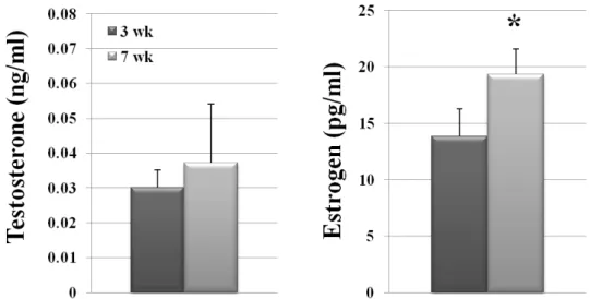
1. Effect of Sexual Maturation on the Levels of Testosterone and 17 β-Estradiol in Female Mice
We measured the levels of two major reproductive hormones, T and E2, in 7-week-old (mature) and 3-week-old (immature) female mice, using ECLIA. The results obtained were shown in Fig. 1.
T level in the mature group was 3.72 × 10—2 ± 0.017 ng/ml, that was not significantly different from 3.02 × 10—2 ± 0.005
Fig. 1. Sexual maturation and steroid hormone level in female mice. The levels of T and E2 were measured by ECLIA in serum of 3-week-old and 7-week-old female mice and are presented as mean ± S.D. (error bars represent SDs). An asterisk (*) indicates a significant difference between the means (P<0.05).
ng/ml of the immature group. In contrast, E2 level in the ma- ture group was 19.35 ± 2.25 pg/ml, that was significantly higher (P<0.05) than 13.84 ± 2.43 pg/ml of the immature group.
2. Effect of Sexual Maturation on the Levels of n-6 PUFA in the Muscle Tissue of Female mice
In order to examine the effects of sexual maturation on fatty acid metabolism, the n-6 PUFA compositions in LMs of im- mature and mature female mice were analyzed by GC and shown in Table 2.
The proportion of n-6 PUFA relative to total fatty acid con- tent in muscle did not significantly differ between immature (23.67 ± 0.78%) and mature group (23.42 ± 1.45%). However,
Table 2. Effect of sexual maturation on n-6 PUFA levels in the muscle tissue of female mice
n-6 fatty acids
Age
3 wk 7 wk
LNA 18.86 ± 1.35b 20.51 ± 1.70a GLA 0.22 ± 0.03b 0.29 ± 0.01a Total 23.67 ± 0.78 23.42 ± 1.45NS The proportions of n-6 PUFA (%, g/100g) in LM tissue sam- ples were determined by GC.
Values are presented as means ± S.D., n = 5.
a,b significant difference between groups (P<0.05).
NS no significant difference between groups (P>0.05).
levels n-6 PUFAs including the LNA and GLA were increased from 18.86 ± 1.35% to 20.51 ± 1.70% and from 0.22 ± 0.03%
to 0.29 ± 0.01%, respectively in mature mice compare to im- mature group.
3. Effect of Sexual Maturation on Expression of FADS2 Gene related to GLA Metabolism in Female Mice
The mRNA expression of FADS2 gene in LM was analyzed using real-time PCR and shown in Fig. 2.
The relative level of FADS2 mRNA significantly increased from 1.00 ± 0.31 to 1.84 ± 0.18 while female mice were sexu- ally matured from 3-week-old to 7-week-old (Fig. 2). The result showed that sexual maturation in female mice should stimulate the mRNA expression of FADS2 gene.
4. Effect of Sexual Maturation on Level of FADS2 Protein for GLA Metabolism in the Female Mice
Also the level of FADS2 protein in LM significantly increa- sed in sexually matured female mice (Fig. 3).
As shown above, mRNA expression of FADS2 gene was significantly increased in sexually matured female mice. To confirm the effect of sexually female maturation on FADS2 gene expression, the FADS2 protein level was determined by western blot analysis and shown in Fig. 3.
The level of FADS2 protein also significantly increased from 9.91 ± 0.63 to 13.43 ± 1.05, while female mice were sexually matured from 3-week-old to 7-week-old. The result proposed
Fig. 2. Sexual maturation and mRNA level of FADS2 in female mice. The gene expression levels of FADS2 in 3-week-old and 7-week-old female mice were measured using real-time PCR and normalized with respect to the β-actin control.
The relative mRNA expression level in 7-week-old female mice is normalized to the level in 3-week-old female mice as mean ± S.D. (error bars represent S.D.). An asterisk (*) indicates a significant difference between means (P<0.05).
that sexual maturation also should increase the FADS2 protein level in the LM of female mice.
DISCUSSION
Sexual maturation increases the levels of steroid hormone E2, which might play a crucial role in regulating the lipid metabolism in muscle tissue (Courant et al., 2010; Salehzadeh et al., 2011). Especially, its functional role is associated with the level and synthesis of fatty acids. Estrogen deficiency increases plasma free fatty acid availability in postmenopausal women (Jensen et al., 1994). Lohner et al. (2013) showed that women with higher E2 level showed more PUFA content in the adipose tissue than the men with higher T levels. These results suggest that sex hormones might affect the composition of fat depots (Extier et al., 2009) and skeletal muscle
Fig. 3. Sexual maturation and protein level of FADS2 in female mice. (A) Expression of FADS2 protein in 3-week-old and 7-week-old mice was determined by western blot analysis.
(B) The FADS2 expression level was normalized to that of α-tubulin. Relative protein levels were quantified using Image J software. FADS2 protein level differed significantly between the 3-week-old group and the 7-week-old groups (P<0.05). An asterisk (*) indicates a significant difference between means (P<0.05).
phenotypes (van den Beld et al., 2000) in the body.
Interestingly, serum E2 level was higher in mature than in immature female mice which is, in turn, possibly related to in- crease in GLA level in muscle tissue. Giltay et al. (2004) also demonstrated that E2 affects both n-3 and n-6 PUFA conversion into through an estrogen receptor-dependent pathway.
E2 may affect GLA synthesis through the expression of FADS2 gene in muscle tissue of female mice. The activity of the FADS2 enzyme was reported to be greater in female rat and humans (Childs et al., 2010). The data presented in our current study showed that higher E2 in mature female mice should increase the level of GLA as well as the expression of
FADS2 gene in LM muscle tissue. Therefore, these results indicate that sexual maturation of female mice might induce higher GLA level by increasing FADS2 gene expression in LM with greater serum E2 concentration.
In female mice, the onset of sexual maturation is associated with the change in level of reproductive steroid hormone. We therefore measured the levels of two major reproductive hor- mones, T and E2 (Fig.1). This result indicates that E2, but not T, level increased in blood of female mice. Our result suggests that sexual maturation did not significantly alter the proportion of total n-6 PUFA in muscle tissue (Table 2). In agreement with our results (Table 2), other studies show that sexual maturation of female mice significantly increases the levels of functional n-6 PUFAs, such as LNA and GLA in the LM (Korotkova et al., 2005). Furthermore, our results implicate that GLA meta- bolism regulated by FADS2 enzyme (de Alaniz and Marra, 2003) might be closely associated with sexual maturation in- duced high estrogen level, which is in agreement with previous study (Childs et al., 2012).
Although further study is necessary to identify precise me- chanism between estrogen treatment and FADS2 related to GLA synthesis in LM, our current study clearly shows that the circulating E2 involved in GLA metabolism in female mice.
REFERENCES
Campbell SE and Febbraio MA. 2001. Effect of ovarian hor- mones on mitochondrial enzyme activity in the fat oxidation pathway of skeletal muscle. Am. J. Physiol. Endocrinol.
Metab. 281:E803-808.
Childs CE, Hoile SP, Burdge GC and Calder PC. 2012. Chan- ges in rat n-3 and n-6 fatty acid composition during preg- nancy are associated with progesterone concentrations and hepatic FADS2 expression. Prostaglandins Leukot Essent Fatty Acids 86:141-147.
Childs CE, Romeu-Nadal M, Burdge GC and Calder PC. 2010.
The polyunsaturated fatty acid composition of hepatic and plasma lipids differ by both sex and dietary fat intake in rats. J. Nutr. 140:245-250.
Courant F et al. 2010. Assessment of circulating sex steroid levels in prepubertal and pubertal boys and girls by a novel ultrasensitive gas chromatography-tandem mass spectrometry method. J. Clin. Endocrinol. Metab. 95:82-92.
Das UN. 2007. Gamma-linolenic acid therapy of human glio-
ma-a review of in vitro, in vivo, and clinical studies. Med.
Sci. Monit. 13:RA119-131.
de Alaniz MJ and Marra CA. 2003. Steroid hormones and fatty acid desaturases. Prostaglandins Leukot Essent Fatty Acids 68:163-170.
Du M et al. 2013. Meat science and muscle biology symposium:
Manipulating mesenchymal progenitor cell differentiation to optimize performance and carcass value of beef cattle. J.
Anim. Sci. 91:1419-1427.
Extier A et al. 2009. Differential effects of steroids on the syn- thesis of polyunsaturated fatty acids by human neuroblastoma cells. Neurochem. Int. 55:295-301.
Folch J, Lees M and Sloane Stanley GH. 1957. A simple me- thod for the isolation and purification of total lipides from animal tissues. J. Biol. Chem. 226:497-509.
Giltay EJ, Gooren LJ, Toorians AW, Katan MB and Zock PL.
2004. Docosahexaenoic acid concentrations are higher in women than in men because of estrogenic effects. Am. J.
Clin. Nutr. 80:1167-1174.
Hausman GJ et al. 2009. Board-invited review: The biology and regulation of preadipocytes and adipocytes in meat ani- mals. J. Anim. Sci. 87:1218-1246.
Horrobin DF. 1992. Nutritional and medical importance of ga- mma-linolenic acid. Prog. Lipid. Res. 31:163-194.
Jensen MD, Martin ML, Cryer PE and Roust LR. 1994. Effects of estrogen on free fatty acid metabolism in humans. Am.
J. Physiol. 266:E914-920.
Korotkova M et al. 2005. Gender-related long-term effects in adult rats by perinatal dietary ratio of n-6/n-3 fatty acids.
Am. J. Physiol. Regul. Integr. Comp. Physiol. 288:R575-579.
Lohner S, Fekete K, Marosvolgyi T and Decsi T. 2013. Gender differences in the long-chain polyunsaturated fatty acid status:
Systematic review of 51 publications. Ann. Nutr. Metab.
62:98-112.
Salehzadeh F, Rune A, Osler M and Al-Khalili L. 2011. Testo- sterone or 17 β-estradiol exposure reveals sex-specific effects on glucose and lipid metabolism in human myotubes. J.
Endocrinol. 210(2):219-29.
Senapati S, Banerjee S and Gangopadhyay DN. 2008. Evening primrose oil is effective in atopic dermatitis: A randomized placebo-controlled trial. Indian J. Dermatol. Venereol. Leprol.
74:447-452.
Stoffel W et al. 2008. Delta6-desaturase (FADS2) deficiency unveils the role of omega3- and omega6-polyunsaturated
fatty acids. Embo. J. 27:2281-2292.
van den Beld AW, de Jong FH, Grobbee DE, Pols HA and Lamberts SW. 2000. Measures of bioavailable serum testo- sterone and estradiol and their relationships with muscle strength, bone density, and body composition in elderly men.
J. Clin. Endocrinol. Metab. 85:3276-3282.
Received April 21, 2015, Revised June 10, 2015, Accepted June 14, 2015