ORIGINAL ARTICLE eISSN 2384-0293 https://doi.org/10.12701/yujm.2017.34.2.238 Yeungnam Univ J Med 2017;34(2):238-241
238 YUJM VOLUME 34, NUMBER 2, DECEMBER 2017
심장 자기공명영상을 이용한 팔로사징 완전 교정술 후 우심실 기능에 대한 연구
장우성1, 최희정2, 이종민3, 김재범1, 김재현1, 장재석4
계명대학교 의과대학 1흉부외과학교실, 2소아과학교실; 3경북대학교 의과대학 영상의학과학교실;
4대구가톨릭대학교 의과대학 흉부외과학교실
Investigation of right ventricle function in patients with tetralogy of Fallot after total correction using cardiac magnetic resonance imaging
Woo Sung Jang1, Hee Joung Choi2, Jong-Min Lee3, Jae Bum Kim1, Jae Hyun Kim1, Jae Seok Jang4 Departments of 1Thoracic and Cardiovascular Surgery and 2Pediatrics, Keimyung University School of Medicine;
3Department of Radiology, Kyungpook National University School of Medicine; 4Department of Cardiothoracic Surgery, School of Medicine, Daegu Catholic University, Daegu, Korea
Background: We investigated the difference in right ventricle (RV) volume and ejection fraction (EF) according to the pulmonary valve (PV) annular extension technique during Tetralogy of Fallot (TOF) total correction.
Methods: We divided patients who underwent the procedure from 1993 to 2003 into two groups according to PV extension technique (group I: PV annular extension, group II: no PV annular extension) during TOF total correction. We then analyzed the three segmental (RV inlet, trabecular and outlet) and whole RV volume and EF by cardiac magnetic resonance imaging (MRI).
Results: Fourteen patients were included in this study (group I: 10 patients, group II: four patients; male: nine patients, female: five patients). Cardiac MRI was conducted after a 16.1 years TOF total correction follow-up period. There was no statistical difference in RV segmental volume index or EF between groups (all p>0.05).
Moreover, the total RV volume index and EF did not differ significantly between groups (all p>0.05).
Conclusion: The RV volume and EF of the PV annular extension group did not differ from that of the PV annular extension group. Thus, PV annular preservation technique did not show the surgical advantage compared to PV annular extension technique in this study.
Keywords: Tetralogy of Fallot; Cardiac MRI; Right ventricle outflow tract function
Copyright ©2017 Yeungnam University College of Medicine
This is an Open Access article distributed under the terms of the Creative Commons Attribution Non-Commercial License (http://creative- commons.org/licenses/by-nc/4.0/) which permits unrestricted non-commercial use, distribution, and reproduction in any medium, provided the original work is properly cited.
Received: November 28, 2017, Revised: December 12, 2017 Accepted: December 12, 2017
Corresponding Author: Woo Sung Jang, Department of Thoracic and Cardiovascular Surgery, Keimyung University School of Medicine, 56 Dalseong-ro, Jung-gu, Daegu 41931, Korea
Tel: +82-53-200-5832, Fax: +82-53-250-8096 E-mail: [email protected]
서 론
팔로사징(tetralogy of Fallot)은 청색증형 선천성 심기형의 가장 흔한 질환으로 팔로사징 완전 교정술 후 외래 경과 관찰 도중 발생할 수 있는 폐동맥판역류(pulmonic regurgita- tion), 폐동맥판협착(pulmonary stenosis), 부정맥 등과 관련 된 만기 심실 기능 저하(late ventricular dysfunction)는 아직
Right ventricle function after tetralogy of Fallot correction
YUJM VOLUME 34, NUMBER 2, DECEMBER 2017 239
까지 해결하지 못하고 있는 문제점으로 남아있다[1]. 최근에 시행된 여러 연구들을 살펴보면 팔로사징 수술 후 시행한 심장 자기공명영상(cardiac magnetic resonance imaging)에 서 우심실류(right ventricle aneurysm) 및 섬유화가 우심실 유출로의 기능을 저하시키고, 이 우심실 유출로의 기능저하 가 전체적인 우심실 기능 및 환자의 운동 능력 저하와 연관이 되어 있는 것으로 보고되고 있다[2-5]. 따라서 본 연구에서는 팔로사징 완전 교정술을 시행한 환자를 대상으로 하여 심장 자기공명영상을 통하여 수술 방법에 따른 우심실 기능의 차 이를 알아봄으로써 팔로사징 완전 교정술의 수술적 방법의 변화를 도모하고자 한다.
대상 및 방법
1. 연구대상
1993년 6월부터 2003년 11월까지 팔로사징 완전 교정술 을 시행한 34명의 환자 중 심장 자기공명영상에 동의한 14명 의 환자들을 대상으로 폐동맥 판막 성형술 방법에 따라 두 그룹으로 나누어 본 연구를 진행하였다(그룹 I: 폐동맥 판막 륜 확장술[pulmonary valve extension technique]을 시행한 그룹, 그룹 II: 폐동맥 판막륜 확장술[pulmonary valve preser- vation technique]을 시행하지 않은 그룹).
2. 수술방법
정중 흉골 절개술 후 동맥 캐뉼라(cannula)는 상행대동맥 에 삽입하였으며, 양쪽 대정맥에 정맥 캐뉼라를 삽입하였다.
주폐동맥 판막륜의 크기 z값이 <-2가 안 되는 경우 폐동맥 판막륜 확장술을 시행하였고, 필요한 경우 폴리테트라플루 오로에틸렌 막(polytetrafluoroethylene membrane, Goretex, WL Gore and Associates, Inc., Flagstaff, AZ, USA) 또는 자가 심낭을 이용하여 단일첨(monocusp) 판막을 삽입하였다. 주 폐동맥 판막륜의 크기 z값이 >-2보다 큰 경우 폐동맥 판막륜 확장술을 시행하지 않았다.
3. 환자 관찰 및 심장 자기공명영상 촬영 기법
1.5 테슬라 심장 자기공명영상 기기를 이용하여 심실 크기, 심실 기능을 평가하기 위해 ECG-gated steady state free-pre- cession cine MR pulse sequence를 다음의 면(plane)을 이용하 여 심실 2-챔버(ventricular 2-chamber [vertical long-axis]), 심실
4-챔버(ventricular 4-chamber [horizontal long-axis]), 단축면 (short-axis plane, perpendicular to the ventricular long-axis plane)을 측정하였다.
4. 분절 심실 구출률 분석
우심실을 3개의 세로 방향, 3개의 수직 방향의 총 9개 분절 로 나누어 분절 심실 구출률을 확장기말 및 수축기에 3D 자료 세트를 이용하여 계산하였다. 그 외 우심실 확장기 말 부피(mL/m2), 우심실 확장기 말 부피지수, 우심실 수축기 말 부피(mL/m2), 우심실 수축기 말 부피 지수, 우심실 심박출률 (%), 좌심실 심박출률(%)을 측정하였다.
5. 통계기법
모든 연속형 변수는 평균±표준편차로 표시하였고, 비연 속형 변수는 빈도 및 퍼센트로 표현하였다. 연속형 변수의 비교는 Mann-Whitney U-test를 이용하여 시행하였고, 비연 속형 변수의 비교는 Fisher’s exact test를 이용하여 분석하였 다. p<0.05인 경우를 통계적으로 유의한 것으로 판정하였다.
모든 통계 분석은 IBM SPSS version 24.0 (IBM Co., Armonk, NY, USA)를 사용하였다.
결 과
이 연구에는 총 14명의 환자가 참여하였고(그룹 I: 10명, 그룹 II: 4명), 남자는 9명, 여자는 5명이었다. 양군 간에 팔로 사징 완전 교정술을 시행한 연령 차이는 보이지 않았다(그룹 I=18.8±7.6개월, 그룹 II=20.2±12.2개월, p=0.805). 환자 는 팔로사징 완전 교정술 후 평균 16.1±3.8년 후에 심장 자 기공명영상을 촬영하였고, 심장 자기공명영상 촬영 연령 역 시 그룹 I에서 17.5±3.7년, 그룹 II에서 15.6±2.4년으로 양 군 간의 차이는 없었다(p=0.379).
수술 중 양군 간에 심폐 순환 시간, 대동맥 겸자 시간의 차이는 보이지 않았다. 양군간의 수술 전 환자 상태 차이를 Table 1에 기술하였다.
양군 간의 우심실 용적을 우심실 유입로(right ventricle inlet), 우심실 기둥(right ventricle trabecula), 유출로(right ventricle outlet) 각각 세 부위에서 비교하였을 때 양군 간의 통계학적 차이는 없었다(각각의 p값: 0.976, 0.940, 0.193).
또한 양군 간의 우심실 심박출률을 비교하여 보았을 때 전체 적인 우심실 심박출률 역시 통계학적 차이를 보이지 않았다
Woo Sung Jang et al.
240 YUJM VOLUME 34, NUMBER 2, DECEMBER 2017
Table 1. Patient characteristics
Group I Group II p-value
Age (year) 18.8±7.6 20.2±12.2 0.805
Body weight (kg) 9.9±0.9 11.5±2.6 0.334
BSA (m2) 0.5±0.0 0.5±0.1 0.593
CPB time (min) 152.4±32.6 121.0±31.3 0.133 ACC time (min) 94.3±27.3 81.0±20.3 0.404 BSA, body surface area; CPB, cardiopulmonary bypass; ACC, aorta cross clamp.
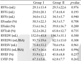
Table 2. Comparison of right ventricle volume and ejection frac- tion by cardiac magnetic resonance imaging
Group I Group II p-value
RVVo (mL) 29.1±13.4 29.5±22.6 0.976
RVVi (mL) 29.0±20.1 17.4±8.4 0.193
RVVt (mL) 34.0±13.2 34.5±5.7 0.940
EFoutlet (%) 50.5±22.3 34.5±5.7 0.708 EFinlet (%) 53.6±16.2 39.8±16.7 0.200 EFTrab (%) 32.2±20.2 65.8±12.7 0.755 RVEDV (mL) 152.0±43.8 134.5±35.1 0.500 RVEDVi (mL/BSA) 93.7±19.6 82.1±13.0 0.308
RVESV (mL) 71.4±33.2 70.6±9.6 0.961
RVESVi (mL/BSA) 43.7±16.6 43.8±4.0 0.996
RVEF (%) 53.9±12.1 45.8±8.8 0.257
LVEF (%) 67.1±5.6 62.8±7.7 0.261
RVVo, right ventricle outlet volume; RVVi, right ventricle inlet volume; RVVt, right ventricle trabecular volume; EFoutlet, right ventricle outlet ejection fraction; EFinlet, right ventricle inlet ejec- tion fraction; EFTrab, right ventricle trabecular ejection fraction;
RVEDV, right ventricle end diastolic volume; RVEDVi, right ven- tricle end diastolic volume index; RVESV, right ventricle end systolic volume; RVESVi, right ventricle end systolic volume in- dex; RVEF, right ventricle ejection fraction; LVEF, left ventricle ejection fraction; BSA, body surface area.
(그룹 I: 53.9±12.1%, 그룹 II: 45.8±8.8%, p=0.257). 우심실 을 세 부위로 나누어 우심실 심박출률을 비교하였을 때도 각 그룹의 우심실 유입로, 우심실 기둥, 우심실 유출로의 통계학 적 차이는 관찰되지 않았다(각각의 p값: 0.200, 0.755, 0.708) (Table 2).
고 찰
팔로사징 완전 교정술 후 우심실 유출로 심장 자기공명영 상 촬영 시 조영 증강 및 국소적인 기능 장애가 가장 많이 관찰 되는 것으로 보고되어 있다[2-5]. 이러한 국소적인 우심실 유출로의 기능장애는 전반적인 우심실 기능, 우심실 심박출
률, 운동 시 최고 심근 산소 소모량에 많은 영향을 끼치고 우심실 기능 저하로 인한 심정지 사망이 발생하는 경우가 보고되고 있다[6-10]. 이의 원인으로 우심실 유출로의 운동 이상증(dyskinesia) 및 섬유화와 전체 우심실 기능 부전과의 관계가 증명된 논문에서 우심실 유출로의 기능장애가 치료 되지 않고 방치되었을 때 우심실 기능 및 운동 능력에 영향을 미칠 수 있다고 보고하고 있다[2-5]. 따라서 최근에 우심실 유출로 및 우심실 전체의 기능을 보존하기 위해 가능하면 폐동맥 판막륜의 절개를 피하고 폐동맥 판막륜 절개 시 우심 실 유출로의 확장을 최소화하려는 노력이 시도되고 있다.
반대로 우심실 기능의 가장 중요한 부분을 차지하는 부분은 우심실 유출로가 아니라 우심실 유입로(right ventricle inlet) 또는 우심실 중격(right ventricle septum)이라는 보고도 있어 팔로사징 완전 교정술 후 손상될 수 있는 우심실 유출로의 기능이 전체 우심실 기능에 미치는 영향은 미미할 것이라는 반대되는 보고도 있다[11,12]. 따라서 팔로사징 완전 교정술 시 폐동맥 판막륜을 포함하여 우심실 유출로까지 절개하여 우심실 유출로를 확장하는 것이 우심실 기능에 미치는 영향 에 대한 평가는 아직까지 논란의 여지가 있고, 지금까지 팔로 사징 완전 교정술 방법에 따른 우심실 기능의 변화, 우심실 유출로 기능의 변화를 비교 분석하는 논문은 없었다.
이 연구에서는 폐동맥 판막륜 확장술 여부와 상관 없이 외 래 경과 관찰 후 시행한 심장 자기공명영상에서 우심실의 용 적 및 기능의 차이를 보이지 않았다. 하지만 우심실 유출로의 심박출량(right ventricle outlet ejection fraction)이 양 군간에 통계학적 차이를 보이지는 않았지만 폐동맥 판막륜 확장술 을 시행한 그룹에서 우심실 유출로의 심박출량 감소를 확인 할 수 있었는데, group II의 수가 적어서 통계학적 차이를 보이지 않았을 것으로 판단된다. 하지만 전체 우심실 심박출 량 차이를 비교했을 때 양군간에 심박출량 차이가 줄어드는 것으로 보아 우심실 유출로 심박출량의 영향이 전체 우심실 심박출량의 차이에 미치는 효과는 그렇게 크지 않을 수 있다 는 것을 간접적으로 시사한다고 볼 수 있다. 이를 뒷받침하는 보고로 우심실의 유입로가 유출로보다 많은 부분 우심실 심 박출량에 더 많은 영향을 끼치고, 우심실의 기둥 부위가 우심 실 심박출량의 중요한 부분을 차지하기 때문에 팔로사징 완 전 교정술 중 폐동맥 판막륜 절개를 가하더라도 우심실 심박 출량에는 영향이 없을 것이라고 말하고 있다[11-12].
결론적으로 이 연구에서는 최근 팔로사징 완전 교정술에 서 시도되고 있는 폐동맥 판막륜 보존 노력에 반하여 기존에 시행되고 있는 폐동맥 판막륜의 z값에 따른 폐동맥 판막륜
Right ventricle function after tetralogy of Fallot correction
YUJM VOLUME 34, NUMBER 2, DECEMBER 2017 241
절개 방법을 적용하여도 관찰 기간 동안 우심실 용적 및 우심 실 심박출량에는 차이를 보이지 않았다.
연구 한계
상기 연구는 연구의 목적과 방법에 비해 수술 증례의 수가 부족하여 일반화된 결론을 내기 어렵다. 따라서 증례의 수를 추가하여 좀 더 심도 깊은 분석이 필요할 것으로 판단된다.
ACKNOWLEDGEMENT
이 연구는 2014년도 경북대학교병원 생명의학연구원 연 구비의 지원으로 이루어졌음.
CONFLICT OF INTEREST
No potential conflict of interest relevant to this article was reported.
ORCID
Woo Sung Jang: https://orcid.org/0000-0001-5805-670X
REFERENCES
1. Saremi F, Ho SY, Cabrera JA, Sánchez-Quintana D. Right ventricular outflow tract imaging with CT and MRI: part 2, function. AJR Am J Roentgenol 2013;200:W51-61.
2. Babu-Narayan SV, Kilner PJ, Li W, Moon JC, Goktekin O, Davlouros PA, et al. Ventricular fibrosis suggested by cardio- vascular magnetic resonance in adults with repaired tetralogy of fallot and its relationship to adverse markers of clinical outcome. Circulation 2006;113:405-13.
3. Oosterhof T, Mulder BJ, Vliegen HW, de Roos A. corrected tetralogy of Fallot: delayed enhancement in right ventricular outflow tract. Radiology 2005;237:868-71.
4. Davlouros PA, Kilner PJ, Hornung TS, Li W, Francis JM, Moon JC, et al. Right ventricular function in adults with re- paired tetralogy of Fallot assessed with cardiovascular mag- netic resonance imaging: detrimental role of right ventricular outflow aneurysms or akinesia and adverse right-to-left ven- tricular interaction. J Am Coll Cardiol 2002;40:2044-52.
5. Wald RM, Haber I, Wald R, Valente AM, Powell AJ, Geva T. Effects of regional dysfunction and late gadolinium enhan- cement on global right ventricular function and exercise capa- city in patients with repaired tetralogy of Fallot. Circulation 2009;119:1370-7.
6. Moore B, Brubaker PH, Stewart KP, Kitzman DW. VE/VCO2 slope in older heart failure patients with normal versus reduced ejection fraction compared with age-matched healthy controls.
J Card Fail 2007;13:259-62.
7. Harrison DA, Siu SC, Hussain F, MacLoghlin CJ, Webb GD, Harris L. Sustained atrial arrhythmias in adults late after re- pair of tetralogy of fallot. Am J Cardiol 2001;87:584-8.
8. Gatzoulis MA, Balaji S, Webber SA, Siu SC, Hokanson JS, Poile C, et al. Risk factors for arrhythmia and sudden cardiac death late after repair of tetralogy of Fallot: a multicentre study. Lancet 2000;356:975-81.
9. Roos-Hesselink J, Perlroth MG, McGhie J, Spitaels S. Atrial arrhythmias in adults after repair of tetralogy of Fallot. Corre- lations with clinical, exercise, and echocardiographic findings.
Circulation 1995;91:2214-9.
10. Niezen RA, Helbing WA, van der Wall EE, van der Geest RJ, Rebergen SA, de Roos A. Biventricular systolic function and mass studied with MR imaging in children with pulmo- nary regurgitation after repair for tetralogy of Fallot. Radio- logy 1996;201:135-40.
11. Geva T, Powell AJ, Crawford EC, Chung T, Colan SD. Evalu- ation of regional differences in right ventricular systolic func- tion by acoustic quantification echocardiography and cine magnetic resonance imaging. Circulation 1998;98:339-45.
12. Bodhey NK, Beerbaum P, Sarikouch S, Kropf S, Lange P, Berger F, et al. Functional analysis of the components of the right ventricle in the setting of tetralogy of Fallot. Circ Car- diovasc Imaging 2008;1:141-7.