Regulation of Wnt/β-catenin Signal Transduction in HT-29 Colon Cancer Cells by a Rhododendron brachycarpum Fraction
Bo Ram Sim1, Young Sun Nam2 and Ja Bok Lee3*
1Beauty care Technology, Dongnam Health University, Cheoncheon-ro 74-gil 50, Jangan-gu, Suwon-si, Gyeonggi-do 16328, Korea
2Department of Beauty Design, 400-5, Namyangjungang-ro, Hwaseong, Gyeonggi-do 18274, Korea
3L.FOUNDER INC., 406, Business Incubation Seoul National University of Science & Technology 232, Gongneung-ro, Nowon-gu, Seoul 01811, Korea
Received April 10, 2019 /Revised July 18, 2019 /Accepted July 22, 2019
The aim of this study was to determine the anticancer activities of an 80% methanol extract and vari- ous fractions of Rhododendron brachycarpum (RB). The n-hexane fraction of RB showed the highest in- hibitory activity (Inhibit concentration 50% = 20.2±1.2 μg/ml) in HT-29 cells. Colony- and sphere- forming abilities were significantly correlated with a decrease in the cell count and size. A TOP/FOP flash reporter assay revealed that the inhibitory activity of the n-hexane fraction of RB (0.22±0.02 fold change) was lower than that of the 80% methanol extract and that of other fractions. The n-hexane and ethyl acetate fractions of RB were predominantly dependent on the expression levels of intra- cellular β-catenin. Western blotting using p-GSK3β with only the n-hexane fraction of RB was con- ducted to examine whether these secondary metabolites reduced β-catenin degradation. Intracellular β-catenin regulation resulted in quantitative changes in the nucleus. In summary, these results demon- strate the potential of the n-hexane fraction of RB as a natural anticancer agent.
Key words : Anti-cancer, β-catenin, p-GSK3β, Rhododendron brachycarpum, Wnt
*Corresponding author
*Tel : +82-2-3296-0886, Fax : +82-2-3296-0889
*E-mail : [email protected]
This is an Open-Access article distributed under the terms of the Creative Commons Attribution Non-Commercial License (http://creativecommons.org/licenses/by-nc/3.0) which permits unrestricted non-commercial use, distribution, and reproduction in any medium, provided the original work is properly cited.
Journal of Life Science 2019 Vol. 29. No. 8. 871~878 DOI : https://doi.org/10.5352/JLS.2019.29.8.871
서 론
우리나라는 식습관이 서구화되면서 암 발병률이 증가하였 고, 국내 대장암 환자수도 빠르게 증가하는 추세다. 국가암 통계에 따르면 국내 대장암 환자수는 최근 180여 개국 중 1위 를 차지하였고, 암발병률은 2위까지 빠르게 증가하였다[16].
혈변, 배변습관의 변화, 복통 및 복부 불편감 등 다양한 증상이 있지만 통증과 같은 증상은 3기 이상의 대장암 환자에서 발병 하기 때문에 쉽게 인지를 못하는 경우가 많다. 표적 약물과 같은 화학요법은 현재 대장암의 표준 치료법으로 치료 효과를 향상시키기 위한 분자 기작에 대한 이해가 필요하다[26].
Wnt 신호전달 시스템은 선충, 초파리 및 포유동물에 이르 기까지 초기발생 또는형태 형성에 필수적 요소이다. 19종류인 Wnt ligand를 형성하여 Frizzled 막 수용체에 결합되면 LRP5/
6와 결합하여 신호전달을 개시한다. β-catenin을 이용한 신호 전달에서 안정성을 조절하여 여러 표적유전자의 발현을 조절 하며, 이 경로의 신호전달은 암발병에 고빈도로 나타난다. 특
히, 대장암에서 Wnt 신호전달 시스템은 주요 종양 저항성을 포함하는 항암 치료를 위한 치료 전략으로 간주되며, 대장암 형성에서 종양구 형성에 주된 매커니즘이다[22, 35]. 텍솔과 같은 1세대 항암치료제를 기반으로 천연물질을 이용한 항암 제가 개발되면서, 천연물을 이용한 다양한 연구가 이루어졌 다. 특히, Wnt 신호조절 작용을 하는 단일물질이 개발되면서 APC mutation 암 종에서 β-catenin 저해를 통해 주된 항암기 전으로 연구되고 있다[12, 19].
홍만병초(Rhododendron brachycarpum, RB)는 높은 산 위에 자라서 최근에는 정원 식물로 약용효과를 갖으며, 상록수가 많아 정원식물로 이용된다. 특히, 홍만병초 잎은 고혈압, 신경 통, 균 억제 등 다양한 반면에 효과를 보이고, 한국의 전통 의학 소재로 이용되고 있다. 일부 논문에서 홍만병초에 포함 된 단일물질 quercetin, quercitrin, avicularin, rhododendron, grayanotoxin I, guaijaverin 및 hyperin이 확인되었고, RB 잎 추출물 및 분획물로부터 항산화제, 항암제, 세포 독성, 유전 독성 및 면역학적 증거 등을 발표되었다[9, 23, 33].
본 연구에서는 홍만병초(지상부) 추출물 및 분획물로부터 항암활성을 분석하고, 대장암세포에서 Wnt/β-catenin 신호전 달 억제를 통해 항암활성을 갖는 기작을 확인하였다.
재료 및 방법 실험재료
홍만병초는 제주도 서귀포시 만병초영농조합에서 구입하
여 본 연구의 시료로 사용하였다. 홍만병초를 4일간 실온 건조 한 뒤 분쇄하여 795.3 g을 48시간 80% 에탄올 1 l에 추출하였 다. 추출물은 여과지(Advantech, circles 300 mm, Japan)에 여 과 후 45℃에서 감압 농축 및 동결건조하였다. 건조 추출분말 (130 g)을 3차 증류수에 섞어 n-hexane (43.35 g), ethyl acetate (42.41 g), n-butanol (29.15 g) 및 water (14.87 g)을 얻어 농축 및 동결건조 후 실험에 이용하였다.
세포배양 및 Cyto-X 분석
Hela, PC-3, AGS (한국세포주은행, Seoul, Korea), HT-29 및 NCI-H1299 (ATCC; manassas, VA, USA) 세포주를 이용하 여 세포의 독성테스트 Cyto-X (LPS solution, Daejeon, Korea) 시약을 이용하였다. 세포를 96 well plate에 1×10⁴ 세포수로 12시간 배양한 뒤 홍만병초 추출물 및 분획물을 1, 10 및 100 μg/ml로 처리하였다. 배양 24시간 후 각 well에 1/10 농도로 Cyto-X 시약을 첨가한 후 호일로 감싸 4시간 배양기에 처리하 였다. Wallac Victor2 plate reader (Perkin Elmer Corp., Ner- walk, CT) 로 450 nm 흡광도에서 측정하여 수치화 하였다.
콜로니형성 억제 실험
콜로니형성 억제 실험은 500개의 세포를 60 mm 세포배양 접시(SPL, Pocheon, Korea)에 처리 후 dimethyl sulfoxide (DMSO) 또는 10 μg/ml로 처리하였다. 10~14일간 배양 후 세 포를 메탄올로 고정 후 trypan blue (Sigma-Aldrich, St. Louis, MO)로 3분간 염색 후 PBS (LPS solution, Daejeon, Korea)로 수세하였다. 염색된 세포수를 카운팅하여 이미지화 하였다.
Sphere 배양법(Sphere forming assay)
HT-29 대장암 세포주 3×10³ 개를 ultra-low attachment plates (Corning Costar, Corning, NY)에 serum-free DMEM- F12 배지(Gibco Laboratories, Grand Island, NY) 를 20 μl/ml B-27 (Invitrogen, Waltham, MA), 20 ng/ml FGF (R&D Systems, Minneapolis, MN)와 20 ng/ml EGF (R&D Systems, Minneapolis, MN)를 첨가하여 접종 후 DMSO 또는 10 μM/
ml로 처리하였다. 10~14일간 배양 후 형성된 구형에 10% for- malin (Sigma Aldrich, St. Louis, MO)을 4%로 처리하여 고정 시켰다. 구형의 숫자 및 사이즈는 Image J software를 통해 분석하였고, 측정은 50 mm 이상의 사이즈만 분석하였다.
TOP/FOP flash 분석법
세포내 Wnt signal을 확인하기 위해 TOP/FOP-flash luci- ferase reporter assay 를 실시하였다. HT-29 세포주를 6-well plate (SPL, Pocheon, Korea)에 3×10⁵ 세포수로 12시간 배양한 뒤 각각 target vector와 pRL-Renilla vector (Control)를 1 μg 용량으로 Terbofect (Thermo Fisher Scientific, Waltham, Massachusetts, USA)시약으로 주입시켰다. 다시 12시간 배양
한 뒤 홍만병초 추출물 및 분획물을 10 μg/ml로 처리하였다.
배양 24시간 후 각 well의 세포를 회수하여 dual-luciferase®
reporter assay kit (Promega, USA)을 이용하여 제공된 실험방 법에 따라 측정하였다.
총단백질 발현분석
HT-29 대장암 세포주를 60 mm 세포배양 접시에 4.5×10⁵ 세포수로 12시간 배양한 뒤 10 μg/ml 홍만병초 추출물 및 분 획물을 24시간 처리하여 세포를 harvest하였다(β-catenin 단 백질은 홍만병초 추출물 및 분획물 12시간 처리). RIPA lysis buffer (GenDEPOT, USA)에 phosphatase inhibitor cocktail과 protease inhibitor cocktail을 1x로 섞어 얼음에 1시간 동안 lysis후 13,000 rpm에서 20분간 원심분리를 실시하였다. 상층 액을 회수하여 Bio-Rad protein assay (Bio-Rad, CA)를 이용하 여 정량 후 10~25 μg의 단백질을 SDS-PAGE gel method를 이용하여 전기영동 하였다. Gel transfer된 단백질을 PBS-T에 희석한 5% skim milk (LPS solution, Daejeon, Korea)에 1시간 동안 blocking한 뒤 1차 항체를 각각 1:1,000의 농도로 12시간 냉장실에 처리하였다. 3회 PBS-T로 세척한 뒤 2차 항체를 각각 1:5,000의 농도로 실온에서 처리 후 다시 3회 PBS-T로 세척하 였다. Amersham ECL Western blotting detection reagent (GE Healthcare)를 이용하여 X-ray film 발색으로 측정하였다.
항체는anti-Cyclin D1, anti-c-myc, anti-β-catenin, anti-p-GSK3 β (Tyr216), anti-GSK3β, anti-LEF1, anti-p-LRP6, anti-β-actin (Santa Cruze, Dallas, TX, USA) 및 anti-Lamin A/C (Ther- moFisher Scientific, Waltham, Massachusetts, USA)를 이용 하였다.
핵단백질 발현분석
HT-29 세포주를 60 mm 세포배양 접시(3개)에 5×10⁵ 세포 수로 12시간 배양한 뒤 10 μg/ml 홍만병초 추출물 및 분획물 을 12시간 처리하여 세포를 harvest하였다. NE-PER™ Nuclear and Cytoplasmic Extraction Reagents (Thermo Scientific, Waltham, MA, USA) 실험법에 맞게 분획하여, Bio-Rad pro- tein assay (Bio-Rad, Richmond, CA)를 이용하여 정량 후 10~25 μg의 단백질을 SDS-PAGE gel method를 이용하여 전 기영동 하였다. Gel transfer된 단백질을 PBS-T에 희석한 5%
skim milk에 1시간 동안 blocking한 뒤 1차 항체를 각각 1:1,000의 농도로 12시간 냉장실에 처리하였다. 3회 PBS-T로 세척한 뒤 2차 항체를 각각 1:5,000의 농도로 실온에서 처리 후 다시 3회 PBS- T로 세척하였다. Amersham ECL Western blotting detection reagent (GE Healthcare, Milwaukee, Wisconsin, USA)를 이용하여 X-ray film 발색으로 측정하였 다. 항체는 anti-β-catenin, anti-LEF1, anti-β-actin, anti-Cyclin D1, anti- GSK-3β 및 anti-p16 (Santa Cruze, Dallas, TX, USA) 를 이용하였고, anti-c-myc, anti-Axin2 및 anti-p-LRP6 (Cell
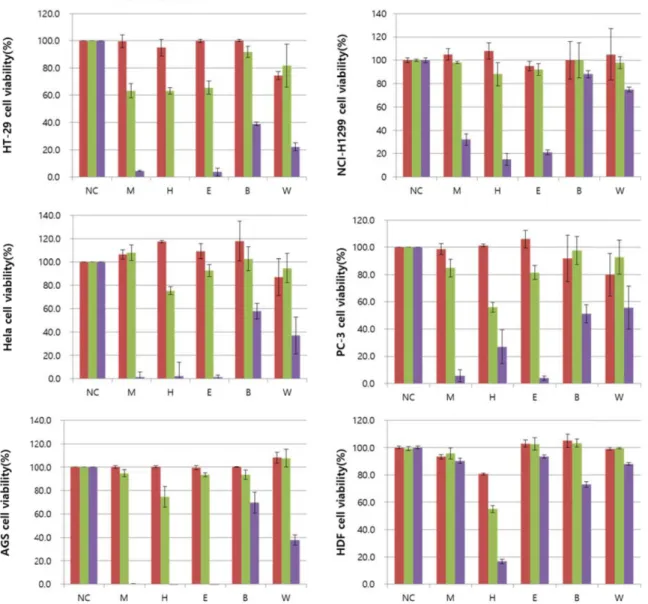
Fig. 1. Cancer cell viability was measured by Cyto-X assay from RB. Hela, PC-3, AGS, HT-29, NCI-H1299 cells were treated with RB extract and fractions for various concentrations. 80% Methanol extract (M), n-hexane fraction (H), ethyl acetate fraction (E), n-butanol fraction (B), aqueous (W).
Signaling Technology, Inc., Danvers, MA, USA)를 이용하였 다.
통계분석
모든 생리활성 실험은 3반복 실험을 통해 수치화 하였으며, 통계분석은 one-way ANOVA로 신뢰구간 p<0.05으로 검정을 실시하였다. 통계프로그램은 Graph Pad Prism 5 software (Graph Pad Software, Inc., La Jolla, CA, USA)를 이용하였다.
결과 및 고찰
홍만병초 추출물 및 분획물의 암세포 및 정상세포 세포독성 본 연구에서는 HT-29, Hela, PC-3, AGS 및 NCI-H1299 5가
지 암세포주와 HDF 1개의 정상세포주를 이용하여 홍만병초 추출물 및 분획물의 항암활성 및 세포독성을 확인하였다. 암 세포의 증식을 억제하는 기전은 다양한 신호를 조절하면서 발생된다. 암세포에 대한 세포 사멸효과를 확인하기 위해 미 토콘드리아에서 배출되는 michondrial dehydrogenase와 반 응하는 Cyto-X 시약을 이용하여 세포의 증식률을 측정하였다.
Cyto-X assay는3-(4,5-dimethylthiazol-2-yl)-2,5-diphenyltetr- azolium bromide (MTT) assay에서 발생되는 tetrazolium salt 를 DMSO (dimethyl sulfoxide)로 용해시키는 과정에서 발생 되는 오차를 줄일 수 있는 시약으로 실험적 오차를 줄이기 위 해 선택하였다.
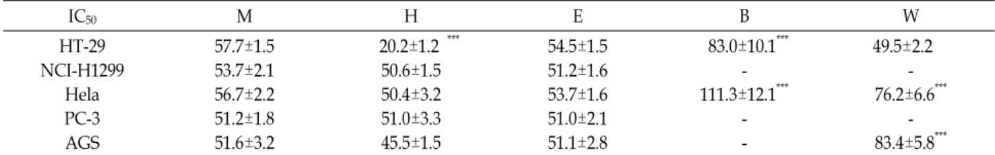
홍만병초 추출물의 암세포 세포사멸은 사멸률 및 half max- imal inhibitory concentration (IC50)은 Fig. 1과 Table 1에 나타
Table 1. Cancer cells induced by the RB extract and fractions when treated at the established IC50 concentrations
IC50 M H E B W
HT-29 NCI-H1299
Hela PC-3 AGS
57.7±1.5 53.7±2.1 56.7±2.2 51.2±1.8 51.6±3.2
20.2±1.2 ***
50.6±1.5 50.4±3.2 51.0±3.3 45.5±1.5
54.5±1.5 51.2±1.6 53.7±1.6 51.0±2.1 51.1±2.8
83.0±10.1***
- 111.3±12.1***
- -
49.5±2.2 - 76.2±6.6***
- 83.4±5.8***
80% Methanol extract (M), n-hexane fraction (H), ethyl acetate fraction (E), n-butanol fraction (B), aqueous (W). Means values ± SD from triplicate experiment are shown. Different letters are significantly different at p<0.05 by Two-way ANOVA. p<0.001 (***).
A
B
Fig. 2. RB extracts and a fraction reduces colony formation and Image J analyze in HT-29 cell. Colony formation (A) and Image J analyze (B) were shown. 80% Methanol extract (M), n-hexane fraction (H), ethyl acetate fraction (E), n-butanol fraction (B), aqueous (W). The red color is counter number.
내었다. 암세포주의 세포사멸 IC50 값은 51.2~57.7 μg/ml으로 나타났다. 특히, HT-29 대장암 세포주에서 홍만병초 n-hexane 분획물의 IC50 값은 20.2±1.2 μg/ml로 가장 큰 세포독성을 보 였다(p<0.001). 홍만병초 n-butanol 분획물은 대조구에 비해 독성을 갖지 않았고, NCI-H1299와 PC-3의 n-butanol 및 water 분획물은 100 μg/ml 처리 농도에서 독성을 갖지 않아 IC50
값을 확인할 수 없었다. 또한, HDF 세포주에서는 n-butanol을 제외한 추출물 및 분획물에서 세포독성이 없음을 확인하였다.
5개의 암세포 중 세포독성이 가장 큰 HT-29 세포주를 이용하 여 추가실험을 실시하였다. 식물유래 추출물 중 비극성 용매 에 용출된 물질들은 난용성인 alkaloid, 배당체, 수지, 식물색 소, tannin 등에 속하는 것으로 보고된다[17]. 특히 천연물 기 원 alkaloid는 항암 약물 개발에 유용한 성격을 갖는다[21].
1세대 대장암 치료 항암제 중 천연물 알칼로이드에서 유래한 vincristine은 microtubule polymerization을 억제하는 약물의 기전이 밝혀졌으며[14], 이외에도 Wnt/β-catenin, apoptosis, necrosis 및 DNA 합성을 억제하는 등 다양한 타겟의 천연물 유래 항암제가 개발되었다[1, 2, 18]. 이처럼 천연물 유래 비극 성 물질들은 다양한 항암활성을 통해 암 예방 또는 치료 효과 를 갖고, 이 같은 원리를 규명할 수 있다. 홍만병초 추출물은 100 μg/ml의 농도에서 항산화 효과, 항염증, 세포보호 및 유방 암, 간암, 폐암세포의 생육억제 활성 효과가 알려져 있다[13, 25]. 본 연구결과에서도 홍만병초 추출물, n-hexane 분획물 ethyl acetate 분획물에서 강한 세포 독성을 보인것은 난용성 인 alkaloid, 페놀 및 플라보노이드의 영향일 것으로 사료된다.
홍만병초 추출물 및 분획물의 암세포 군집형성 및 암세포 Sphere 억제
세포독성이 가장 크게 나타난HT-29 세포주를 이용하여 암 세포 군집형성 정도 및 sphere 측정법을 이용하여 Fig. 2와 Fig. 4에 나타내었다. Cyto-X와 같은 세포독성 측정법과 다르 게 군집형성도 및 sphere도는 암세포의 장기간 증식을 조절하 는 기전을 확인하는 방법 임상에서 치료법과 유사한 실험법이 다. 암세포 군집형성은 세포수와 세포사이즈의 상관계수 0.646 으로 서로 경향성을 보였으며(p=0.165, data not shown), 이는 세포독성과 유의적인 결과를 보였다(p<0.05). 특히, 홍만병초 n-hexane 분획물에서는 강한 세포독성으로 암세포 군집형성
을 확인할 수 없었고, 이 같은 결과는 장시간 분획물의 노출이 암세포의 성장을 저해한다는 것을 알 수 있었다. 암세포sphere 에서는 50 mm 이상의 사이즈는 홍만병초 추출물 및 n-hexane 분획물에서 확인되지 않았으며, 이는 apoptosis 및 G2/M ar- rest에 의한 결과로 볼 수 있다(Fig. 3). Ethyl acetate 분획물에 서 40 mm정도의 암세포구만 확인할 수 있었다.
대장암 세포의 종양형성능은 군집 또는 종양구를 형성하는 특징을 갖는다. 특히, β-catenin은 cyclin D1을 조절하여 대장 암 생성 및 구형화에 큰 영향을 미치는 것으로 보고된 바 있다 [31]. 상기 연구에서 보여준 종양 세포의 억제는 자가사멸 및 종양형성 단백질의 억제 등 다양한 원인에 의해 사멸한 것으 로 사료된다. 이는 홍만병초 n-hexane 분획물에서 효과적인
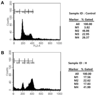
A
B
Fig. 3. RB n-hexane fraction activates cell cycle through the in- hibition of cell viability in HT-29 cell. DMSO treated cell (A) and RB n-hexane fraction treated cell (B) in HT-29 cell. Cells were incubated with PI added (5 μg/ml) at 37℃. The area parameter histogram was used to de- termine the percentage of cells in apoptosis (M1), G1 (M2), S (M3), G2/M (M4) phases.
Fig. 4. RB extract and fractions reduces 3D sphere culture in HT-29 cell. 80% Methanol extract (M), n-hexane fraction (H), ethyl acetate fraction (E), n-butanol fraction (B), aqueous (W). The red color is counter number.
Fig. 5. RB n-hexane fraction reduces TOP/FOP flash reporter gene expression in HT-29 cell. 80% Methanol extract (M), n-hexane fraction (H), ethyl acetate fraction (E), n-butanol fraction (B), aqueous (W). Different letters are significantly different at p<0.05 by ANOVA. p<0.05 (*).
억제를 보였고, β-catenin의 조절에 의한 대장암 억제활성을 보인 것으로 보이며, 이에 대장암 억제에 영향을 준 β-catenin 레벨을 확인하는 추가적인 연구가 필요할 것으로 사료된다.
홍만병초에 존재하는 플라보노이드 중 quercetin은 SW480 대 장암세포주의 β-catenin레벨을 조절할 것으로 알려졌으며, 직 장암세포에서도 동일한 암세포 억제 매커니즘이 보고되었다 [1, 4, 30]. 따라서 본 연구에서도 플라보노이드 계열의 물질이 Wnt/β-catenin레벨을 조절을 통해 암세포 억제에 밀접한 관 련이 있음을 알 수 있었다.
홍만병초 추출물 및 분획물의 Wnt 전사 활성
TOP/FOP flash reporter assay는 Wnt 신호에 의한 전사 활성화가 β-catenin 및 TCF 결합을 유도하여 전사 활성에 영 향을 주는지 확인하는 실험법이다. FOP flash는 thymidine Kinase (TK) 최초 프로모터에서 역방향으로 3 개의 카피가 뒤 따르는 TCF 결합 부위를 mutation 시켜 제작된 음성 대조 군 역할을 하며, TOP flash는 TCF 결합 부위를 포함하여 Wnt 신호에 반응하여 정량화를 가능하게 한다.
Wnt 전사활성은 Fig. 5와 같이 홍만병초 n-hexane 분획물 에서만 유의한 감소를 보였으며(p<0.05), 추출물 및 다른 분획 물에서는 유의한 변화를 확인할 수 없었다.
HT-29 대장암 세포주는 APC mutation (c.2557G>T, c.4666_
4667insA), BRAF mutation (c.1799T>A), PIK3CA mutation (c.1345C>A), SMAD4 mutation (c.931C>T) 및 TP53 mutation (c.818G>A)을 갖고 있다[3, 8, 28, 29, 32]. 특히 APC는 Wnt 신호 전달 경로의 길항제 역할을 하는 종양 억제 단백질로 세포 이동 및 부착, 전사 활성화 및 세포 사멸과 같은 다른 과정에도 관여한다. HT-29 세포는 APC 유전자의 변이로 Wnt 신호를 자극하여 악성 종양으로 진행되는 유전성 대장 용종증 (FAC)을 유발한다. 이때, 조절되지 못하는 Wnt 신호전달이 무분별한 관련 유전자를 발현시켜 단백질을 생성하고 암종의 위험성을 증대시킨다. 식물에서 생성되는 2차 대사산물인 ste- roid alkaloids는 대부분 배당체로서 존재하며 식물의 종류 따 라 solanum alkaloid와 veratrum alkaloid가 알려져 있다. 가 지속에서 분리한 tomatine은 alkaloid계열의 물질로 HT-29 대 장암세포에서 세포독성을 보이며, 배당체의 형태가 더 활성이 높다고 보고하였다[34]. APC mutation된 대장암세포에서도 alkaloid계열의 약물이 β-catenin mRNA 레벨을 조절했다는 연구도 있었다[23]. 따라서, 홍만병초 내 존재하는 alkaloid계 열의 물질은 세포내 β-catenin을 감소시키는 것으로 보여지며, 홍만병초에 존재하는 alkaloid계열의 추가 연구가 필요할 것 으로 사료된다.
A B
C
Fig. 6. RB n-hexane fraction activates p-GSK3β expression through the inhibition of β- catenin in HT-29 cell. The total protein ex- pression (A) and nuclear protein expre- ssion (B) and Western blot expression lev- el (C) were detected protein antibodies.
Protein levels were quantified from three independent expreiments using Image J software. The cells were analyzed by im- munoblotting with a β-actin was used as a loading control. 80% Methanol extract (M), n-hexane fraction (H), ethyl acetate fraction (E), n-butanol fraction (B), aque- ous (W). Western blot were using three different test to the average value of mean and SD.
홍만병초 추출물의 Wnt 신호 단백질 분석
Wnt 신호는 세포 성장 조절, 세포 운명 결정, 기관 형성 및 종양 발생에 관여하는 단백질에 관여한다. Wnt ligand는 frizzled 수용체를 통해 disheveled에 신호를 보내고, glycogen synthase kinase-3β (GSK-3β) 활성을 조절한다. 이것은 GSK-3 β가 인산화되었을 때 ubiquitin-mediated proteolysis를 목표 로 GSK-3β 기질인 cytosolic β-catenin의 안정화와 축적을 가 져온다. 안정화된 β-catenin은 핵으로 전위하여 TCF / LEF 전사 조절 인자와 복합체를 형성하여 TCF 반응 유전자의 전사 를 활성화시키는 역할을 한다[4]. 대장암의 대다수는 Wnt/β- catenin 신호전달 경로를 활성화시키는 변이에 의해 시작된다.
APC, axin, CK1α 및 GSK3로 구성된 복합체에서 β-catenin인 산화를 유도함으로써 ubiquitination과 proteasomal 분해를 유도합니다. 특히, TCF/LEF 전사인자와 상호작용하는 타겟 은 LGR5, AXIN2, MYC 및 CCND1이 관여하며, Mitogenic β- catenin 표적 유전자는 MYC 및 CCND1 과 같은 세포분열과 일부 대장암에서 Axin2의 조절이 주요 기전으로 확인된 바 있다[5].
홍만병초 n-hexane 분획물은 Fig. 6A와 같이 GSK-3β의 인 산화는 증가하였고 β-catenin의 감소가 확인되었다. GSK-3β 는 인산화 타입에 따라서 Ser9 (inactive type)과 Tyr216 (active type) 으로 구분된다. 본 연구에서 이용된 GSK-3β는 Ser9 인산 화된 타입으로 홍만병초 n-hexane 분획물의 처리가 GSK-3β 를 비활성화 시킨 것으로 보여진다. 반면, GSK-3β를 비활성화 형태임에도 β-catenin의 감소가 확인된 것은 홍만병초 n-hex- ane 분획물의 특정 물질이 β-catenin의 인산화에 영향을 미쳐 분해시킨 것으로 판단된다. 본 연구에서 Wnt 신호전달의 하위 발현 인자인 c-myc, Cyclin D1, VEGF, MMP-7, cx43, Axin2 및 fibronectin 등 다양한 인자 중 Cyclin, c-myc 및 Axin2를
확인하였다. 세포내 c-myc 발현은 홍만병초 추출물보다 n- hexane 분획물에서 더 효과적으로 감소되어 Wnt 신호전달이 효과적으로 억제된 것을 확인할 수 있었다. 핵 내 존재하는 β-catenin의 핵으로 이동을 확인하기 위해 Fig. 6B와 같이 세포 단백질을 핵단백질과 분리하여 실험하였고, 핵의 β-catenin 양 을 확인한 결과 홍만병초 n-hexane 분획물에서만 조절한다는 것을 확인하였다. 또한, Wnt 신호전달의 하위 인자인c-myc 타겟만 조절되는 것을 관찰하였다.
홍만병초 n-hexane 분획물의 Fig. 3B에서 G2/M 정체가 유 지되면서 재순환 양상을 보이지 않았고, 이는 세포의 사멸을 유도한 것으로 판단된다. 이 같은 결과는 Fig. 2A에서 장시간 노출 시 암세포 군집을 전혀 확인할 수 없는 것을 볼 수 있었 다. G2/M기의 조절은 Cyclin B/cdc2 복합체의 의해 이루어지 며, cdc25C의 작용억제에 의하여 유지되며, 이들은 세포질에 서 핵으로 이동에 의해 세포분열기를 진행시킨다. 특히 G2기 의 정체가 진행됨에 따라 Cyclin B1의 점진적인 증가 등으로 인하여 염색체의 복제가 완료되지 않은 상태에서 M기로 진행 한 결과로 설명된다. 다만 G2/M 정체현상의 지속적 유지는 세포노화 현상이 확인되고 apoptosis와 관련성이 있다는 보고 가 있다[15]. 세포노화와 관련된 western blot 결과에서 Cyclin D1의 통계적 유의한 증가는 확인할 수 없었고, Cyclin D/cdk4 복합체에 특이적인 저해제로 작용하는 p16의 발현 증가도 확 인할 수 없었다(Fig. 6A). 이 같은 결과는 G2/M 정체현상을 통해 세포의 죽음을 유도하는 것은 아님을 뒷받침해 준다.
천연물질에서 다양한 Wnt 억제제를 개발하기 위해서 GSK 과발현, axin 과발현 및 flezzled 수용체의 결합 억제제를 찾고 자 하는 연구가 지속되고 있다[20]. 최근 항암 표적치료제는 단백질 저해제를 통해 암 세포의 신호 전달 과정을 조절하는 단백질 간의 상호작용 신호전달을 조절하여 항암치료의 효과
를 얻는 시도가 진행된다. 암치료에서 단백질 저해제는 표적 치료제로 접근성이 뛰어나고, 다양한 확장성 및 여러 후보 물 질을 동시에 개발할 수 있는 장점을 갖는다[6]. 또한 kinase inhibitor에 비해 부작용이 적어 표적단백질 및 다양한 표적을 타겟할 수 있다. 홍만병초를 이용한 세포실험에는 protein ty- rosine phosphatase 1B (PTP1B)를 저해하여 인슐린의 작용을 촉진시킴으로 당뇨병의 증상인 혈당상승 강하 효과를 갖고, A549, AGS, Hep3B 및 MCF7 세포주에서 암세포 억제활성은 본 연구의 결과와 매우 유사하였다[10]. 반면, 정상세포(HEL 299)에서 1 mg/ml의 고농도에서 약한 DNA 손상이 확인된 것으로 보아 저농도에서 정상세포 독성이 없는 것을 알 수 있다[7]. APC/β-catenin/GSK3β의 복합체에서 GSK3β 인산 화를 통해 β-catenin의 발현이 조절되었고, 이는 홍만병초 n- hexane 분획물에 존재하는 물질이 복합단백질간 조절을 통해 대장암세포주의 성장을 억제하는 기전으로 보여진다.
일부 천연물질 중 n-hexane 분획물에서 E-phytol, cyclo- artenol, stigmasterol, β-sitosterol, campesta-5, 7, 22-trien-3β- ol, stigmasta-4-22-dien-3-one 및ketosteroid류 물질들이 확인 되었고[27], 이중 cycloartenol, stigmasterol 및 β-sitosterol은 Wnt 억제제로 확인된 바 있다[11, 24]. 이 세가지 물질은 Rhododendron속에서 확인되는 물질로 홍만병초에도 존재할 것으로 보이며, 이 같은 물질에 의해 Wnt 신호전달의 억제된 것으로 사료된다. 또한, 만병초 내 존재하는 난용성인 alkaloid 계열의 단일물질 또는 이의 배당체가 HT-29 세포내 β-catenin 을 감소시키고, 핵으로 이동하는 β-catenin을 감소시켜 Wnt/
β-catenin 신호를 억제한 것으로 확인되었다. 따라서 홍만병초 지상부의 생리활성 물질은 향후 분리 및 정제 연구가 필요할 것으로 생각되며, 본 연구는 향후 홍만병초를 이용한 항암활 성의 기초 자료를 제시하고자 한다.
References
1. Amado, N., Predes, D., Moreno, M., Carvalho, I., Mendes, F. and Abreu, J. 2014. Flavonoids and Wnt/β-catenin signal- ing: potential role in colorectal cancer therapies. Int. J. Mol.
Sci. 15, 12094-12106.
2. Amado, N. G., Fonseca, B. F., Cerqueira, D. M., Neto, V.
M. and Abreu, J. G. 2011. Flavonoids: potential Wnt/beta- catenin signaling modulators in cancer. Life Sci. 89, 545-554.
3. Barberi-Heyob, M., Védrine, P. O., Merlin, J. L., Millon, R., Abecassis, J., Poupon, M. F. and Guillemin, F. 2004. Wild- type p53 gene transfer into mutated p53 HT29 cells im- proves sensitivity to photodynamic therapy via induction of apoptosis. Int. J. Oncol. 24, 951-958.
4. Barker, N. and Clevers, H. 2006. Mining the Wnt pathway for cancer therapeutics. Nat. Rev. Drug Discov. 5, 997.
5. Bernkopf, D. B., Daum, G., Brückner, M. and Behrens, J.
2018. Sulforaphane inhibits growth and blocks Wnt/β-cat- enin signaling of colorectal cancer cells. Oncotarget 9, 33982.
6. Bjornsti, M. A. and Houghton, P. J. 2004. The TOR pathway:
a target for cancer therapy. Nat. Rev. Cancer 4, 335.
7. Byun, K. S., Lee, Y. W., Jin, H. J., Lee, M. K., Lee, H. Y., Lee, K. J., Heo, M. Y., Yu, C. Y. and Lee, J. H. 2005.
Genotoxicity and cytotoxicity in human cancer and normal cell lines of the extracts of Rhododendron brachycarpum D. Don leaves. Kor. J. Med. Crop Sci. 13, 199-205.
8. Chiu, S. J., Hsaio, C. H., Tseng, H. H., Su, Y. H., Shih, W.
L., Lee, J. W. and Chuah, J. Q. Y. 2010. Rosiglitazone enhan- ces the radiosensitivity of p53-mutant HT-29 human color- ectal cancer cells. Biochem. Biophys. Res. Commun. 394, 774- 779.
9. Choi, J. S., Young, H. S., Park, J. C., Choi, J. H. and Woo, W. S. 1986. Flavonoids from the leaves of Rhododendron brachycarpum. Arch. Pharm. Res. 9, 233-236.
10. Choi, Y. H., Zhou, W., Oh, J., Choe, S., Kim, D. W., Lee, S. H. and Na, M. K. 2012. Rhododendric acid A, a new ur- sane-type PTP1B inhibitor from the endangered plant Rhododendron brachycarpum G. Don. Bioorg. Med. Chem.
Lett. 22, 6116-6119.
11. Clouse, S. D. 2002. Brassinosteroid signal transduction: clar- ifying the pathway from ligand perception to gene ex- pression. Mol. Cell 10, 973-982.
12. Cruz-Lozano, M., González-González, A., Marchal, J. A., Muñoz-Muela, E., Molina, M. P., Cara, F. E., Brown, A. M., García-Rivas, G., Hernández-Brenes, C. and Lorente, J. A.
2018. Hydroxytyrosol inhibits cancer stem cells and the metastatic capacity of triple-negative breast cancer cell lines by the simultaneous targeting of epithelial-to-mesenchymal transition, Wnt/β-catenin and TGFβ signaling pathways.
Eur. J. Nutr. 1-13.
13. Ghimeray, A. K., Lee, H. Y., Kim, Y. H., Ryu, E. K. and Chang, M. S. 2015. Evaluation of antioxidant and anti-in- flammatory effect of Rhododendron brachycarpum extract used in skin care product by In Vitro and In Vivo test.
Technol. Invest. 6, 105.
14. Himes, R. H., Kersey, R. N., Heller-Bettinger, I. and Samson, F. E. 1976. Action of the vinca alkaloids vincristine, vinblas- tine, and desacetyl vinblastine amide on microtubules in vitro. Cancer Res. 36, 3798-3802.
15. Jeong, S. K., Jeong, M. H., Jang, J. Y., Soon, J. W., Nam, B.H., Jeong, M. Z., Lim, Y. J., Jang, B. G., Youn, S. M., Lee, H. S., Hur, W. J. and Yang, K. M. 2003. Regulatory mecha- nism of radiation-induced cancer cell death by the change of cell cycle. Kor. Soc. Radiat. Oncol. 21, 306-314.
16. Jung, K. W., Won, Y. J., Kong, H. J., Oh, C. M., Cho, H.
S., Lee, D. H. and Lee, K. H. 2015. Cancer statistics in Korea:
incidence, mortality, survival, and prevalence in 2012.
Cancer Res. Treatment 47, 127.
17. Kang, J. R. and Yoo, S. M. 2009. A study on effect on skin whitening action in Taraxacum platycarpum fractions.
Asaian J. Beauty Cosmotol. 7, 199-206.
18. Kraupp, B. G., Ruttkay-Nedecky, B., Koudelka, H., Bukow- ska, K., Bursch, W. and Schulte-Hermann, R. 1995. In situ detection of fragmented DNA (TUNEL assay) fails to dis- criminate among apoptosis, necrosis, and autolytic cell death:
초록:홍만병초 분획물에 의한 HT-29대장암 세포의 Wnt/β-catenin 신호전달 조절
심보람1․남영선2․이자복3*
(1동남보건대학교 뷰티케어과, 2신경대학교 뷰티디자인학과, 3㈜엘파운더)
본 연구에서는 Rhododendron brachycarpum (RB, 홍만병초)의 80% 메탄올 추출물 및 분획물의 항암 활성을 규명 하고자 하였다. RB n-hexane 분획물은 HT-29 세포에서 가장 높은 활성 저해를 보였다(IC50 = 20.2±1.2 μg/ml).
더욱이, 콜로니와 구형 형성은 수와 크기는 유의적으로 감소시켰다. RB의 n-hexane 분획물에서(0.22±0.02 fold change) TOP / FOP 플래시 리포터 억제 활성은 추출물 및 다른 분획물 보다 낮게 나타났다. n-hexane 및 ethyl acetate 분획물은 세포 내 β-catenin의 발현을 조절하였다. 2 차 대사 산물이 β-catenin 분해를 감소시킬 수 있는지 여부를 조사하기 위해 Western blot을 실시한 결과 n-hexane 분획물에서 p-GSK3β를 조절하였으며, 세포내 β- catenin은 핵에서 정량적인 변화를 가져왔다. 이러한 결과는 RB의 n-hexane 분획물로부터 천연 항암 물질을 포함 하고 있음을 보여줍니다.
a cautionary note. Hepatology 21, 1465-1468.
19. Lanier, M., Schade, D., Willems, E., Tsuda, M., Spiering, S., Kalisiak, J., Mercola, M. and Cashman, J. R. 2012. Wnt in- hibition correlates with human embryonic stem cell car- diomyogenesis: a structure–activity relationship study based on inhibitors for the Wnt response. J. Med. Chem. 55, 697-708.
20. Leow, P. C., Tian, Q., Ong, Z. Y., Yang, Z. and Ee, P. L.
R. 2010. Antitumor activity of natural compounds, curcumin and PKF118-310, as Wnt/β-catenin antagonists against hu- man osteosarcoma cells. Invest. New Drugs 28, 766-782.
21. Lu, J. J., Bao, J. L., Chen, X. P., Huang, M. and Wang, Y.
T. 2012. Alkaloids isolated from natural herbs as the anti- cancer agents. Evid. Based Complement Altern. Med. 2012, 1-12.
22. Nile, A. H., Mukund, S., Stanger, K., Wang, W. and Han- noush, R. N. 2017. Unsaturated fatty acyl recognition by Frizzled receptors mediates dimerization upon Wnt ligand binding. PNAS 114, 4147-4152.
23. Ohishi, K., Toume, K., Arai, M. A., Sadhu, S. K., Ahmed, F. and Ishibashi, M. 2015. Coronaridine, an iboga type alka- loid from Tabernaemontana divaricata, inhibits the Wnt sig- naling pathway by decreasing β-catenin mRNA expression.
Bioorg. Med. Chem. Lett. 25, 3937-3940.
24. Park, H. Y., Toume, K., Arai, M. A., Koyano, T., Kowithaya- korn, T. and Ishibashi, M. 2014. β-Sitosterol and flavonoids isolated from Bauhinia malabarica found during screening for Wnt signaling inhibitory activity. J. Nat. Med. 68, 242- 245.
25. Rhim, T. J. and Choi, M. Y. 2011. The antioxidative effects of Rhododendron brachycarpum extracts. Kor. J. Plant Resour. 24, 456-460.
26. Russo, M., Siravegna, G., Blaszkowsky, L. S., Corti, G., Crisafulli, G., Ahronian, L. G., Mussolin, B., Kwak, E. L., Buscarino, M. and Lazzari, L. 2016. Tumor heterogeneity and lesion-specific response to targeted therapy in colorectal cancer. Cancer Res. 6, 147-153.
27. Saludes, J. P., Garson, M. J., Franzblau, S. G. and Aguinaldo,
A. M. 2002. Antitubercular constituents from the hexane fraction of Morinda citrifolia Linn.(Rubiaceae). Phytother.
Res. 16, 683-685.
28. Samowitz, W. S., Thliveris, A., Spirio, L. N. and White, R.
1995. Alternatively spliced adenomatous polyposis coli (APC) gene transcripts that delete exons mutated in attenu- ated APC. Cancer Res. 55, 3732-3734.
29. Sanmamed, M. F., Fernández-Landázuri, S., Rodríguez, C., Zárate, R., Lozano, M. D., Zubiri, L., Perez-Gracia, J. L., Martín-Algarra, S. and González, A. 2015. Quantitative cell-free circulating BRAFV600E mutation analysis by use of droplet digital PCR in the follow-up of patients with mel- anoma being treated with BRAF inhibitors. Clin. Chem. 61, 297-304.
30. Shan, B. E., Wang, M. X. and Li, R. Q. 2009. Quercetin inhibit human SW480 colon cancer growth in association with in- hibition of cyclin D1 and survivin expression through Wnt/
β-catenin signaling pathway. Cancer Invest. 27, 604-612.
31. Tetsu, O. and McCormick, F. 1999. β-Catenin regulates ex- pression of cyclin D1 in colon carcinoma cells. Nature 398, 422.
32. Woodford-Richens, K., Rowan, A., Gorman, P., Halford, S., Bicknell, D., Wasan, H., Roylance, R., Bodmer, W. and Tomlinson, I. 2001. SMAD4 mutations in colorectal cancer probably occur before chromosomal instability, but after di- vergence of the microsatellite instability pathway. PNAS 98, 9719-9723.
33. Yang, J., Kim, M. O., Kwon, Y. S. and Kim, M. J. 2017.
Antioxidant activity, α-glucosidase inhibitory activity and chemoprotective properties of Rhododendron brachycarpum leaves extracts. Curr. Pharm. Biotechnol. 18, 849-854.
34. Yun, H. J., Jung, J. H., Hyun, S. K., Kim, B. W. and Kwon, H. J. 2014. Isolation and identification of a novel anticancer compound from solanum nigrum. J. Life Sci. 24, 234-241.
35. Zhan, T., Rindtorff, N. and Boutros, M. 2017. Wnt signaling in cancer. Oncogene 36, 1461.