52
DOI: 10.4046/trd.2010.69.1.52
ISSN: 1738-3536(Print)/2005-6184(Online) Tuberc Respir Dis 2010;69:52-55
CopyrightⒸ2010. The Korean Academy of Tuberculosis and Respiratory Diseases. All rights reserved.
사지마비로 장기 침상안정 환자에서 폐결핵 발생부위 변화 1예
순천향대학교 의과대학 구미병원
1호흡기내과학교실,
2영상의학교실
황헌규1, 정은정1, 임건일1, 양승부2, 임한혁2
A Case of Atypical Distribution of Pulmonary Tuberculosis in Bedridden Patient with Quadriplegia
Hun Gyu Hwang, M.D.
1, Eun Jung Jung, M.D.
1, Gune Il Lim, M.D.
1, Seung Boo Yang, M.D.
2, Han Hyeok Im, M.D.
21
Respiratory Division, Department of Internal Medicine,
2Department of Radiology, Soonchunhyang University Gumi Hospital, Soonchunhyang University School of Medicine, Gumi, Korea
Pulmonary tuberculosis has intermediate prevalence in Korea. It is known that tuberculosis infection predominantly involves the upper lobes, based on the fact that multiplication of Mycobacterium tuberculosis is favored in areas with decreased pulmonary blood flow, impaired lymphatic drainage, and high oxygen tension. We report this case of a 40-year-old man who was brought to our hospital with hemoptysis and dyspnea. Prior to admission, the patient had been in a bedridden state for 15 years due to an injury of the cervical spine 4∼5. A 3-Dimensional computed tomography showed predominantly longitudinal distribution of centrilobular nodules along the anterior chest wall, in the left lung. MTB-PCR and AFB culture of bronchial washing fluid revealed pulmonary tuberculosis.
This case shows that long-standing supine posture and decreased motion of the anterior chest wall may change the distribution of preferential infection site of Mycobacterium tuberculosis in the lung, resulting in a ventral predominance of tuberculosis infection in the quadriplegic patient.
Key Words: Tuberculosis; Quadriplegia; Tomography, X-Ray Computed
Address for correspondence: Hun Gyu Hwang, M.D.
Respiratory Division, Department of Internal Medicine, Soonchunhyang University Gumi Hospital, 250, Gongdan- dong, Gumi 730-706, Korea
Phone: 82-54-468-9040, Fax: 82-54-463-7504 E-mail: [email protected]
Received: Apr. 7, 2010 Accepted: Apr. 26, 2010
서 론
결핵의 유병률은 한국에서 10만 명당 123명으로 중등 도인 것으로 알려져 있다1. 높은 산소분압을 선호하는 결 핵균의 특성과 상엽에 혈액순환감소, 환기량의 차이, 림프 선 배액 장애로 상엽의 첨부와 후구역, 하엽의 첨부에 결 핵이 호발하는 것으로 알려져 있다2,3. 고령이나 당뇨 등을 가진 환자에서 발생한 폐결핵이 비전형적인 분포를 보인 다는 것은 알려져 있으나, 사지마비로 인한 장기간 침상안
정상태에서 발생한 폐결핵의 분포에 대하여는 발표된 연 구가 없다.
경추손상으로 인한 사지마비상태로 15년간 앙와위자세 로 지내던 환자에서 폐결핵이 발생하였는데 흉부단층촬 영사진에서 폐의 전방 흉부쪽에 결핵병변이 분포하는 양 상을 보였다. 이러한 결핵의 비특이적 분포양상에 대하여 문헌고찰과 함께 보고하는 바이다.
증 례
환 자: 이○○, 40세, 남자 주 소: 기침, 호흡곤란, 객혈
과거력: 결핵, 당뇨, 고혈압, 간염 등의 과거력은 없었으 며, 과거 흡연자로 17갑년이었다. 1994년에 경추 4∼5번 손상으로 사지마비상태가 된 이후로 15년간 앙와위 자세 로 지냈다.
Case Report
Tuberculosis and Respiratory Diseases Vol. 69. No. 1, Jul. 2010
53
Figure 1. Chest X-ray shows (A) peribronchial patchy infiltration in left upper and lower lung fields on admission, and (B) much cleared lung field after antituberculosis treatment for 9 months.Figure 2. Sagittal section of lung setting shows (A) patchy and centrilobular infiltration prominently in anterior chest wall of left lung field, and (B) much cleared lung field after antituberculosis treatment for 9 months.
현병력: 기침, 호흡곤란과 객혈을 주소로 응급실을 통 해 내원하였다.
진찰 소견: 내원 당시 혈압은 99/70 mm Hg, 맥박은 99/분, 호흡수는 22회/분, 체온은 36.4oC이었으며, 급성병 색소견 관찰되었고, 정신상태는 명료하였고, 결막은 창백 하지 않았고, 좌엽쪽으로 수포음이 들렸다.
검사실 소견: 내원 당시 말초혈액 검사상 백혈구 12,310/mm3 (다핵구 74.7%, 림프구 16.9%, 호산구 2.9%), 혈색소 12.5 g/dL, 헤마토크릿 38.6%, 혈소판 272,000/
mm3이었다. CRP는 4.26 mg/dL이었다. 단백은 7.7 g/dL, 알부민은 3.9 g/dL이었고, 객담 검체에서 항산균도말검사 와 배양검사는 음성이었다. 입원 시 동맥혈검사는 pH 7.472, pCO2 27.3 mm Hg, pO2 50.8 mm Hg, HCO3 19.5
mmol/L, 산소포화도 88.9%이었다. B형 간염 바이러스 음 성, C형 간염 바이러스 음성, 인간면역결핍바이러스 음성 이었고, α-FP 0.8 ng/mL (0∼8.0), CEA 0.76 ng/mL (0∼
5.0)이었다.
기관지경검사에서 활동성 출혈은 보이지 않았다. 좌측 기관지세척액 항상균 도말검사에서 Ziehl-Neelsen 염색한 결과 음성이었으나, 결핵균 중합효소연쇄반응(MTB-PCR) 에서 양성소견을 보였고, 배양검사에서 결핵균이 동정되 었다. 기관지내시경상 좌폐상엽 기관지에서 혈전이 관찰 되었고, 활동성 출혈은 보이지 않았다. 괴사나 협착, 종괴 는 보이지 않고, 부종과 염증성 소견을 보여 이 부위에서 시행한 조직검사에서 만성 염증을 보였으나, 육아종(gra- nuloma)은 관찰되지 않았으며, 기관지내시경하 세척액으
HG Hwang et al: Atypical distribution of tuberculosis in CT finding
54
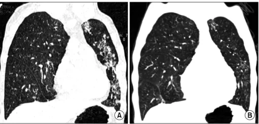
Figure 3. Coronal section of lung setting shows (A) patchy and centrilobular infiltration in line along anterior chest wall of left lung field, and (B) much cleared lung field after antituberculosis treatment for 9 months.
로 시행한 병리세포검사에서 악성세포는 보이지 않았다.
우측흉수에서 시행한 검사에서 ADA 14.50 U/min, pH 7.5, 중성구 88%, 림프구 11%, 단백 4.3 g/dL, 알부민 2.2 g/dL, LDH 621 IU/L, Glucose 107 mg/dL로 단순 부폐렴 삼출액(uncomplicated parapneumonic exudates)소견을 보였다. 객담배양검사에서 Klebsiella pneumoniae균이 동정되었다.
방사선학적 검사: 내원 시 시행한 단순 흉부 X-선 사진 (Figure 1)에서 좌상엽과 좌하엽 폐의 기관지주변에 침윤 소견을 보였다. 삼차원 흉부단층촬영의 시상면(Figure 2), 관상면(Figure 3)에서 폐실질 내에 전방흉벽면을 따라 종 단으로 분포하는 centrilobular nodule이 관찰되었다.
치료 및 경과: 기관지동맥 조영술에서 좌측폐의 하엽부 위에 혈관분포의 증가를 보였으나, 좌측늑간동맥조영에 서는 이상소견을 보이지 않았다. 우측폐의 상늑간동맥 부 위에 혈관분포의 증가소견을 보였다. 각각의 이상 부위에 대하여 색전술을 시행하여 지혈하였다. 기관지세척액으 로 시행한 검사에서 결핵에 합당한 결과를 보여 항결핵제 로 9개월간의 치료를 시행하였으며 약제내성이나 약제 부 작용은 보이지 않았다.
고 찰
결핵은
Mycobacterium tuberculosis
균에 의하여 공기 중으로 전염되는 질환으로 개발도상국 등에서 질환발생과 사망률의 주요 원인이 되고 있다4,5. 세계보건기구(World Health Organization, WHO) 2008년도 통계에 따르면 한국에서 결핵의 유병률은 연 10만 명당 123명으로 중등도 정도이다1.
활동성 결핵의 흉부단층촬영 소견에 따른 진단은 폐실 질의 이상소견 양상과 공동의 유무, 기관지파급, cen- trilobular nodule이나 tree-in-bud 양상 등에 기초한다.
Jeoung과 Lee5가 발표한 자료에 따르면 활동성 폐결핵을 가진 환자의 80%, 비활동성 폐결핵을 가진 환자의 89%에 서 고해상도 흉부단층촬영으로 정확한 감별이 가능하였다.
그러나, 영상학적으로 비전형적인 폐결핵의 양상을 보 이는 경우는 진단에 어려움을 초래할 수 있다. 면역저하 가 있거나 당뇨가 있는 활동성 폐결핵의 경우 결핵병변에 다발성의 공동이나 비분절성 분포와 같은 비전형적인 양 상을 보이는 것으로 알려져 있다5-7. 영상학적 소견으로 노 인의 결핵에서 비특이적 소견은 공동이 흔하지 않은 것으 로 알려져 있다. 이는 세포면역의 기능저하를 반영하는 것으로 보이며 따라서 조직의 파괴도 적다. 이러한 이유 때문에 노인에서 객혈이 적을 것으로 보인다3,8.
높은 산소분압을 선호하는 결핵균의 특성과, 림프선의 배액장애, 상엽첨부에 혈행순환의 감소 등의 이유로 상엽 의 상분절과 후분절, 하엽의 상분절에 결핵균의 증식이 호발하는 것으로 알려져 있다2,3,9,10.
기립자세에서는 폐첨부에 폐혈류가 크게 감소하는 것 으로 알려져 있으며11,12, West와 Dollery11는 동위원소검 사법으로 국소적인 혈류와 폐환기를 측정하여 제1늑간부 위 산소분압은 132 mm Hg였고, 5번째 늑간부위는 89 mm Hg로 폐의 부위에 따른 산소분압 감소를 확인하였 다. 동물실험에서 고농도에서 균의 감염이 증폭되고, 저
Tuberculosis and Respiratory Diseases Vol. 69. No. 1, Jul. 2010
55
산소에서 균감염이 억제되는 소견을 보였다. 박쥐는 거꾸로 매달려서 쉬기 때문에 폐기저부에 결핵이 발생하는 것 으로 알려져 있다13. 본 증례에서 경추손상으로 사지마비 가 되어 15년간 침상안정상태를 하며 후방흉부면과 비교 하여 전방흉부면에 접한 종단면 부위의 혈류가 크게 감소 하였을 것으로 보인다. 폐환기도 전방흉부면을 따라서 상 대적으로 증가하여 산소분압이 증가하고 결핵균이 증식 하기 좋은 환경으로 변화되었을 것으로 사료된다.
정상적인 환자의 경우 전방 흉곽은 움직임이 많고, 후 방흉곽은 움직임이 적으나 이 환자와 같이 경추손상으로 호흡근육이 움직일 수 없는 경우에 전방의 흉곽을 따라서 분포하는 림프액의 배액장애가 상대적으로 컸을 것으로 추정된다. 뇌경색으로 요양병원에서 1∼2년의 침상안정 으로 치료중, 호흡기증상으로 본원에 전원되어 결핵이 발 견될 경우에, 본 증례와 같은 영상학적 분포를 경험한 적 이 없다. 뇌경색 환자와 본 증례의 사지마비 환자에서 영 상학적 차이를 초래한 요인은 전방흉부의 움직임 유무에 따른 림프액 배액 장애 여부일 것으로 사료된다. 사지마 비나 뇌경색 등으로 침상안정 후에 이러한 폐결핵의 분포 변화를 위해 얼마간의 기간이 필요한 지에 대하여는 추가 적인 연구가 필요할 것으로 사료된다.
결핵이 초기에 발견되어 치료가 이루어지면, 감염력을 소실하여 결국에 완치에 도달하게 된다. 따라서 감염된 환 자를 정확하게 진단하는 것이 환자의 치료뿐만 아니라 지 역사회 대중감염을 예방하기 위한 중요한 수단이 된다5,6. 사지마비로 인한 침상안정환자에서 폐결핵 발생 시 분포 양상에 대하여 숙고한다면 폐결핵을 영상학적으로 초기 에 진단하고, 적절한 치료를 바로 시행할 수 있을 것으로 보인다.
참 고 문 헌
1. World Health Organization. TB prevalence and in- cidence: Republic of Korea [Internet]. Geneva: World Health Organization; c2010 [cited 2010 April 1]. Avail-
able from: http://www.who.int/countries/kor/en.
2. Meylan PR, Richman DD, Kornbluth RS. Reduced intra- cellular growth of mycobacteria in human macrophages cultivated at physiologic oxygen pressure. Am Rev Re- spir Dis 1992;145:947-53.
3. Perez-Guzman C, Torres-Cruz A, Villarreal-Velarde H, Vargas MH. Progressive age-related changes in pulmo- nary tuberculosis images and the effect of diabetes. Am J Respir Crit Care Med 2000;162:1738-40.
4. Cegielski JP, Chin DP, Espinal MA, Frieden TR, Rodri- quez Cruz R, Talbot EA, et al. The global tuberculosis situation. Progress and problems in the 20th century, prospects for the 21st century. Infect Dis Clin North Am 2002;16:1-58.
5. Jeong YJ, Lee KS. Pulmonary tuberculosis: up-to-date imaging and management. AJR Am J Roentgenol 2008;
191:834-44.
6. Lee KS, Im JG. CT in adults with tuberculosis of the chest: characteristic findings and role in management.
AJR Am J Roentgenol 1995;164:1361-7.
7. Chan CH, Woo J, Or KK, Chan RC, Cheung W. The effect of age on the presentation of patients with tuber- culosis. Tuber Lung Dis 1995;76:290-4.
8. Umeki S. Comparison of younger and elderly patients with pulmonary tuberculosis. Respiration 1989;55:75-83.
9. Houben EN, Nguyen L, Pieters J. Interaction of patho- genic mycobacteria with the host immune system. Curr Opin Microbiol 2006;9:76-85.
10. MacGregor RR. Tuberculosis: from history to current management. Semin Roentgenol 1993;28:101-8.
11. West JB, Dollery CT. Distribution of blood flow and ventilation-perfusion ratio in the lung, measured with radioactive carbon dioxide. J Appl Physiol 1960;15:
405-10.
12. Anthonisen NR, Milic-Emili J. Distribution of pulmonary perfusion in erect man. J Appl Physiol 1966;21:760-6.
13. Goodwin RA, Des Prez RM. Apical localization of pul- monary tuberculosis, chronic pulmonary histoplasmo- sis, and progressive massive fibrosis of the lung. Chest 1983;83:801-5.