ted transoral approach and retromandibular approach for surgical treatment of mandibular subcondyle fracture
7
Dept. of oral and maxillofacial surgery, Gachon university Gil medical center Woo-Yul Lee*, Jin-Yong Cho, Sung-Won Yang
Comparison of the clinical results between endoscopically assisted transoral
approach and retromandibular approach for surgical treatment of mandibular subcondyle fracture
Comparison of the clinical results between endoscopically assisted transoral approach and retromandibular approach for surgical treatment of mandibular subcondyle fracture
Dept. of oral and maxillofacial surgery, Gachon university Gil medical center Woo-Yul Lee*, Jin-Yong Cho, Sung-Won Yang
Purpose : Aim of this study is to describe and compare clinical results and complications epending on the surgical approaches for the mandibular subcondyle fracture
Materials and methods : The patients who had been diagnosed as the mandibular subcondyle fracture and underwent open reduction and internal fixation from May 2009 to December 2014 were included. They were divided into two groups depending on the surgical approaches; endoscopically assisted transoral approach and retromandibular approach. Association between the pre- operative fracture classification and post-operative results was reviewed depending on the surgical approaches.
Results : The number of patients selected in this study was 33. Eighteen patients (male 7, female 11) underwent open reduction and internal fixation via retromandibular approach and fifteen patients (male 12, female 3) underwent open reduction and internal fixation via endoscopically assisted transoral approach. The mean age, follow up period, and operation time were 44.29 15.19 years, 9.97 7.82 months, and 161 89.44 minutes. Post-operative results were all “good” state in the retromandibular approach group regardless of the fracture classification but two patients in the endoscopically assisted transoral approach group underwent re-operation due to “poor” results. The fracture types of two were classified as displacement and lateral override at the same time.
There was no statistically significant difference between two groups. Three patients in the retromandibular approach group had experienced facial nerve palsy (17%) temporarily. No one showed malocclusion in this study. There was no significant difference on the complications such as temporomandibular disorder, local infection, and condyle resorption depending on the surgical approaches.
Conclusion : In this study, there was no significant difference on the complications between the two groups but retromandibular approach has advantage over endoscopically assisted transoral approach in case of the severely displaced subcondyle fracture.
Key words : Mandibular fracture, Surgical approach, Complication ABSTRACT
Corresponding Author Jin-Yong Cho
Dept. of oral and maxillofacial surgery, Gachon university Gil medical center, [email protected]
Ⅰ. Introduction
Mandibular subcondyle fracture is a break from the mandibular notch to the posterior border of the mandible and constitutes 26-57% of all mandible fractures1). It can be treated with closed reduction(CR) or open reduction(OR). The reduction method would be determined by the various factors such as the conditions of the fracture, the surgeon's preference, the clinical symptoms, and the needs of the patient. It has been a debate about the reduction methods of mandibular subcondyle fracture because of potential of the complications and treatment results2, 3). Generally, when the displacement of the fractured bone is severe and the change in the vertical dimension is observed, open reduction is considered for the first choice of the treatment.
Approach methods for the open reduction are including transoral approach, submandibular approach, retromandibular approach and rhytidectomy approach. Retromandibular approach(RMA) is relatively easy and can provide the surgeons with better vision of the surgical field but can damage to the facial nerve and make a facial scar4). According to Manisali et al.5), it is about 30% chances to encounter the branches of the facial nerve during the retromandibular approach and the cadaveric study also reported it about 40% chances.
Recently, owing to the development of the endoscopic technique, endoscopically assisted transoral approach(EATA) has been also expanded to minimize the complications such as nerve damage and scarring. There is no risk of
facial nerve damage and scarring via EATA on the operation but it has a difficulty to manipulate and fix the fractured bone.
The purpose of this study is to compare the post-operative clinical results and complications and also to suggest the suitable approach depending on the conditions of the mandibular subcondyle fracture.
Ⅱ. Materials and methods
The patients who were diagnosed with mandibular subcondyle fracture and underwent open reduction and internal fixation(ORIF) from May 2009 to December 2014 were included in this study. The medical records, radiographs, and computed tomography(CT) scans were collected for analysis. The present study was performed under the principles of the Declaration of Helsinki and ethical approval by the University of Gachon institutional review board(IRB No.
GCIRB2016-301) was obtained before the beginning of the study.
Inclusion criteria were containing 1) mandibular unilateral subcondyle fracture, 2) no systemic disease affecting on the results, 3) more than 3 months follow up period after the surgery, and 4) post-operative radiograph and/or CT scans available. Exclusion criteria were containing 1) concomitant fractures affecting on the complications(e.g., bilateral subcondyle frac tures, condyle head fracture, panfacial fracture, and comminuted fracture), and 2) cases using submandibular or preauricular approach in
ted transoral approach and retromandibular approach for surgical treatment of mandibular subcondyle fracture combination.
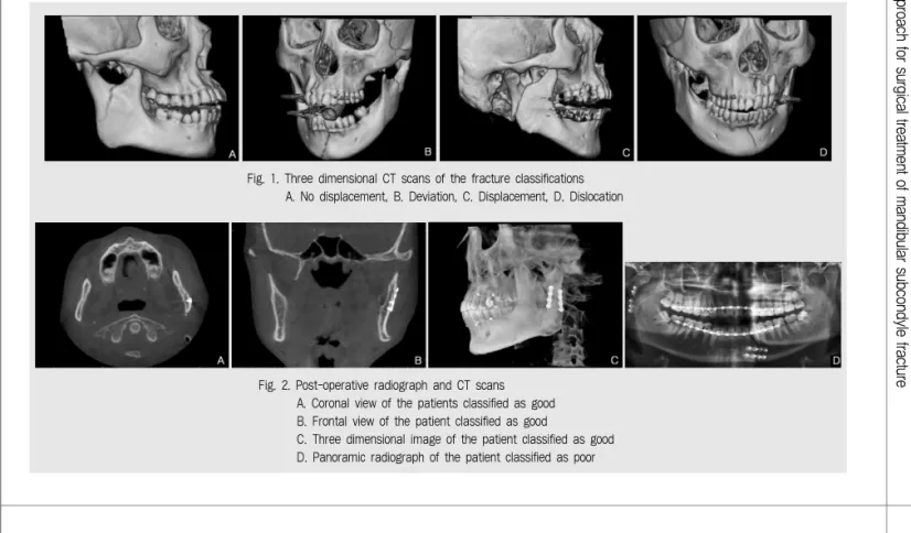
The fracture types were classified as no displacement, deviation, displacement, and dislocation according to the system of MacLennan6). If only fracture line was observed without displacement, it was classified as no displacement. Deviation was defined as the displaced proximal part keeping the contact with distal part of fractured mandible. When the displaced proximal part was separated from the distal part but was within the joint, it was classified as displacement. When the displaced proximal part was separated and out of the joint, it was defined as dislocation. Additionally, based on the location of the displaced proximal part, the fracture type was divided into no displacement, medial override, and lateral override(Fig. 1).
The result of reduction was evaluated with post-operative radiographs and CT scans after
surgery. Three-dimensional(3D) image was reconstructed using InVivodental software (Anatomage, San Jose, CA, USA) to improve accuracy of the evaluation. When the interfragmentary gap was less than 2 mm, it was classified as good, and when the gap was more than 2 mm, it was classified as poor(Fig. 2).
Pre and post-operative radiograph and CT data were examined by one surgeon. Five cases were randomly selected and examined at intervals to assess the intrarater reliability. The resulting intraclass correlation coefficient was 0.88, suggesting high intrarater reliability for the evaluation protocol.
Evaluation of the facial nerve damage and local infection
Facial nerve damage was clinically evaluated.
After the operation, the surgeon instructed the
Fig. 2. Post-operative radiograph and CT scans A. Coronal view of the patients classified as good B. Frontal view of the patient classified as good
C. Three dimensional image of the patient classified as good D. Panoramic radiograph of the patient classified as poor Fig. 1. Three dimensional CT scans of the fracture classifications
A. No displacement, B. Deviation, C. Displacement, D. Dislocation
patient to act the facial muscles for evaluation of motor reflex; raise the eyebrow and wrinkle the forehead(frontalis, orbicularis oculi) for the temporal branch, close the eyelid tightly
(orbicularis oculi) for the zygomatic branch, blow the cheek and smile(orbicularis oris, buccinators, and zygomaticus) for the buccal branch, depress the mouth corner(depressor angulioris and depressor labii inferioris) for the marginal mandibular branch, and taut the neck skin(platysma) for the cervical branch. When muscle function was improved, it was considered as recovery of the facial nerve.
If continuous swelling, pain, and fever were checked after the operation, white blood cell and C-reactive protein were examined for diagnosis of infection.
Surgical procedures
Retromandibular approach (RMA)
The surgery was undergone via transparotid method. A skin incision was performed on the posterior area of the mandibular ramus
approximately 2 cm in length. The parotid sheath was exposed and incision was made on it. After then, dissection of parotid gland was executed in parallel with the expected direction of the facial nerve. If the facial nerve was shown, it was carefully retracted in a superioinferior manner to secure the surgical field. The fractured site was exposed after making an incision at the pterygomandibular sling.
The fractured bone was placed in the proper position and maxillomandibular fixation(MMF) was implemented with wires after occlusion was guided and internal fixation was executed with plates and screws(Fig. 3). Finally, layer by layer suture was executed with a conventional method.
Endoscopically assisted transoral approach (EATA)
After making an incision at the vestibule from the first molar to 1cm above the occlusion plane, periosteal dissection was executed on the lateral side of the ramus to expose fractured site. With a 30 degree endoscopy(KarlStorz, Tuttlingen,
Fig. 3. Intra-operative clinical pictures of RMA
A. After the drawing for the anatomy, B. After fixing the fractured bone
ted transoral approach and retromandibular approach for surgical treatment of mandibular subcondyle fracture Germany), the fractured site was found and
explored to evaluate the fracture state.
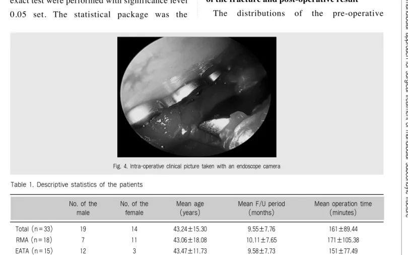
The mesial part was reduced to the anatomically normal position. After that, the MMF was implemented with wires after occlusion was guided. In order to perform drilling and screwing, a stab incision of about 5 mm was made on the buccal skin for trocar insertion and then, internal fixation was executed with plates and screws(Fig. 4). Finally, primary closure was executed with a conventional method.
Statistical analysis
For comparisons of clinical results depending on the variables, the chi-square test and Fisher’s exact test were performed with significance level 0.05 set. The statistical package was the
Statistical Package for Social Sciences(SPSS), version 20.0(IBM, Chicago, IL, USA).
Ⅲ. Results
Fifty four patients were selected at the first step and 33 patients(Male : 19, Female : 14) met the inclusion criteria. The patients were divided into two groups depending on the surgical approaches; RMA and EATA. The descriptive statistics such as the number of patients, sex, age, follow up periods, and operation times were described in Table 1.
The association between the classifications of the fracture and post-operative result
The distributions of the pre-operative
Fig. 4. Intra-operative clinical picture taken with an endoscope camera Table 1. Descriptive statistics of the patients
Total (n=33) 19 14 43.24±15.30 9.55±7.76 161±89.44
RMA (n=18) 7 11 43.06±18.08 10.11±7.65 171±105.38
EATA (n=15) 12 3 43.47±11.73 9.58±7.73 151±77.49
No. of the No. of the Mean age Mean F/U period Mean operation time
male female (years) (months) (minutes)
classification and post-operative result were described(Table 2). There was no statistically significant difference between RMA and EATA on the post-operative result depending on the classification of the fracture.
The association between the mediolateral displacement and post-operative result
The distributions of the pre-operative mediolateral displacement and post-operative result were describe (Table 3). There was
statistically difference between RMA and EATA on the post-operative result with lateral override displacement.
The number and type of plates used
A metal or absorbable plate was used to fix the fractured bone and the number of the plated used was varied. These variables depended on the conditions of the fracture, surgeon’s preference, and the needs of the patients(Table 4).
* By Fisher’s exact test
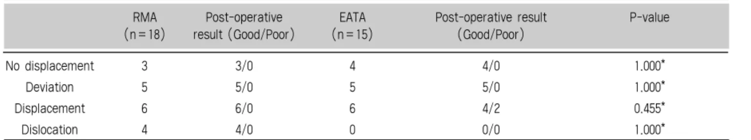
Table 2. Classifications of the fracture type and post-operative result
No displacement 3 3/0 4 4/0 1.000*
Deviation 5 5/0 5 5/0 1.000*
Displacement 6 6/0 6 4/2 0.455*
Dislocation 4 4/0 0 0/0 1.000*
RMA Post-operative EATA Post-operative result P-value
(n=18) result (Good/Poor) (n=15) (Good/Poor)
* By Fisher’s exact test
Table 3. Distributions of mediolateral displacement and post-operative result
No displacement 3 3/0 5 5/0 1.000*
Medial override 10 10/0 8 8/0 1.000*
Lateral override 5 5/0 2 0/2 0.048*
RMA Post-operative EATA Post-operative result P-value
(n=18) result (Good/Poor) (n=15) (Good/Poor)
Table 4. The type and number of the plate used
†plate fracture occurred in one case from each group
Type of the plate Metal Absorbable Metal Absorbable
17 1 13 2
RMA (n=18) EATA (n=15)
No. of the plates used One Two Three Four One Two Three Four
8 9† None 1 7 7† 1 None
RMA (n=18) EATA (n=15)
ted transoral approach and retromandibular approach for surgical treatment of mandibular subcondyle fracture The comparison of the complication rate
depending on the surgical approaches
Two patients in the EATA group had experienced malocclusion. They underwent reoperation to solve the problem. After the reoperation, occlusion of the patients became favorable.
The numbers of the patients showing temporomandibular disorder(TMD) in the RMA and EATA group were six and three, respectively.
The symptoms of TMD were clicking sound, pain, and opening limitation. All symptoms are solved with TMD treatment protocol within 3 months.
Other three patients had experienced facial nerve palsy after the operation. All patients were
taking steroid medication as treatment. As time had passed, the symptoms had been improved in all patients. Two patients showed symptoms of local infection so incision and drainage with antibiotic therapy was performed for elimination of the symptoms.
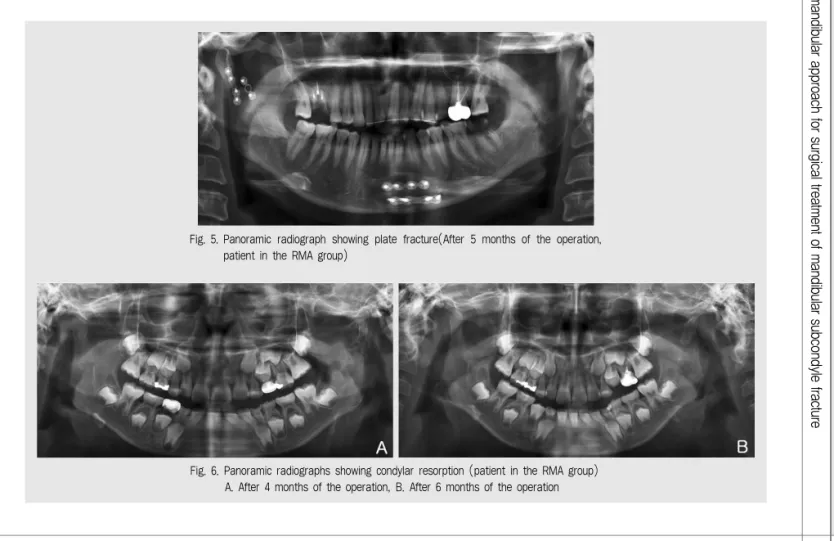
One case of plate fracture was observed from each group and in the 2 cases, one of two metal plates was fractured(Fig. 5) and one condylar resorption case was observed from the RMA group(Fig. 6). The patients did not undergo any treatment because specific signs and symptoms did not appear. There was no statistically significant difference on complications depending on the surgical approaches(Table 5).
Fig. 6. Panoramic radiographs showing condylar resorption (patient in the RMA group) A. After 4 months of the operation, B. After 6 months of the operation Fig. 5. Panoramic radiograph showing plate fracture(After 5 months of the operation,
patient in the RMA group)
Ⅳ. Discussion
The purposes of the treatment for mandibular subcondyle fracture are as follows; 1) maximum mouth opening is more than 40mm without pain, 2) the movement of the mandible is not limited in all directions, 3) occlusion is normally guided as the preoperative state, and 4) no facial asymmetry exists after the surgery1). To achieve these purposes, various surgical approaches had been suggested. In this study, it was researched which approach is more suitable for the mandibular subcondyle fracture to minimize the post-operative complications and to obtain the outstanding results.
When an extraoral approach is conducted for the mandibular subcondyle fracture, submandibular and retromandibular approach are most used7). Submandibular approach is relatively easy to perform and safe. However, scarring is more standing out and retraction damage to the facial nerve could arise more easily and vision of the surgical field is poorer8).
RMA provides the surgeon with clear vision of the surgical field so that more precise reduction
and shortening of the surgery time could be accomplished and post-operative results are generally favorable. In contrast, EATA is laborious to perform ORIF due to limited vision and lack of space of the surgical field and so it is more time-consuming and post-operative results could depend on the skill and experience of the surgeon. However, it is free from facial nerve damage and skin scarring. In this study, the operation time of EATA is less than that of RMA on the contrary to other studies but there is no no significant difference between two approaches. It is thought due to the difference of operator skill proficiency and difficulty of the operation. This is one of the limitations of the retrospective study.
Some authors reported that EATA was rather limited to vision of the surgical field compared with extraoral approaches and limited to precise reduction of the fractured bone9, 10). In this study, the post-operative results of all patients operated with RMA were good but 2 of 15(13.3%) patients operated with EATA had poor post-operative results. With post-operative radiograph, the fractured bone was not properly fixed in 2 Table 5. Complications after the surgery
Abbreviations: CI, confidence interval; TMD, temporomandibular disorder;
* By Fisher’s exact test, †Re-ORIF was executed
Malocclusion 1 2† 0.38 (0.03 - 4.68) 0.579*
TMD symptoms 6 3 2.00 (0.40 - 9.91) 0.458*
Facial nerve damage 3 0 Cannot be calculated 0.233*
Local infection 1 1 0.82 (0.05 - 14.39) 1.000*
Condyle resorption 1 0 Cannot be calculated 1.000*
Plate fracture 1 1 0.82 (0.05 - 14.39) 1.000*
RMA (n=18) EATA (n=15) Odd ratio (CI) P-value
ted transoral approach and retromandibular approach for surgical treatment of mandibular subcondyle fracture patients via EATA. It was because of limited
vision of the surgical field. Thus, two patients should undergo re-ORIF.
If the fractured condylar part is severely displaced and/or is deviated in medial override, it would be quite difficult to operate via EATA.
Undt et al11). recommended that if the condylar part is deviated in medial override with 14 degrees or more inclined and the vertical dimension is decreased more than 5%, an extraoral approach needs to be considered.
Schneider et al.12) and Bhagol et al.13) also recommended an extraoral approach in case of more than 10 degrees inclined and 2mm or more vertical dimension decrease.
When comparing of two approaches, one clear difference was the facial nerve damage. All patients(n=15) operated with EATA did not experience nerve disturbance at all because it was not likely to damage the nerve as long as paying attention of using a trocar. On the other hand, 3 of 18 patients(16.7%) operated with RMA experienced temporary weakness of the facial nerve. These results were consistent with the results reported in the previous studies14, 15).
Many articles described that if an extraoral approach is undergone under appropriate procedures, it is possible to minimize the nerve damage and perform a faster operation16). According to Ellis et al.8), the rate of facial nerve weakness was varied from 0 to 41%. Most cases were temporary and resolved within 6 weeks.
Choi et al.17) reported that the rate of neurologic complication was 20%(5 of 25 patients) via preauricular approach and all of the cases
involved the buccal or zygomatic nerve branch and all resolved within 3 months. Another author18)reported that there was one case(3%) of temporary facial nerve weakness out of the 35 patients and it resolved within 2 weeks. Raveh et al.19)suggested that facial nerve damage is caused chiefly by excessive traction of the retractors or by electrocauterization of the vessels adjacent to the facial nerve.
We assumed that when subcondyle fracture occurs, adjacent muscles, ligaments, TMJ disk, and retrodiscal tissue are somewhat changed.
Therefore, although ORIF for the fracture is appropriately performed with any approach, TMD could occur after the operation. In this study, 6(33.3%) patients in the RMA group and 3 patients(20.0%) in the EATA group presented TMD signs and symptoms after the operation.
Incidence rate of TMD was no statistically significant difference between two groups as assumed. All patients were treated with TMD treatment protocol(medication and physical therapy).
The limitation of this study is that 1) post- operative CT scan for 8 patients did not exist so the post-operative reduction states were evaluated only by radiographs for them, 2) MMF period comparison was not presented because the MMF period of some patients had not been recorded, 3) mouth opening length also was not presented since many cases were recorded as
“within normal range” instead of the exact length.
1. Peter WB, Barry LE, Rainer S. Maxillofacial trauma and Esthetic facial reconstruction. 2nd ed.
Philadelphia: Churchill Living stone 2011.
2. Brandt MT, Haug RH. Open versus closed reduction of adult mandibular condyle fractures: a review of the literature regarding the evolution of current thoughts on management. J Oral Maxillofac Surg 2003;61:1324-1332.
3. Villarreal PM, Monje F, Junquera LM, Mateo J, Morillo AJ, Gonzalez C. Mandibular condyle fractures: determinants of treatment and outcome. J Oral Maxillofac Surg 2004;62:155-163.
4. Delvin MF, Hislop WS, Carton ATM: Open reduction and internal fixation of fractured mandibular condyles by a retromandibular approach:surgical morbidity and informed consent. Br J Oral Maxillofac Surg 2002;40:23-25.
5. Manisali M, Amin M, Aghabeigi B, Newman L.
Retromandibular approach to the mandibular condyle: a clinical and cadaveric study. Int J Oral Maxillofac Surg 2003;32(3):253-256.
6. MacLennan WD. Consideration of 180 cases of typical fractures of the mandibular condylar process.
Br J Plast Surg 1952;5:122-128.
7. Jensen T, Jensen J, Norholt SE, Dahl M, Lenk-Hansen L, Svensson P. Open reduction and rigid internal fixation of mandibular condylar fractures by an intraoral approach: a long-term follow-up study of 15 patients. J Oral Maxillofac Surg 2006;64:1771-1779.
8. Ellis E, McFadden D, Simon P, Throckmorton G.
Surgical complications with open treatment of mandibular condylar process fractures. J Oral Maxillofac Surg 2000;58:950-958.
9. Park JM, Jang YW, Kim SG, Park YW, Rotaru H, Baciut G, et al. Comparative study of the prognosis of an extracorporeal reduction and a closed treatment in mandibular condyle head and/or neck fractures. J Oral Maxillofac Surg 2010;68:2986-2993.
10. Schon R, Gutwald R, Schramm A, Gellrich NC, Schmelzeisen R. Endoscopy-assisted open treatment of condylar fractures of the mandible:
extraoral vs intraoral approach. Int J Oral Maxillofac Surg 2002;31:237-243.
11. Undt G, Kermer C, Rasse M, Sinko K, Ewers R.
Transoral miniplate osteosynthesis of condylar neck fractures. Oral Surg Oral Med Oral Pathol Oral Radiol Endod 1999;88:534-543.
12. Schneider M, Erasmus F, Gerlach KL, Kuhlisch E, Loukota RA, Rasse M, et al. Open reduction and internal fixation versus closed treatment and mandibulomaxillary fixation of fractures of the mandibular condylar process: a randomized, prospective, multicenter study with special evaluation of fracture level. J Oral Maxillofac Surg 2008;66:2537-2544.
13. Bhagol A, Singh V, Kumar I, Verma A. Prospective evaluation of a new classification system for the management of mandibular subcondylar fractures. J Oral Maxillofac Surg 2011;69:1159-1165.
14. Schon R, Gutwald R, Schramm A, Gellrich NC, Schmelzeisen R. Endoscopy-assisted open treatment of condylar fractures of the mandible:
extraoral vs intraoral approach. Int J Oral Maxillofac Surg 2002;31:237-243.
15. Schmelzeisen R, Cienfuegos-Monroy R, Schon R, Chen CT, Cunningham L, Jr., Goldhahn S. Patient benefit from endoscopically assisted fixation of condylar neck fractures-a randomized controlled trial. J Oral Maxillofac Surg 2009;67:147-158.
16. Paeng JY, Ok YJ, Myoung H, et al. Endoscopic- assisted open reduction and internal fixation (EAORIF) for condylar fracture. J Korean Assoc Oral Maxillofac Surg 2006;32:474-481.
17. Choi BH, Yoo JK. Open reduction of condylar neck fractures with exposure of the facial nerve. Oral Surg Oral Med Oral Pathol Oral Radiol Endod 1999;88:292-296.
18. Narayanan V, Kannan R, Sreekumar K.
Retromandibular approach for reduction and fixation of mandibular condylar fractures: A clinical experience.
Int J Oral Maxillofac Surg 2009;38:835-839.
19. Raveh J, Vuillemin T, Ladrach K. Open reduction of the dislocated, fractured condylar process:
indications and surgical procedures. J Oral Maxillofac Surg 1989;47:120-126.
참 고 문 헌