YUJM 2013;30(2):136-40
제 2형 당뇨병에서 발생한 막증식성 사구체신염 1예
김동현, 이장원, 정민석, 이승현, 민병철, 김현주 왈레스 기념 침례병원 내과
A Case of Membranoproliferative Glomerulonephritis in a Patient with Type 2 Diabetes Mellitus
Dong Hyun Kim, Jang Won Lee, Min Suk Jung, Seung Hyun Lee, Byung Cheol Min, Hyun Ju Kim
Department of Internal Medicine, Wallace Memorial Baptist Hospital, Busan, Korea
Diabetic nephropathy (DN) is a common complication and the leading cause of end-stage renal disease (ESRD) in diabetic patients. The occurrence of non-diabetic renal disease (NDRD) in diabetic patients has been increasingly recognized in recent years. Generally, renal injuries in DN are deemed difficult to reverse, whereas some NDRDs are often treatable and even remittable. Thus, the diagnosis of NDRD in patients with diabetes mellitus (DM) via a kidney biopsy would be significant for its prognosis and therapeutic stra- tegy. According to recent studies, the most common NDRD is IgA nephropathy in type 2 diabetic patients, and some cases of minimal change disease and membranous glomerulonephritis have been reported in Korea. However, membranoproliferative glomerulonephritis (MPGN) is an uncommon condition in diabetic patients. To our knowledge, there has been no case yet of MPGN, except in a child with type 1 DM. We present an unusual case of a 27-year-old woman who had type 2 DM with MPGN, as confirmed via a kidney biopsy.
Key Words: Diabetes mellitus, Membranoproliferative glomerulonephritis, Proteinuria
Received: May 28, 2013, Revised: July 4, 2013, Accepted: July 11, 2013
Corresponding Author: Hyun Ju Kim, Department of Inter- nal Medicine, Wallace Memorial Baptist Hospital, 200, Geumdan-ro, Geumjeong-gu, Busan 609-728, Korea Tel: 82-51-580-1259, Fax: 82-51-583-7114
E-mail: [email protected]
서 론
2011년 우리나라 신대체 요법의 현황에 따르면 당뇨에 의한 말기신부전의 비율이 45.4%로 가장 주된 원인인 동시 에 점차 증가되는 추세로 보고되고 있다.1 당뇨병 환자에서 의 단백뇨는 대부분 당뇨병성 신병증을 시사하는 것으로 받 아들여진다. 그러나 당뇨병 환자에서 신기능의 저하와 단백 뇨 발생의 경우 당뇨병성 망막증이나 신병증을 동반하지 않 은 경우에는 비당뇨병성 신병증을 고려해 보는 것이 필요하 다. 해외와 국내의 후향적 조사에 따르면 환자군의 각 53%와 42%에서 비당뇨병성 신병증이 보고되었다.2,3
이러한 비당뇨병성 신병증을 진단하는 것은 치료 가능한 질병을 가진 환자에게 적절한 치료 방안을 제공하고 말기 신부전으로 진행을 예방하는데 도움이 될 수 있으므로, 치료 와 예후에 영향을 미치는 중요한 인자이다.
국내에서 당뇨병 환자에서 발생한 미세변화신병증, 막성 사구체신염의 사례는 보고된 바 있으나, 막증식성 사구체신 염의 증례는 제 1형 당뇨병 소아에서 발생한 사례 이외 제 2형 당뇨병 환자에서는 따로 보고된 바 없다. 이에 저자들은 제 2형 당뇨병 환자에서 검진상 우연히 발견된 단백뇨로 인 해 시행한 신생검에서 당뇨병성 신병증이 아닌 막증식성 사 구체신염이 확인된 증례를 경험하였기에 보고하는 바이다.
증 례
환 자: 여자, 27세, 159 cm, 64 kg 주 소: 검사 상 우연히 발견된 단백뇨
현병력: 3년 전부터 제 2형 당뇨병을 진단받고, 경구 혈당
Fig. 1. (A) Light microscopy shows that the glomeruli are markedly increased size and severely hypercellular mainly involving mesan- gial cells. Mesangial matrix is also increased. (B) Glomerulus with mesangial interposition into the capillary wall produces a double-contour or tram-track appearance of basement membranes (arrow) (PAS stain, ×400).
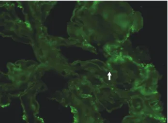
Fig. 2. Immunofluorescent staining shows diffuse linear and partly granular peripheral straining of IgG is bound to the glomerular basement membrane (arrow).
강하제(metformin) 복용과 더불어 지속형 인슐린(Insuline glar- gine) 아침 식전 16단위로 조절하고 있으며, 2개월 전부터 고지질혈증으로 Atorvastatin 20 mg을 복용 중이었다. 외래 에서 시행한 단회뇨 단백/크레아티닌 비(spot urine protein/
creatinine ratio) 2.26으로 단백뇨를 보여 신생검을 하기 위해 입원하였다.
수술력: 2010년 외상으로 인한 우측 3, 4번째 중수골 골절 로 내고정술
가족력: 외조모 당뇨.
사회력: 음주와 흡연은 없었음.
진찰 소견: 혈압 130/90 mm Hg, 맥박 70회/분, 호흡 20회/
분, 체온 36.4℃였다. 별다른 병색을 보이지 않았으며, 의식은 명료하였다. 안과에서 시행한 안저 소견상 당뇨병성 변화를 나타내는 소견은 없었다. 경부의 특이 소견은 관찰되지 않았 으며, 호흡음은 정상이었고, 심잡음은 들리지 않았다. 복부 는 편평하고 압통과 반발통은 없었다. 양측 하지에 함요 부종 은 관찰되지 않았다.
검사실 소견: 시행한 일반 혈액학 검사에서 혈색소 11.3 g/dL, 헤마토크릿 32.9%, 백혈구 7,100/mm3(중성구 66.6%, 림프구 27.3%), 혈소판 214,000/mm3였으며, 혈청 생화학 검 사에서는 혈중요소질소(BUN) 17.6 mg/dL, 크레아티닌(Cr) 0.6 mg/dL였고, 총 단백질과 알부민은 각각 6.1 g/dL, 3.8 g/dL 였다. 요 검사상 단백뇨 4+, 고배율 현미경 시야에서 백혈구 0-2개, 적혈구 30-50개였다. 입원 전 시행한 단회뇨 단백/크 레아티닌 비 2.26, 입원 후 시행한 24시간 소변 검사상 단백질 1,829 mg이었다. 혈청 C3 108 mg/dL (참고치 90-180), C4 38.1 mg/dL (참고치 10-40)로 정상범위였다. IgG/IgA/IgM는 각각 838/101/238 (mg/L)였다. 류마티스 인자(RF) 197 IU/
mL로 양성 소견 보였으나, 항 CCP 항체(Anti-CCP Ab)는 7 U/mL로 음성이었다. 항핵항체(ANA), 항호중구 항체(c-ANCA, p-ANCA), 항 dsDNA 항체는 음성이었다. 혈청 단백 전기영 동검사에서는 알부민의 경한 감소와 소변 단백 전기영동 검 사에서는 알부민뇨를 보였다. 혈청 한냉글로불린(cryoglo- bulin)은 음성이었다. 당화혈색소(HbA1c)는 6.1%였고, 공복 시 혈당 103 mg/dL, 혈청 C-peptide 1.14 ng/mL였다. HBsAg, HBsAb, anti-HCV, HIV Ab는 모두 음성이었다. Total choles- terol/triglyceride/HDL cholesterol/LDL cholesterol은 각각 146/73/43/86 (mg/dL)였다.
방사선 검사 소견: 복부 초음파 검사 상 복부 내 장기의 병변은 관찰되지 않았으며, 양측 신장의 크기는 비교적 정상 소견을 보였다.
조직 병리 소견: 광학 현미경 검사에서 모두 38개의 사구 체가 관찰되었으며, 이 중 2개(약 5%)의 사구체에서 경화 (global sclerosis)가 관찰되었으며, 나머지 사구체는 크기가 현저하게 증가되어 있고, 메산지움 증식 소견을 보였다(Fig. 1A).
모세혈관벽이 두 겹으로 보이는 특징적인 소견이 자주 관찰 되어 막증식성 사구체신염의 소견에 합당하였다(Fig. 1B).
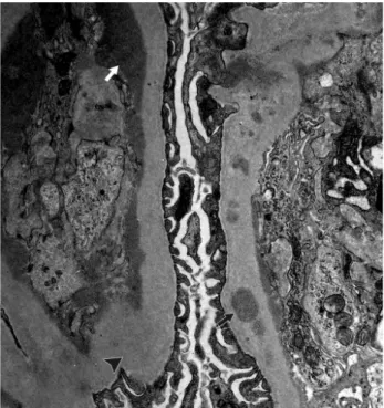
면역 형광 현미경 검사에서 사구체기저막을 따라 IgG, IgM, C3, C1q와 kappa, lamda chain 주변의 불규칙한 미만성 면역 형광 반응이 관찰되었다(Fig. 2). 전자 현미경 검사에서는 주로 내피하 전자고밀도 물질의 침착이 관찰되어 이상의 병리 소견 에 의해 제 1형 막증식성 사구체신염으로 진단하였다(Fig. 3).
치료 및 경과: 입원 후 매일 losartan potassium 50 mg을 복용하면서 경과 관찰 중이다. 약 3개월 후 시행한 단회뇨 단백/크레아티닌 비는 2.44로 아직 단백뇨의 감소는 보이지 않고 있다.
Fig. 3. Electron microscopy shows massive subendothelial depo- sits (white arrow) and subepithelial and intramembranous deposits (black arrow). The glomerular basement membrane is moderately thickened with irregular contours (arrowhead). Epithelial foot processes show focal effacement (electron microscopy, ×10,000).
고 찰
당뇨병은 말기 신부전의 가장 흔한 원인으로 당뇨병 환자 에게서의 신기능의 저하와 단백뇨를 보일 경우 일반적으로 당뇨병성 신병증을 고려하게 된다.1 그러나 본 증례와 같이 비교적 짧은 유병 기간이나 동반된 당뇨 망막병증이 없을 경 우 비당뇨병성 신병증의 발생을 생각하여야 하며, 이러한 경우 신조직 검사를 이용하여 정확한 진단을 하고, 이에 따라 효과적인 면역억제제 사용 등을 통하여 환자의 증상과 예후 를 개선할 수 있으므로 신병증의 원인에 대한 감별이 중요하 다 할 수 있겠다.
일반적으로 당뇨병 환자에서 신생검에 대해서 표준화된 적응증이 없으나, (1) 당뇨 발생 5년 이내의 거대알부민뇨의 발생, (2) 당뇨병성 망막병증 없는 거대알부민뇨의 발생, (3) 심한 현미경적 혈뇨, (4) 신기능의 급격한 악화, (5) 단백뇨의 급속한 악화, (6) 당뇨병성 사구체경화증 이외 다른 신질환 이 의심되는 경우를 들 수 있다.4
막증식성 사구체신염은 혈관사이질 모세관 토리콩팥염 (mesangiocapillary glomerulonephritis)으로 불리기도 하는 질환으로 조직학적으로 확진된 사구체신염의 약 7-10%를
차지한다고 한다. 전 연령대에 걸쳐서 발생하나 주로 소아기 때 흔하며, 무증상 혈뇨에서 부터 신증후군과 급속진행성 사 구체신염(rapidly progressive glomerulonephritis)까지 다양 한 임상 양상을 보인다. 광학 현미경 상으로는 혈관사이질의 세포과다, 모세혈관내 증식, 모세혈관벽 재형성 등이 관찰되 며, 전자 현미경 상으로 분류하여 면역 조직학적 복합체가 내피 하에 침착(subendothelial deposits) 할 경우를 제1형, 내 피층과 상피층(subepithelial deposits)에 침착할 경우를 제3 형, 그리고 사구체 기저막 내에 고밀도 물질의 침착을 보일 때(dense deposit disease) 제2형으로 나누게 된다.
병인에 따라서는 원인을 모르는 특발성(1차성)과 만성감염, 자가면역질환, 이상단백혈증(dysproteinemia) 등이 원인이 되는 2차성으로 나눌 수 있다.5,6 대부분의 경우 2차성이며, 특발성의 경우 국내 후향적 분석에서 약 10%를 차지한다고 보고된 바 있다.7
이에 따라 막증식성 사구체 신염의 진단 과정에서 2차성 질환의 원인이 될만한 질환을 감별하고 배제하는 것이 필요 하다. 감염에 의한 원인의 대표적인 예로 만성 B형과 C형 간염, HIV 감염, 심내막염, 내장농양 및 Mycoplasma 감염 등이 있고, 자가면역성 질환으로는 전신홍반 루푸스, 피부경 화증, 쇼그렌 증후군, 기타 백혈병이나 림프종 및 이상단백혈 증 등이 원인 질환이 될 수 있겠다.5,8 우리나라에서는 B형 간 염 바이러스와 루푸스 신염에 의한 2차성 막증식성 사구체 신염이 많았으며, 외국에서는 C형 간염 바이러스에 의한 경 우가 흔한 것으로 알려져 있다.7,9
진단 과정에서 이러한 질환을 찾기 위해서 면밀한 병력청 취 및 이학적 검사와 더불어 HCV, HBV, HIV에 대한 혈청학 적 검사, 각종 보체(C3, C4, CH50)와 ANA, ANCA, RF 등 자가면역항체 검사와 혈청 및 소변 단백전기영동 검사 등이 필요하다.8
특발성 막증식성 사구체신염의 특징 중 하나로 저보체 혈증이 있는데, 혈청 C3의 감소가 전체의 77% 가량에서 관찰된다. 이는 순환 면역 복합체와 자가항체인 신염인자 (nephritic factor, NeF)에 의한 이화 작용(catabolism)의 증가 와 분해산물들에 의한 음성되먹임(negative feedback)에 따른 합성의 감소에 기인하는 것으로 알려져 있다. 제1형 막증식 성 사구체신염의 경우 면역복합체 형성에 따른 보체계의 고 전경로(classical pathway) 활성화에 따라 대게 정상 또는 저 하된 혈청 C3와 감소된 C4 농도를 나타내는 반면, 제2형 막증식성 사구체신염은 보체계의 대체경로(alternative path- way)가 활성화되어 혈청 C3의 감소와 정상농도의 C4를 보
이는 경우가 많다. 제 3형 막증식성 사구체신염은 대체경로 와 말단경로(terminal pathway)의 활성화가 함께 일어난다고 한다. 제 2형 막증식성 사구체신염에서는 C3NeF가 약 80%
에서 양성을 보여 감별에 도움이 될 수 있으나, 아직 제한적 으로 쓰이고 있다.10-12
특발성 막증식성 사구체신염의 예후는 진단 당시의 신증 후군, 크레아티닌의 상승, 고혈압의 유무, 조직 검사상 반월체 (crescents), 세뇨관간질 손상(tubulointerstitial injuries)의 동반 유무가 관여한다고 되어 있다. 보체의 감소 등은 예후 에 큰 영향을 미치지 못한다고 보고되어 있다. 평균 9세의 소아에서 시행한 후향적 분석에서는 비신증후군 범위의 단 백뇨와 정상 혈압을 보일 경우 좋은 예후를 보였다는 보고 도 있다.13-15
막증식성 사구체 질환의 치료는 2차성일 경우에는 치료 가능한 원인 질환의 치료를 우선시 한다. 예를 들어 만성 B형과 C형 간염의 경우에는 항바이러스 치료, 세균성 심내 막염의 경우 항생제 치료, 백혈병과 림프종의 경우 항암제 치료를 하게 되며, 이에 따라 기존질환의 호전과 더불어 신기 능의 회복과 단백뇨의 감소를 기대할 수 있다고 한다.16-18 특발성의 경우 환자군이 많지 않고, 대부분의 연구가 후향 적으로 진행되어 치료법이 아직 확립되어 있지 않으나, 부신 피질호르몬의 격일 치료와 디피리다몰(dipyridamole), 와파린 및 사이클로포스파마이드(cyclophosphamide)의 병합치료 등 이 질병의 진행을 지연시킬 수 있다고 보고되었다.19,20
최근의 권고에 따르면 신기능이 정상이고, 일일 단백뇨 3 g 이하의 경우에는 3개월마다 24시간 소변 단백질 검사 또는 단회뇨 단백/크레아티닌 비와 함께 크레아티닌 청소율을 측 정하여 추적 검사하고, 안지오텐신 전환효소 억제제(ACEI) 또는 안지오텐신 수용체 차단제(ARB)를 처방하여 고혈압의 조절과 단백뇨의 감소를 유도하도록 권고하고 있으며, 일일 단백뇨 3 g 이상의 경우에는 소아의 경우에는 격일제 부신피 질호르몬 치료를 시행하고, 성인의 경우에는 아스피린과 디 피리다몰의 병합요법을 추천한다. 만일 치료에 반응이 없을 경우나 신기능의 급격한 악화가 보일 경우 부신피질호르몬 과 사이클로포스파마이드 또는 mycophenolate mofetil 병합 요법 같은 면역억제 치료를 고려할 수 있다.8,21
본 증례는 제 2형 당뇨의 짦은 유병기간을 가진 환자가 외래 검사상 당뇨병성 망막병증 등이 없이 우연히 발견된 단백뇨로 인하여 시행한 신생검에서 막증식성 사구체신염을 진단한 1예의 보고이다. 제 2형 당뇨병 환자에게서 단백뇨로 인하여 시행한 검사 상 막증식성 사구체신염을 증례로 보고
한 예는 조사한 바로는 국내에서는 없었으며, 후향적 조사로 시행한 당뇨병 환자에서의 막증식성 사구체신염 발생은 해 외에서 51명의 환자 중 1예, 45명의 환자 중 1예가 보고 되었 으며, 국내에서는 74명의 환자 중 1예를 보고된 바 있었다.
이는 상대적으로 IgA 신병증이나 막성사구체신염, 미세변화 콩팥병증 등에 비해서 드문 발생을 보였다.3,22,23
성인에서의 막증식성 사구체신염은 1차성 보다 만성감염 과 자가면역 질환 등 2차성의 원인이 흔하다고 알려져 있어, 증례의 환자의 경우 진단 당시 다른 원인 질환 감별을 위한 검사를 함께 시행하였으나, 특이 소견 보이지 않아 특발성 막증식성 사구체신염으로 판단하였다.7 이에 환자는 진단 당시 정상 신기능을 보이고 신증후군 범위의 단백뇨(소변 단백질
>3.5 g/일)는 발생하지 않아 안지오텐신 수용체 차단제인 losartan potassium을 처방하여 외래에서 경과 관찰 중이다.6 보고마다 다르지만 제 1형 막증식성 사구체신염의 경우 진단 5년 후 약 49%의 환자에서 사망 또는 말기 신질환으로 발전 가능성 등 불량한 예후를 보일 수 있으므로 환자의 경우 3개월 마다 신기능 및 단백뇨에 대한 추적 검사를 시행할 예정이다.24 본 증례는 비교적 짧은 유병기간을 가진 제 2형 당뇨병 환자에서 당뇨병성 망막병증 없이 정기 검사상 발견된 단백 뇨로 인하여 시행한 신장 조직 검사 상 제 1형 막증식성 사구체신염을 진단하였고, 이에 안지오텐신 수용체 차단제 를 처방하여 외래에서 경과 관찰 중인 예로 추후 정기적인 추적 관찰 및 치료 방안 수립이 필요할 것으로 사료된다.
참고문헌