RESEARCH ARTICLE
Received: July 15, 2016, Revised: August 23, 2016, Accepted: August 29, 2016 ISSN 1598-4478 (Print) / ISSN 2233-7679 (Online)
†
Correspondence to: Youn-Soo Shim
Department of Dental Hygiene, Sun Moon University, 70 Sunmoon-ro 221beon-gil, Tangjeong-myeon, Asan 31460, Korea Tel: +82-41-530-2740, Fax: +82-41-530-2726, E-mail: [email protected]
Copyright © 2016 by Journal of Dental Hygiene Science
Comparison between Qraypen TM Imaging and the Conventional Methods of Visual Inspection and Periapical Radiography for Proximal Caries Detection in Primary Molars: An In Vivo Study
So-Youn An, So-Young Park 1 , and Youn-Soo Shim 2†
Department of Pediatric Dentistry, Wonkwang University Daejeon Dental Hospital, Daejeon 35233, Korea
1 Department of Dental Hygiene, Vision College of Jeonju, Jeonju 55069, Korea
2 Department of Dental Hygiene, Sun Moon University, Asan 31460, Korea
유구치 인접면 우식 병소 진단에 있어 Qraypen
TM
과 시진 및 구내 치근단 방사선의 비교안소연ㆍ박소영
1
ㆍ심연수2†
원광대학교 대전치과병원 소아치과,
1전주비전대학교 치위생과,
2선문대학교 치위생학과
The purpose of this study was to evaluate the efficacy of the newly-developed Qraypen
TM(All In One Bio, Korea) system for the diagnosis of early proximal caries by comparing it with the conventional methods of visual inspection and periapical radiography. This study was carried out from July 2015 to April 2016 targeting 32 children aged 7∼12 years who visited Y-Dental Clinic for school oral health examinations. Two investigators selected and examined a total of 153 primary molars that had not undergone restorative treatment. Comparisons were carried out between visual inspections, readings of posterior periapical radiography images, and readings of Qraypen
TMimages. This study revealed that the percentage of interproximal surfaces of primary molar teeth without caries incidence was 83.7% using Qraypen
TMimaging and 84.9% using visual inspection and periapical radiography. The differences between the two methods were not statistically significant. Thus, Qraypen
TMis expected to be a useful and convenient auxiliary diagnostic device that can facilitate the detection of hidden proximal caries in primary molars.
Key Words: Dental caries, Early diagnosis, Primary molar, Proximal caries lesion, Qraypen
TMIntroduction
Recently, the potential of growing child’s dental caries is the most common chronic disease that occurs in the elementary school time
1)
. The 1981 World Health Organi- zation was set to 'Health for all' spirit of the world take over an average 12-year-old caries experience in permanentteeth index (decayed, missing, and filled teeth index, DMFT index) the oral health goal of making less than 3 almata declared in oral health sector
2)
.As a result, although the 1980 survey, with 51% achi- eving these goals in 107 countries, in the 2004 survey was 74% among the 139 countries to achieve the target
3)
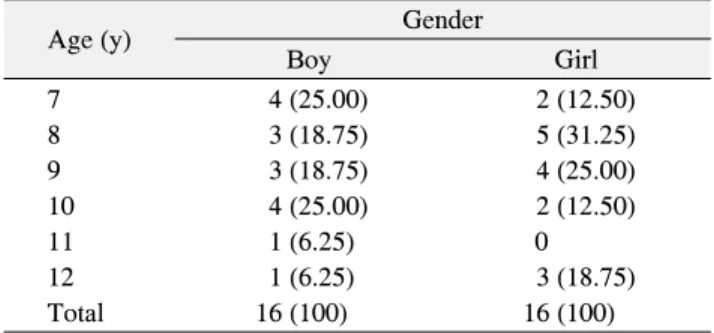
. According to the National Oral Health Status ReportTable 1. Age Distribution of Study Subjects
Age (y) Gender
Boy Girl
7 4 (25.00) 2 (12.50)
8 3 (18.75) 5 (31.25)
9 3 (18.75) 4 (25.00)
10 4 (25.00) 2 (12.50)
11 1 (6.25) 0
12 1 (6.25) 3 (18.75)
Total 16 (100) 16 (100)
Values are presented as n (%).
conducted in 2015, the DMFT index in Korean children at the age of 12 was 1.9
4)
. Dental caries is a multifactorial disease and so far, there have been many theories reported regarding its pathogenesis and currently it is known to be caused by a combinatorial effect of the host, bacteria, diet and time factors5)
.The progression of proximal caries in the primary molar is very fast. It is because the accumulation of plaque is easy due to the large contact area on the adjacent surface of the primary molar, it is less mineralized than the permanent teeth and the thickness of enamel and dentin layers is thin
6)
. It is widely recognized that progression of an incipient carious lesion can be arrested and remine- ralized. This knowledge has resulted in the development of several diagnostic techniques for early detection and quantification of carious lesions. However, different from smooth surface caries and occlusal caries of which lesions are exposed, proximal caries is difficult to be detected by visual inspection and with a probe, which makes the early detection difficult and lesions are usually found only after being quite advanced7)
. If the marginal ridge of the primary molar has not been destroyed yet or the color of the tooth structure has not been changed due to the progression of proximal caries, it is very difficult even for skilled pediatric dentists to determine the progression of caries accurately only by visual inspection alone8)
. Visual inspection shows a relatively high specificity, but a low sensitivity in the detection of proximal caries9,10)
.American Academy of Pediatric Dentistry has reco- mmended to perform bitewing radiography every 6∼12 months in case of pediatric patients with high caries activity, even though proximal caries is not confirmed by visual inspection
11)
. However, in case of children who have small mouths and are hard to control their behavior, there are limitations in taking a radiograph on a regular basis appropriate for the detection of proximal caries.While the need for development of convenient and reliable devices which can help the detection of proximal caries is emerging, Qraypen
TM
(All In One Bio, Seoul, Korea) was developed domestically in 2015.The purpose of this study is to evaluated the efficacy of the newly-developed Qraypen
TM
system for the diagnosis of early proximal caries by comparing it with the conven-tional methods of visual inspection and periapical radiography, to identify potential problems in its clinical application to present an improvement plan for Qray- pen
TM
in the future and to provide baseline research data for methods for the prevention of dental caries and remi- neralization of early dental caries.Materials and Methods
1. Study subjects and equipment
1) Study subjects
The study was reviewed and approved by the Ethics Committee of the College of Dentistry of Wonkwang University, Daejeon, Korea (IRB no. W1505/002-001).
This study was carried out from July 2015 to April 2016 targeting 32 children aged 7∼12 years who visited Y-Dental Clinic for school oral health examinations. The gender distribution was 16 boys and 16 girls, they were matched each same grade, and the age distribution was 7 to 12 years old (mean age, 8.5 years old) (Table 1).
2) Equipment for study (1) Oral examination
Mirrors, probes, tweezers, and compressed air sprayer (3 way syringe) were used.
(2) Radiography
In order to facilitate occlusal photography, tap (Omnitap;
Matricom, Tokyo, Japan) was fixed perpendicular to the film for photographing using conventional radiography devices used for oral radiography (Vatech, Hwaseong, Korea) and pediatric oral film (Kodak, Rochester, NY, USA).
Table 2. ICDAS II Criteria about Visual Inspection and Qraypen
TMImage on Interproximal Surfaces
Code ICDAS II criteria about visual inspection ICDAS II criteria about Qraypen
TMinspection
0 Sound Sound tooth surface
1 First visual change in enamel (seen only after prolonged air drying or restricted to the confines of a pit or fissure)
Slight fluorescence change
2 Distinct visual change in enamel Distinct fluorescence change 3 Localized enamel breakdown (without clinical visual signs
of dentinal involvement)
Visible enamel breakdown with a distinct fluorescnec change
4 Underlying dark shadow from dentin Poorly delineated distinct fluorescence change with or without enamel breakdown
5 Distinct cavity with visible dentin Cavitation visible with distinct fluorescence change (5 and 6) 6 Extensive distinct cavity with visible dentin Collapsed with 5
ICDAS: International Caries Detection and Assessment System.
(3) Digital oral imaging equipment
Qraypen
TM
System, digital oral imaging equipment was used.2. Study methods
1) Methods for examination: oral examination
The subjects were seated in a chair for dental treatment in Y-Dental Clinic located in Seongdong-gu, Seoul and oral examinations were conducted twice using mirrors, probes, tweezers and compressed air sprayer (3 way syringe).After removing saliva, etc. from the tooth surface using compressed air, tooth examination was performed by visual inspection. All teeth were subject to the exami- nations and the two examiners performed independent oral examinations on the same child. Each tooth was divided into occlusal, buccal, lingual, mesial and distal surfaces and the evaluation criteria which were the examination (International Caries Detection and Assessment System, ICDAS II) and the examination table of 2010 National Survey. In this study, two dentists performed the exami- nations independently, and all the examiners for clinical studies went through sufficient preliminary discussion and trainings for the criteria for the readings of visual inspec- tion, periapical radiographs and Qraypen
TM
. Thirty photos taken with QraypenTM
were prepared by a third party and a preliminary training was performed for the two examiners who were to conduct the clinical research. The intraclass correlation coefficient value was 0.92 before training and once the intraclass correlation coefficient value of 0.98 was obtained, the clinical study was started (Table 2).2) Methods for examination: radiography
Periapical radiography was conducted by a skilled dentist in Y-Dental Clinic. The conditions for radiography are as follows: the horizontal and the vertical angles were adjusted to make the center line of the radiation pass through the adjacent surface perpendicularly and the radiation dose and time for the radiography were 70 kv, 8 mA and 0.24 seconds, respectively. The first and second primary molars and the first permanent molar were to be included. The photographed films were placed on the fluorescent screen for a radiographic reading in the dark room and the same film was read by two examiners independently.
3) Methods for examination: photographing of the Qraypen TM image
Two examiners created the criteria of readings on the first and second primary molars, and after they conducted independent readings accordingly on the same image, the results were recorded in the chart (Fig. 1).
3. Statistical methods
All statistics were performed using PASW Stastistics ver. 18.0 (IBM Co., Armonk, NY, USA). Statistical signi- ficance was determined at p<0.05.
Results
This study revealed that the percentage of interproximal surfaces of primary molar teeth without caries incidence was 83.7% using Qraypen
TM
imaging and 84.9% using visual inspection and periapical radiography. So, the per-Fig. 1. Radiography and photographing of the Qraypen
TMimage.
Table 3. Distribution of Non-Cavitated and Cavitated Caries on Interproximal Surfaces
ICDAS Visual
inspection
Periapical radiography
Qraypen inspection Code 0 130 (84.96) 130 (84.96) 128 (83.67)
Code 1 0 0 3 (1.96)
Code 2 2 (1.31) 0 5 (3.27)
Code 3 3 (1.96) 4 (2.61) 1 (0.65)
Code 4 7 (4.58) 7 (4.58) 6 (3.92)
Code 5 9 (5.88) 10 (6.54) 9 (5.88)
Code 6 2 (1.31) 2 (1.31) 1 (0.65)
Total 153 (100) 153 (100) 153 (100)
Values are presented as n (%)
centage with caries incidence of interproximal surfaces primary molar teeth were very small. And their com- parization were no statically coincidence.
The distribution of non-cavitated and cavitated caries on interproximal surfaces of selected and examined a total of 153 primary molars is as follows (Table 3).
Discussion
Early dental caries is a progressive disease in which decalcification and demineralization occur simultaneously and it is difficult to be identified by visual inspection, thus requires a special equipment for its diagnosis
12)
. Of the existing oral examination methods, the method using a probe rather destroys the intact surface layer of the enamel surface, leading to a side-effect of promoting cavity for- mation, and even if its presence or absence is recognized,the degree of its progression and its state cannot be deter- mined accurately, and radiography also causes a concern for radiation exposure
13,14)
. Since the early 1990s, several methods for the detection of occlusal caries have been introduced; some were only research tools, whereas others have been used in dental practice. These techniques include, among others, confocal microscopy15)
, fiber-optic tran- sillumination (FOTI)16)
, digital fiber optic transillumi- nation (DIFOTI)17)
, light-induced fluorescence (quanti- tative light-induced fluorescence, QLF), laser fluorescence (DIAGNOdent)18)
. Studies comparing emerging technologies with conventional methods have shown mixed results19)
.Qraypen
TM
was developed at All in one bio in the last year. It was approved by the Korea Federal Drug Admini- stration and was developed based on the principle of QLF.If blue visible light with 405 nm-wave length is irradiated, red fluorescence can be detected in the carious area where porphyrin, a bacterial metabolite, is present and the area where the old plaque or tartar is present
20)
. This fluore- scence maybe observed through a yellow highpass filter that excludes the tooth-scattered light. When enamel demineralization takes place, minerals will be replaced mainly by water, which results in a reduction of light res- orption by the enamel, and the intensity of the fluores- cence will decrease consequently. The demineralized region will appear darker than the surrounding sound tooth structure. Studies on QLF have also shown great potential to detect and measure early mineral loss21)
, whereas limited information on this is available for other methods.It has been suggested that some types of detection aids may augment visual examination. Considering that no studies have investigated how data provided by multiple diagnostic methods would influence the perception of caries status and subsequent treatment-planning decisions.
Oh et al.
22)
suggested that Q-ray view can be a promising device for conducting and educating the dental hygiene process better.Interproximal caries lesions develop between the con- tacting proximal surfaces of two adjacent teeth. They first appear clinically as opaque regions and are caused by the loss of enamel translucency at the outer most enamel between the contact point and the top of the free gingival margin
23)
. Owing to the large size of the proximal surfaces of posterior teeth and the subtle mineral loss initially presented by lesions on these surfaces, proximal caries on posterior teeth are usually difficult to identify on radio- graphs24)
. The early and accurate diagnosis of a proximal caries lesion enables immediate operative therapy, thereby preventing extensive tooth loss25)
.Using QLF, early demineralization lesions can be eval- uated quantitatively in the occlusal and smooth surfa- ces
18)
. However, there used to be limitations in detecting proximal caries in the enamel layer using the early QLF device. The conventional oral camera-type QLF has been evolved multiple times and in 2011, a digital camera-type QLF-D equipped with a special light filter and a digital image sensor was developed. However, there are a few precautions to take when you use QraypenTM
in the clinical practice. When QraypenTM
is actually used in the clinical practice, red fluorescence can also be observed at the non-carious area such as tartar or plaque build-up area, therefore, in order to obtain an accurate result, it is better to perform the examination after removing the tartar or plaque through flossing and oral prophylaxis. In addition, when using QraypenTM
, because the dental light of strong intensity may affect the observation result, the fluore- scence should be observed by irradiating the light of QraypenTM
to the teeth after turning off the dental light26)
. Thus, QraypenTM
is expected to be a useful and con- venient auxiliary diagnostic device that can facilitate the detection of hidden proximal caries in primary molars.However, this study was limited in that the number of
subjects was restricted to students from one elementary school in Seoul, and that there was no long term follow-up data.
Summary
The results of this study showed that Qraypen
TM
was effective for the detection of proximal caries in the primary molar progressed up to the dentin layer without destruction of the marginal ridge. However, the distinct red fluorescence was visible only when the lesions were so mature enough for proximal caries to be progressed to the dentin layers.In order to verify the relationship between the degree of progression of carious lesions and the red fluorescence, additional studies accompanied by histological examina- tions of extracted teeth are needed in the future.
요 약
2015년 실시한 국민구강실태조사에 따르면 우리나라 만 12세 아동들의 우식경험영구치지수(DMFT index)는 1.9로 주요 OECD 국가들의 평균인 1.6에 거의 근접한 것으로 조 사되었다. 본 연구의 목적은 인접면 우식증의 진단에 있어 새로 개발된 Qraypen
TM
의 효능을 기존의 방법인 시진 및 구 내 치근단 방사선 사진과 비교 평가하고, 임상 적용 시의 문제점을 파악하여 차후 Qraypen
TM
에 필요한 개선안을 제시함과 아울러 치아우식증의 예방 및 초기 우식증 재광화 방 법에 대한 기초 연구자료를 마련하고자 하였다. 학교 구강 검진을 목적으로 내원한 학령기의 혼합치열을 가진 32명의 어린이들을 대상으로 구강검진 2회, 구치부 치근단 방사선 필름 판독 2회 그리고 구치부 인접면 Qraypen
TM
이미지 판독 2회를 실시하고 비교한 결과 Qraypen
TM
영상은 변연융선이 파괴되지 않은 유구치의 인접면 우식증의 탐지에 효과 적이었다. 또한 방사선 촬영 결과와 비교해 보니 차이가 없 음을 확인하였다. 그러나 인접면 우식증이 상아질까지 진행 되어 병소가 성숙하여야 뚜렷한 붉은색 형광을 관찰할 수 있었기에 우식 병소의 진행 정도와 붉은색 형광 발생의 관 련성을 확인하기 위해서는 향후 발치된 치아를 사용하여 조 직학적 검사를 병행한 추가적인 연구가 필요할 것으로 생각 된다. 본 연구 결과 Qraypen