정향에서 분리한 질염 유발 병원균 억제 물질 Eugenol의 특성
박상희1, 고희선1, 윤재우2, 김현수1*
Characterization of Eugenol an Inhibitory Material to Vaginitis-inducing Pathogen, Isolated from Syzygium aromaticum (L.) Merrill et Perry
Sang-Hee Park1, Hee-Sun Ko1, Jaewoo Yoon2, and Hyun-Soo Kim1*
Received: 22 November 2019 / Revised: 16 December 2019 / Accepted: 18 December 2019
© 2019 The Korean Society for Biotechnology and Bioengineering
Abstract: Vaginitis is a disease related to women's immunity and is largely divided into bacterial vaginosis (BV) and vul- vovaginal candidiasis (VVC). In this study, we investigate Korean medicinal plants that have antimicrobial activity against vaginitis-inducing strains without inhibiting useful strains of bacteria. The antimicrobial activity results of the Syzygium aromaticum (L.) Merrill et Perry (SA) methyl alco- hol extract were confirmed for C. albicans ATCC 28367, C.
gleum KCTC 2904, and S. paucimobilis KCTC 2834 but not L. coleohominis KCTC 21007. Eugenol, a substance that shows antimicrobial activity, was separated from SA by refer- ence to prior research. To study the characteristics of isolated eugenol from SA (IESA), we measured the antioxidant activ- ity of IESA and stimulated vaginal epithelial cells (VK2/
E6E7 ATCC CRL-2616) with LPS and C. albicans to con- firm the effect of inhibiting stimulation. IESA showed higher antioxidant activity than commercial eugenol; the results of immunoblotting showed that IESA inhibited p38 activation at 60~90 minutes. According to the results of ELISA, the amount of TNF-α was 48.32% lower than that of C. albicans ATCC 28367 when treating together with IESA. Therefore, IESA could be developed as a therapeutic agent for BV and VVC.
Keywords: eugenol, Syzygium aromaticum (L.) Merrill et Perry, VK2/E6E7 ATCC CRL-2616, bacterial vaginosis, vul- vovaginal candidiasis
1. INTRODUCTION
질염은 여성이 감기만큼 자주 걸리는 질환으로 면역력이 약 해지면 나타나는 질병 중 하나이다. 또한 재발률이 높은 질 병이기에 여성은 항상 질염의 위험에 노출되어있다. 질염은 크게 세균성 질증과 칸디다성 질염, 트리코모나스 질염으로 나뉜다 [1]. 세균성 질증치료제로는 metronidazole, tinidazole 과 clindamycin이 사용되며, 칸디다성 질염에는 miconazole 과 terconazole이 사용되는데 이는 항생제의 일종으로 질병 을 치료하지만 항생제 내성균을 야기함과 동시에 질 내 유용 균에도 영향을 미친다 [2]. 이에 부작용이 적은 천연물에 대 한 관심이 증가하여 한약재 추출물에 대한 항균활성 연구가 많이 보고되고 있다. 한약재 추출물은 인체 내에서 식균작용 을 활성화하고, 항체의 생성을 촉진시키는 등 면역력을 증가 시켜 질병을 예방하고 치료한다 [3].
본 연구의 재료인 정향 (Syzygium aromaticum (L.) Merrill et Perry)은 도금양과 (Myrtaceae)에 속하는 식물로 약용부위 는 꽃봉오리 (clove)이다. Clove에는 18% 이상의 정유가 함 유되어 있으며, 31가지의 성분이 존재하는데 그 중 eugenol 이 가장 큰 비율을 차지한다. Eugenol이라는 이름은 정향의 옛 학명인 Eugenia caryophyllata에서 유래되었다. Eugenol은 연한 노란색을 띄며 향료로 사용되고, 방부제 및 마취제로 사용되며 항산화, 항염증 활성에 효과가 있다 [4-6].
본 실험에는 세균성 질증을 유발하는 세균이 질 상피 세포
1계명대학교 자연과학대학 기초과학부 생명과학전공
1Major in Biological Sciences, Faculty of Basic Sciences, College of Natural Science, Keimyung University, Daegu 42601, Korea
Tel: +82-53-580-5284, Fax: +82-53-580-5284 e-mail: [email protected]
2계명대학교 약학대학
2College of Pharmacy, Keimyung University, Daegu 42601, Korea
Research Paper
에 부착되어 biofilm을 형성하고, 칸디다성 질염을 유발하는 Candida albicans가 질 상피 세포에서 biofilm을 형성하여 숙 주에 감염된다는 연구결과를 토대로 질 상피 세포주 (VK2/
E6E7 ATCC CRL-2616) 를 사용하였다 [7-10].
질염유발균주로 알려진 Chryseobacterium gleum, Sphingomonas paucimobilis, Proteus mirabilis는 그람음성균이다. 그람음성 균의 특징 중 하나인 lipopolysaccharide (LPS)를 세포에 처리 하면 LPS에 의한 자극에 반응하여 티로신에 의한 급성인산 화 반응이 일어나 p38이 발현된다. p38은 38 kDa의 단백질 중 p38 MAPK (Mitogen-activated protein kinase) family로 분 류된 단백질이다 [11,12]. p38 MAPK 경로는 세포의 신호전 달을 조절하는 세 가지 주요 MAPK 경로 중 하나이다. 세포 외자극에 의해 p38 MAPK가 phosphorylation되어 활성화되 는데 이를 pp38이라한다 [13]. p38 MAPK의 활성화는 전사, 단백질 합성, 세포막 수용체 발현, 세포주기 조절단백질 발 현 및 세포의 사멸을 조절한다 [12]. 또한, p38 MAPK는 류마 티스 관절염, 크론병, 알츠하이머 등 중요한 질병에 관련되 어 있고 cytokine 신호전달, 염증반응, 산화스트레스 반응 등 의 생리적 작용에 중요한 역할을 한다. 따라서 p38 MAPK 저 해제는 질병 치료의 목적으로 사용될 가능성 높다 [14-19].
칸디다성 질염을 유발하는 균주인 C. albicans는 인체 내 공생균이자 기회감염균이다. 건강한 사람에게는 염증반응 을 일으키지 않지만 면역력이 약해진 환자에게 주로 감염양 상이 나타난다 [20, 21]. 칸디다성 질염으로 유발된 반응은
TNF-α (Tumor necrosis factor-α)에 의한 자극과 유사하다는 연구결과가 있으며 세포 내 cytokine 연구가 활발히 이루어 져 있다 [22, 23].
따라서 본 연구에서는 정향을 비롯한 한약재로부터 질염 유발균주에 대한 항균활성 유무를 확인하여 우수한 효과의 추출물을 선발하였고, 그람음성균의 자극유발원인 LPS에 의해 자극을 받은 질 상피 세포주에 처리하여 p38 MAPK의 억제확인과 칸디다성 질염의 원인균인 C. albicans로부터 질 상피 세포주로의 자극억제효과를 확인하고자 하였다.
2. MATERIALS AND METHOD
2.1. 실험재료 2.1.1. 사용한 한약재
본 연구에 사용된 한약재는 Table 1에서 보는 바와 같이 감초 외 22개를 사용하였다. 감초, 감잎, 구기자, 금은화, 뽕잎, 치 자, 칡, 칡순과 헛개나무는 국내산이며, 그라비올라잎과 정 향은 인도네시아산으로 ‘자연초’에서 구매하였다. 국내산 꽈리, 신이화, 약쑥과 베트남산 연자육은 대구 약령시장에서 구매하였다. 국내산 구아바잎은 천연약초, 중국산 연교는 ‘한 첩’, 국내산 오배자는 ‘산청한방약초’, 중국산 황련은 ‘심홍 물산’에서 구매하여 사용하였다. 구매한 한약재는 실온에서 보관하여 사용하였다.
Table 1. List of Korean medicinal plants used in antimicrobial activity
Korean Scientific name Effective part
감초 Glycyrrhiza uralensis Fisch. Root
구기자 Lycium chinense Mill. Fruit
금은화 Lonicera japonica Thunb. Flower buds
꽈리 Physalis alkekengi var. franchetii. Calyx
신이화 Magnolia kobus DC. Flower buds
약쑥 Artemisia princeps Pamp. Leaves
연교 Forsythia viridissima Lindl. Fruit
연자육 Nelumbo nucifera Gaertn. Seeds
오배자 Rhus chinensis Mill. Insect gal
정향 Syzygium aromaticum (L.) Merr. et Perr. Flower buds
치자 Gardenia jasminoides Ellis. Fruit
칡 Pueraria thunbergiana (Sieb. et Zucc.) Benth. Root
칡순 Pueraria thunbergiana (Sieb. et Zucc.) Benth. Sprout
헛개나무 Hovenia dulcis Thumb. Trunk
황련 Coptis chinensis F. Root
감잎 Diospyros kaki Thumb. Leaves
구아바잎 Psidium guajava L. Leaves
그라비올라잎 Annona muricata L. Leaves
뽕잎 Morus alba L. Leaves
삼백초 Saururus chinensis Baill. Leaves
은행잎 Ginkgo biloba L. Leaves
측백나무잎 Platycladus orientalis L. Leaves
황칠나무잎 Dendropanax morbifer H.Lev. Leaves
2.1.2. 균주 및 사용배지
실험에 사용된 균주는 KCTC (Korean Collection for Type Cultures)에서 Lactobacillus coleohominis KCTC 21007, Chryseobacterium gleum KCTC 2904, Sphingomonas paucimobilis KCTC 2834와 Proteus mirabilis KCTC 2510을 분양받았으며, ATCC (American Type Culture Collection)에서 Candida albicans ATCC 28367을 분양받아 사용하였다. L. coleohominis KCTC 21007은 Lactobacilli MRS broth, C. gleum KCTC 2904와 P.
mirabilis KCTC 2510 은 nutrient broth (Difco Co., USA), S.
paucimobilis KCTC 2834는 R2A broth (Difco Co., USA), C.
albicans ATCC 28367은 YM broth (Difco Co., USA)에 배양 하였다. L. coleohominis KCTC 21007과 P. mirabilis KCTC 2510은 35
oC, 나머지 3균주는 28
oC에서 배양하여 실험에 사 용하였다.
2.1.3. 세포 및 사용배지
VK2/E6E7 ATCC CRL-2616 은 여성의 질 상피 세포주로 ATCC에서 분양받아 사용하였다. Keratinocyte serum free medium (KSFM, Gibco BRL, USA)에 recombinant epidermal growth factor (Gibco BRL, USA) 5 μg/L, bovine pituitary extract (Gibco BRL, USA) 50 mg/L, 1% antibiotics (Gibco BRL, USA) 와 0.4 mM calcium chloride (Sigma aldrich Co., USA)를 첨가하여 생육배지로 사용하였고, 37
oC, 5% CO
2의 조건에서 배양하였다.
2.2. 한약재 추출
한약재는 ethyl alcohol과 methyl alcohol로 추출하였다. 추출 용매 100 mL당 한약재 10 g을 첨가하여 rotary shaking incubator (Gaon Science, Korea)에서 25
oC, 150 rpm의 조건으 로 24시간 추출 후, rotary vacuum evaporator (EYELA, Japan) 로 농축하였다. 농축물은 추출 용매와 동일한 용매에 10 mg/
mL 의 농도로 용해 후 4
oC 에 보관하여 실험에 사용하였다.
2.3. 항균활성 측정
질염유발균주 및 질 내 유용균에 대한 한약재 추출물의 항균 활성을 검토하기위해 혼합평판법으로 배지를 제조하였다.
Paper disc (Ø = 6 mm, Toyo Roshi Kaisha, Ltd., Japan) 에 한약 재 추출물을 20 μL 첨가하여 혼합평판법으로 제조된 배지에 얹어 배양하였다. 균주에 대한 한약재 추출물의 항균력 유무 및 측정은 inhibitory zone의 유무 및 크기를 통해 확인하였다.
2.4. 정향에서 eugenol 추출
정향에서 eugenol의 추출은 Jang의 방법 [24]을 참고하였다.
정향을 methyl alcohol에 추출하여 rotary vacuum evaporator 로 농축하였다. Hot water로 suspension 후 n-hexane으로 분획 추출하였고, 질염유발균주에 항균활성을 나타낸 n-hexane 8 번 분획을 silica gel column chromatography로 hexane: acetone = 9 : 1의 비율로 조정한 전개용매를 이용하여 추출하였다. 추 출물의 HPLC peak 확인을 위하여 전개용매는 methyl alcohol
: H
2O = 6 : 4의 비율로 조정하고, flow rate를 0.8 mL/min로 설정하였다. 추출물은 10 mg/mL 농도로 조정 후 10 μL injection하여 HPLC를 수행하였다. n-hexane 8번 분획은 구 입한 commercial eugenol (Sigma aldrich Co., USA)의 HPLC pattern과 비교하였다.
2.5. 항산화 활성 측정
2.5.1. DPPH free radical 소거능
안정한 radical인 DPPH free radical의 소거능을 측정해 정향 에서 분리한 eugenol (isolated eugenol from Syzygium aromaticum (L.) Merrill et Perry, IESA)의 항산화활성을 검토 하였다. DPPH free radical 소거능 실험은 Blois의 방법 [25]
을 참고하였다. IESA는 DPPH (Sigma aldrich Co., USA)와 1 : 1의 비율로 혼합하여 실온에서 30분간 반응시켰다. UV- visible spectrophotometer (Mecasys Co., Ltd., Korea)로 517 nm에서 흡광도를 측정하였으며, 양성대조구는 butylated hydroxyanisole (BHA, Sigma aldrich, Co., USA), 음성대조구 는 methyl alcohol로 측정하였다. DPPH free radical 소거능은 다음의 식으로 계산하였다.
DPPH free redical 소거능 (%) =
Con: methyl alcohol + DPPH reagent의 흡광도 S
1: IESA + DPPH reagent의 흡광도
S
2: IESA + methyl alcohol의 흡광도
2.5.2. ABTS free radical 소거능
ABTS free radical 소거능 실험은 양이온 ABTS
+가 IESA에 의해 환원되어 소거되는 정도를 측정하기 위해 수행하였다.
ABTS free radical 소거능 실험은 Re의 방법 [26]을 사용하였 다. IESA 10 μL와 ABTS radical cation reagent 1 mL를 1분간 실온에서 반응하여 UV-visible spectrophotometer로 734 nm 에서 흡광도를 측정하였다. 양성대조구는 BHA, 음성대조구 는 PBS로 측정하였다. ABTS free radical 소거능은 다음의 식으로 계산하였다.
ABTS free radical 소거능 (%) =
Con: methyl alcohol + ABTS radical cation reagent의 흡광도 S
1: IESA + ABTS radical cation reagent의 흡광도
S
2: IESA + PBS의 흡광도
2.6. 세포실험 2.6.1. 세포배양
VK2/E6E7 ATCC CRL-2616은 KSFM을 이용하여 1×10
5cells/mL로 조정 후 cell culture dish (Ø = 100 mm, Corning Inc., USA) 에 10 mL seeding하였다. VK2/E6E7 ATCC CRL- 2616은 37
oC, 5% CO
2의 조건에서 배양하였으며, 2일마다 계 대배양하여 실험에 사용하였다.
Con–(S1–S2) ---Con ×100
Con–(S1–S2) ---Con ×100
2.6.2. 세포독성 (MTT assay)
VK2/E6E7 ATCC CRL-2616에 대한 IESA의 독성 유무 및 농도에 따른 세포생존률을 확인하기 위해 MTT assay를 수 행하였다. 96 well microplate (Corning Inc., USA)에 세포를 seeding하여 24시간 배양한 후, IESA를 24시간 처리하였다.
MTT (Sigma aldrich Co., USA) solution을 첨가 후 4시간 반 응시켰고, 반응액을 제거한 후 DMSO (Sigma aldrich Co., USA)와 ethyl alcohol을 1 : 1 비율로 혼합하여 well 당 100 μL 씩 분주 후, iMark™ microplate absorbance reader (Bio-Rad Laboratories, Inc., USA)로 490 nm에서 흡광도를 측정하였다.
2.7 Western blotting 2.7.1. LPS 처리
질염유발균주 4종 중 C. gleum KCTC 2904, S. paucimobilis KCTC 2834와 P. mirabilis KCTC 2510은 Gram (-) 균으로, Gram (-) 균이 가지는 LPS를 자극원으로써 VK2/E6E7 ATCC CRL-2616에 처리하였다. LPS 처리농도와 조건은 Lee의 방 법 [27]을 참고하였다. Cell culture dish (Ø = 100 mm, Corning Inc., USA)에 1×10
5cells/mL의 세포를 seeding하여 24시간 배양한 후, IESA를 세포에 3시간 처리하였다. IESA 처리 후 10 μg/mL 의 LPS (Sigma aldrich Co., USA)를 30분, 60분, 90 분, 120분 처리하였다.
2.7.2. Immunoblotting
LPS를 처리한 세포는 lysis하여 단백질을 회수하였다. SDS PAGE 를 수행하기 위해 회수한 단백질은 PowerPac™ HC Power Supply (Bio-Rad Laboratories, Inc., USA)와 electro- phoresis unit (Hoefer, Inc., USA)으로 전개하였다. 전개 후 Trans-Blot
®Turbo™ blotting system (Bio-Rad Laboratories, Inc., USA)에서 nitrocellulose membrane (GE Healthcare Life science, USA) 에 transfer하였다. Transfer된 membrane은 blocking후 1차 항체 (human anti-p38 MAPK, human anti- phospho-p38 MAPK; Cell signaling technology Inc., USA)를 membrane에 처리하여 4
oC에서 rocker shaker (CR 100;
FINEPCR
®, Korea)로 overnight shaking하였다. 1차 항체를 제거 후, 2차 항체 (rabbit anti-p38 MAPK, rabbit anti- phospho-p38 MAPK; Cell signaling technology Inc., USA)를 membrane에 처리하여 2시간동안 실온에서 shaking하였다. 2 차 항체를 제거 후, TBST로 washing하였으며 단백질 발현확인 을 위해 clarity™ western ECL substrate (Bio-Rad Laboratories, Inc., USA) 를 처리하여 ImageQuant LAS 4000 (GE Heal- thcare Bio-Science Corp., USA)으로 측정하였다.
2.8. ELISA
2.8.1. Candida albicans ATCC 28367 처리
C. albicans 는 칸디다성 질염을 일으키는 원인균으로 VK2/
E6E7 ATCC CRL-2616에 자극을 주기 위해 처리하였다. Cell culture dish에 세포를 1×10
5cells/mL로 seeding하여 24시간
배양하였다. 세포가 배양된 dish에 C. albicans ATCC 28367 (1×10
5CFU/mL)과 IESA (500 nM)를 첨가하여 6시간, 12시 간 배양하였다. 배양액은 회수하여 원심분리 후 상등액을 사 용하였다.
2.8.2. Human TNF-α immunoassay
Human TNF-α immunoassay는 C. albicans ATCC 28367에 의 해 자극된 VK2/E6E7 ATCC CRL-2616에 IESA를 처리하였 을 때 자극에 대한 반응을 확인하기 위해 수행하였다.
Human TNF-α quantikine ELISA kit (R&D systems
®, USA)를 사용하여 TNF-α를 정량하였다.
2.9. 통계분석
모든 실험은 3회 반복하였으며, 본 실험의 모든 데이터 결과 는 EXCEL 2016 (Microsoft, USA)으로 통계처리 하였다.
Error bar는 표준편차 값을 표시하였으며 p<0.05인 경우 통계 적으로 유의하다 판정하였다.
3. RESULTS AND DISCUSSION
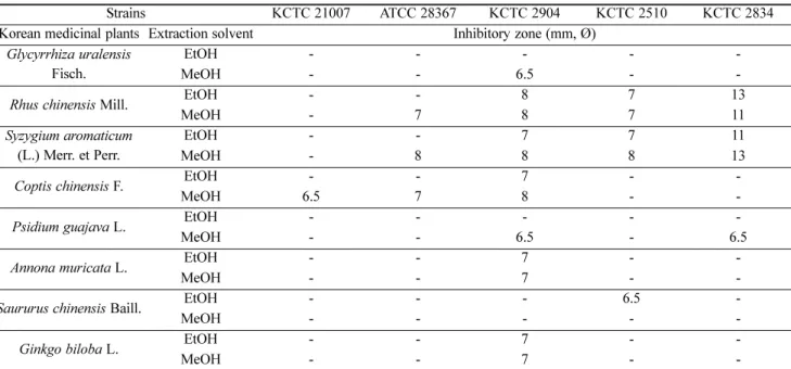
3.1. 한약재 추출물의 항균활성
질 내 유용균인 L. coleohominis KCTC 21007은 억제하지 않 으면서 질염유발균주인 C. albicans ATCC 28367, C. gleum KCTC 2904, P. mirabilis KCTC 2510과 S. paucimobilis KCTC 2834는 억제하는 한약재를 탐색하기 위하여 항균활성을 수 행하였고, 그 결과는 Fig. 1에 나타내었으며 inhibitory zone의 크기를 측정하여 Table 2에 나타내었다. 감초 methyl alcohol 추출물의 항균활성 결과 C. gleum KCTC 2904에서 6.5 mm 의 inhibitory zone이 확인되었다. 오배자 ethyl alcohol 추출물 의 항균활성은 C. gleum KCTC 2904에서 8 mm, P. mirabilis KCTC 2510 에서 7 mm, S. paucimobilis KCTC 2834에서 13 mm 의 inhibitory zone이 확인되었으며, 오배자 methyl alcohol 추 출물의 항균활성은 C. albicans ATCC 28367에서 7 mm, C.
gleum KCTC 2904에서 8 mm, P. mirabilis KCTC 2510에서
7 mm, S. paucimobilis KCTC 2834에서 11 mm의 inhibitory
zone 이 확인되었다. 정향 ehtyl alcohol 추출물의 항균활성 결
과 C. gleum KCTC 2904에서 7 mm, P. mirabilis KCTC 2510
에서 7 mm, S. paucimobilis KCTC 2834에서 11 mm의 inhi-
bitory zone이 확인되었으며, 정향 methyl alcohol 추출물의
항균활성은 C. albicans ATCC 28367에서 8 mm, C. gleum
KCTC 2904 에서 8 mm, P. mirabilis KCTC 2510에서 8 mm,
S. paucimobilis KCTC 2834에서 13mm의 inhibitory zone이
확인되었다. 황련 ethyl alcohol 추출물의 항균활성은 C. gleum
KCTC 2904에서 7 mm의 inhibitory zone이 확인되었으며, 황
련 methyl alcohol 추출물의 항균활성은 L. coleohominis
KCTC 21007 에서 6.5 mm, C. albicans ATCC 28367에서
7 mm, C. gleum KCTC 2904에서 8 mm의 inhibitory zone이
확인되었다. 구아바잎 methyl alcohol 추출물의 항균활성은
C. gleum KCTC 2904와 S. paucimobilis KCTC 2834에서 각 각 6.5 mm의 inhibitory zone이 확인되었다. 그라비올라잎 ethyl alcohol, methyl alcohol 추출물은 C. gleum KCTC 2904
에서 각각 7 mm의 inhibitory zone이 확인되었다. 삼백초 ethyl alcohol 추출물은 P. mirabilis KCTC 2510에서 6.5 mm 의 inhibitory zone이 확인되었다. 은행잎 ethyl alcohol,
Fig. 1. Antimicrobial activity of Korean medicinal plant extracts against L. coleohominis KCTC 21007 (A), C. albicans ATCC 28367 (B), C.gleum KCTC 2904 (C), S. paucimobilis KCTC 2834 (D), and P. mirabilis KCTC 2510 (E). a~w: methyl alcohol extracts, a'~w': ethyl alcohol extracts. a: Glycyrrhiza uralensis Fisch., b: Lycium chinense Mill., c: Lonicera japonica Thunb., d: Physalis alkekengi var. franchetii, e:
Magnolia kobus DC., f: Artemisia princeps Pamp., g: Forsythia viridissima Lindl., h: Nelumbo nucifera Gaertn., i: Rhus chinensis Mill., j:
SA, k: Gardenia jasminoides Ellis., l: Pueraria thunbergiana (Sieb. et Zucc.) Benth. (root), m: Pueraria thunbergiana (Sieb. et Zucc.) Benth.
(sprout), n: Hovenia dulcis Thumb., o: Coptis chinensis F., p: Diospyros kaki Thumb., q: Psidium guajava L., r: Annona muricata L., s:
Morus alba L., t: Saururus chinensis Baill., u: Ginkgo biloba L., v: Platycladus orientalis L., w: Dendropanax morbifer H.Lev.
methyl alcohol 추출물은 C. gleum KCTC 2904에서 각각 7 mm의 inhibitory zone이 확인되었다. 황련 methyl alcohol 추출물은 질 내 유용균주인 L. coleohominis KCTC 21007에 대해 항균활성을 나타내어 제외하였으며, 항균스펙트럼이
가장 넓은 정향 methyl alcohol 추출물과 오배자 methyl alcohol 추출물 중 inhibitory zone의 크기가 더 크게 측정된 정향 methyl alcohol 추출물을 선택하여 실험에 사용하였다.
한편, 질염치료와 관련하여 정향 추출물을 연구한 사례는 거 Table 2. Antimicrobial activity of ethyl alcohol and methyl alcohol extracts (10 mg/mL) of Korean medicinal plants
Strains KCTC 21007 ATCC 28367 KCTC 2904 KCTC 2510 KCTC 2834
Korean medicinal plants Extraction solvent Inhibitory zone (mm, Ø) Glycyrrhiza uralensis
Fisch.
EtOH - - - - -
MeOH - - 6.5 - -
Rhus chinensis Mill. EtOH - - 8 7 13
MeOH - 7 8 7 11
Syzygium aromaticum (L.) Merr. et Perr.
EtOH - - 7 7 11
MeOH - 8 8 8 13
Coptis chinensis F. EtOH - - 7 - -
MeOH 6.5 7 8 - -
Psidium guajava L. EtOH - - - - -
MeOH - - 6.5 - 6.5
Annona muricata L. EtOH - - 7 - -
MeOH - - 7 - -
Saururus chinensis Baill. EtOH - - - 6.5 -
MeOH - - - - -
Ginkgo biloba L. EtOH - - 7 - -
MeOH - - 7 - -
EtOH : ethyl alcohol, MeOH : methyl alcohol, KCTC 21007 : L. coleohominis, ATCC 28367: C. albicans, KCTC 2904: C. gleum, KCTC 2510 : P.
mirabilis, KCTC 2834: S. paucimobilis.
Fig. 2. HPLC pattern analysis (a) Fraction number 8 of n-hexane, (b) Commercial eugenol. Column: HAIsil C
185 micron, 250 ×
4.6 mm, Mobile phase; methyl alcohol : H
2O = 6:4, Flow rate; 0.8 mL/min, Detection; 254 nm, Injection; 10 µL.
의 없는 것으로 나타났다.
3.2. 정향에서 분리한 eugenol과 commercial eugenol의 HPLC pattern 분석
Jang의 방법 [24]에 따라 분리한 n-hexane 8번 분획 (순도;
96%)과 commercial eugenol (순도; 95%)은 methyl alcohol로 10 mg/mL의 농도를 조정하였다. Mobile phase는 methyl alcohol과 H
2O를 6 : 4의 비율로 조정하여 사용하였으며, flow rate 는 0.8 mL/min의 속도로 설정 후 HPLC에 10 μL를 주입하여 분석하였다. Fig. 2에서 보는 바와 같이 HPLC 분석 결과 n-hexane 8번 분획은 R
t(retention time) 17.438분, com- mercial eugenol 은 R
t17.638 분에 높은 peak가 확인되었다.
HPLC pattern에서 peak가 동일한 시간에 검출되어 선행논문 에 따라 분리한 8번 분획은 eugenol로 확인되었다.
3.3. 항산화활성
3.3.1. DPPH free radical 소거능 결과
항산화활성은 n-hexane 8번 분획과 commercial eugenol을 함
께 검토하였다. Fig. 3에서 보는 바와 같이 IESA의 DPPH radical scavenging activity가 commercial eugenol의 DPPH radical scavenging activity보다 높은 항산화활성을 나타내었 으며, 이는 n-hexane 8번 분획의 순도가 높은 결과로 추정되 었다. 25 μg/mL BHA의 DPPH radical scavenging activity는 66.72 ± 1.69%이며 60 μM IESA의 DPPH radical scavenging activity는 66.31 ± 1.82%로, DPPH radial 소거능 실험의 positive control인 BHA와 IESA를 비교하였을 때, 60 μM의 IESA 는 25 μg/mL BHA와 유사한 정도의 DPPH radical scavenging activity를 나타냄을 확인하였다.
3.3.2. ABTS free radical 소거능 결과
Fig. 4에서 보는 바와 같이 10 μM의 IESA를 제외하고 com- mercial eugenol보다 ABTS radical 소거능 활성이 높게 확인 되었다. 125 mg/mL BHA의 ABTS radical scavenging activity 는 41.25 ± 0.32%이며 90 μM IESA의 ABTS radical scavenging activity 는 42.76 ± 1.91%로, positive control인 BHA와 IESA를 비교하였을 때, 90 μM의 IESA가 125 mg/mL BHA와 유사한
Fig. 4. ABTS radical scavenging activity (a) Positive control; Butylated hydroxyanisole (BHA), (b) Comparison of isolated eugenol from SA (IESA) and commercial eugenol. Data values were expressed as mean ± SD.
Fig. 3. DPPH radical scavenging activity (a) Positive control; Butylated hydroxyanisole (BHA), (b) Comparison of isolated eugenol
from SA (IESA) and commercial eugenol. Data values were expressed as mean ± SD.
정도의 ABTS radical scavenging activity를 나타냄을 확인하 였다.
3.4 세포독성
IESA의 세포에 영향을 미치는 농도 검토 및 cell viability가 90 ± α%가 가능한 IESA의 농도 설정을 위해 MTT assay를 수행하였다. IESA를 100 nM, 200 nM, 300 nM, 400 nM, 500 nM, 600 nM, 700 nM, 800 nM, 900 nM과 1,000 nM의 농도로 24시간 처리하였다. 그 결과 Fig. 5에서 보는 바와 같 이 500 nM의 IESA를 처리하였을 때 cell viability가 89%로 측정되었다.
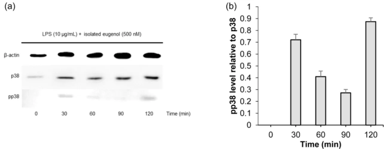
3.5. Western blotting
질 상피 세포주인 VK2/E6E7 ATCC CRL-2616에 500 nM IESA 를 처리 후, LPS를 시간 별로 처리한 결과는 Fig. 6에 나 타내었다. Fig. 6(a)에서 보는 바와 같이 pp38의 발현량은 60~90분 사이에 감소하였다가 120분에 다시 증가하였음을 알 수 있다. Fig. 6(b)는 p38 발현량에 대한 pp38 발현량의 비 를 나타낸 것으로 처리시간이 30분일 때 pp38/p38의 값이 0.7 ± 0.04, 60 분일 때 0.37 ± 0.04, 90분일 때 0.23 ± 0.02로 감
Fig. 5. Cell viability of VK2/E6E7 ATCC CRL-2616 by IESA.Cell viability was examined using MTT assay. Data values were expressed as mean ± SD.
Fig. 7. The secretion of TNF-α from VK2/E6E7 ATCC CRL-2616 as assessed by ELISA. (a) Treated with C. albicans ATCC 28367, (b) Treated with 500 nM IESA and C. albicans ATCC 28367. Data values were expressed as mean±SD.
Fig. 6. Western blot analysis of time-dependent p38 phosphorylation in VK2/E6E7 ATCC CRL-2616. (a) Time-dependent activation of p38 and pp38 in response to LPS (10 μg/mL) after treatment with 500 nM IESA, (b) Bar graph expressed as the pp38 level relative to p38.
Data values were expressed as mean ± SD.
소추세를 보이다 120분 때 0.94 ± 0.03으로 증가하는 것을 볼 수 있다. 이러한 결과를 통해, LPS를 처리한 후 30분부 터 p38의 활성화가 시작되었고 60~90분 사이에 p38의 활성 이 억제되었다가 120분부터 다시 활성화가 시작됨을 알 수 있다. IESA는 LPS로 세포에 자극을 주었을 때, LPS 처리시 간이 60~90분 사이에 p38의 활성화를 억제하는 것으로 사 료된다.
3.6 ELISA
칸디다성 질염을 유발하는 원인균인 C. albicans로 질 상피 세포에 자극을 유발하였을 때, IESA가 미치는 영향을 확인 하기 위해 수행하였다. Fig. 7(a)는 VK2/E6E7 ATCC CRL- 2616에 C. albicans ATCC 28367을 처리한 결과이고, Fig.
7(b)는 C. albicans ATCC 28367과 500 nM IESA를 처리하였 을 때 TNF-α의 양을 측정한 결과이다. C. albicans ATCC 28367을 12시간 처리 시 TNF-α의 양은 822.625 ± 20 pg/mL 로 측정되었으며, C. albicans ATCC 28367과 IESA를 12시간 처리 시 TNF-α의 양은 425.125 ± 20 pg/mL로 측정되었다.
IESA를 처리하지 않았을 때보다 처리하였을 때 TNF-α의 양 이 48.32% 감소하였다. 이러한 결과를 통하여 IESA가 C.
albicans ATCC 28367로 유발된 TNF-α를 억제하는 효과가 있음을 확인했다.
4. CONCLUSION
질염은 여성의 면역력과 관련된 질환으로 크게 세균성 질증 과 칸디다성 질염이 있다. 항생제의 사용은 질 내 유용균의 수를 감소시키고, 항생제내성균의 출현을 야기한다. 이에 본 연구는 질 내 유용균인 L. coleohominis KCTC 21007은 억제 하지 않으면서 질염유발균주인 C. albicans ATCC 28367, C.
gleum KCTC 2904, Proteus mirabilis KCTC 2510 과 S.
paucimobilis KCTC 2834에 대해 항균력을 가지는 한약재를 탐색하였고, 한약재로부터 유효물질을 분리하여 추출물질 의 특성을 확립하고자 하였다. 정향 methyl alcohol 추출물의 항균활성 결과로 L. coleohominis KCTC 21007은 억제하지 않으면서 질염유발균주인 C. albicans ATCC 28367에서 7 mm, C. gleum KCTC 2904에서 8 mm, S. paucimobilis KCTC 2834에서 13 mm의 inhibitory zone이 확인되었다. 정향 methyl alcohol 추출물에서 항균력을 가진 물질은 Jang의 방 법 [24]에 따라 분리하였고, 정향에서 분리한 n-hexane 8번 분획은 commercial eugenol과 HPLC pattern을 비교하여 retention time이 동일한 peak의 유무를 확인 후 eugenol로 판 단하여 사용하였다. 정향에서 분리한 eugenol (IESA)은 DPPH free radical 소거능, ABTS radical 소거능 실험을 수행 한 결과 commercial eugenol보다 항산화 활성이 높게 측정되 었다. 질 상피 세포주인 VK2/E6E7 ATCC CRL-2616은 500 nM의 IESA를 처리하였을 때 89%의 세포 생존율을 나타내 었다. Gram (-)균이 가지는 자극유발원인 LPS를 처리하여
회수한 단백질로 immunoblotting을 수행한 결과, pp38/p38의 값은 처리시간이 30~60분에 감소하였고, 이를 통해 p38의 활성화가 IESA에 의해 억제되었음을 알 수 있다. 칸디다성 질염으로 유발된 자극에 대한 IESA의 반응을 확인하기 위해 Human TNF-α ELISA를 수행하였으며, 그 결과 TNF-α양은 세포에 C. albicans ATCC 28367만 처리하였을 때 보다 IESA 를 함께 처리하였을 때 48.32%의 감소가 나타났다. 이에 IESA의 세균성 질증 및 칸디다성 질염의 치료제로서의 가능 성을 확인하였다.
Acknowledgement
이 논문은 2019년도 정부 (교육부)의 재원으로 한국연구재 단의 지원을 받아 수행된 기초연구사업입니다 (NRF-2017 R1D1A1B03036147).
REFERENCES
1. Van Der Pol, B. (2010) Diagnosing Vaginal Infections: It’s Time to join the 21st Century. Curr. Infect. Dis. Rep. 12: 225-230.
2. Kim, T. (2008) Treatment and management of sexually transmit- ted diseases. J. Korean Med. Assoc. 51: 884-896.
3. Jang, J., D. H. Kang, J. Yoon, and H. S. Kim (2018) Separation and Purification of Antimicrobial Substance from Syzygium aro- maticum Merrill et Perry for Treatment of Microbial Vaginosis.
KSBB J. 33: 285-292.
4. Cortés-Rojas, D. F., C. R. F. de Souza, and W. P. Oliveira (2014) Clove (Syzygium aromaticum): a precious spice. Asian Pac. J.
Trop. Biomed. 4: 90-96.
5. Bhuiyan, M. N. I., J. Begum, and F. Akter (2010) Constituents of the essential oil from leaves and buds of clove (Syzigium caryo- phyllatum (L.) Alston). Afr. J. Pharm. Phatmacol. 4: 451-454.
6. Sell, A. B. and E. A. Carlini (1976) Anesthetic action of methyleu- genol and other eugenol derivatives. Pharmacology. 14: 367-377.
7. Swidsinski, A., W. Mendling, V. Loening-Baucke, S. Swidsinski, Y. Dörffel, J. Scholze, H. Lochs, and H. verstraelen (2008) An adherent Gardnerella vaginalis biofilm persists on the vaginal epi- thelium after standard therapy with oral metronidazole. Am. J.
Obstet. Gynecol. 198: 97.e1-6.
8. Norrington, D. W., J. Ruby, P. Beck, and P. D. Eleazer (2008) Observations of biofilm growth on human dentin and potential destruction after exposure to antibiotics. Oral Surg. Oral Med.
Oral Pathol. Oral Radiol. Endod. 105: 526-529.
9. Machado, D., J. Castro, A. Palmeira-de-Oliveira, J. Martinez-de- Oliveira, and N. Cerca (2016) Bacterial vaginosis biofilms: chal- lenges to current therapies and emerging solutions. Front. Micro- biol. 6: 1528.
10. Harriott, M. M., E. A. Lilly, T. E. Rodriguez, P. L. Fidel, and M. C.
Noverr (2010) Candida albicans forms biofilms on the vaginal mucosa. Microbiology. 156: 3635-3644.
11. Han, J., J. D. Lee, L. Bibbs, and R. J. Ulevitch (1994) A MAP
kinase targeted by endotoxin and hyperosmolarity in mammalian cells. Science. 265: 808-811.
12. Malcolm, K. C. and G. S. Worthen (2003) Lipopolysaccharide stimulates p38-dependent induction of antiviral genes in neutro- phils independently of paracrine factors. J. Biol. Chem. 278:
15693-15701.
13. Goldsmith, E. J., M. H. Cobb, and C. I. Chang (2004) Structure of MAPKs. Methods Mol. Biol. 250: 127-144.
14. Kumar, S., J. Boehm, and J. C. Lee (2003) p38 MAP kinases: key signalling molecules as therapeutic targets for inflammatory dis- eases. Nat. Rev. Drug Discov. 2: 717-726.
15. Pargellis, C. and J. Regan (2003) Inhibitors of p38 mitogen-acti- vated protein kinase for the treatment of rheumatoid arthritis. Curr.
Opin. Investig. Drugs. 4: 566-571.
16. Hommes, D., B. Van Den Blink, T. Plasse, J. Bartelsman, C. Xu, B. Macpherson, G. Tygat, M. Peppelenbosch, and S. Van Deventer (2002) Inhibition of stress-activated MAP kinases induces clinical improvement in moderate to severe Crohn's disease. Gastroenter- ology. 122: 7-14.
17. Johnson, G. V. and C. D. Bailey (2003) The p38 MAP kinase sig- naling pathway in Alzheimer's disease. Exp. neurol. 183: 263-268.
18. Bachstetter, A. D. and L. J. Van Eldik (2010) The p38 MAP kinase family as regulators of proinflammatory cytokine production in degenerative diseases of the CNS. Aging Dis. 1: 199-211.
19. Wilms, H., P. Rosenstiel, J. Sievers, G. Deuschl, L. Zecca, and R.
Lucius (2003) Activation of microglia by human neuromelanin is NF-kappaB dependent and involves p38 mitogen-activated pro- tein kinase: Implications for Parkinson’s disease. FASEB J. 17:
500-502.
20. Basma, A. A., Z. Zuraini, and S. Sasidharan (2011) A transmis- sion electron microscopy study of the diversity of Candida albi- cans cells induced by Euphorbia hirta L. leaf extract in vitro.
Asian Pac. J. Trop. Biomed. 1: 20-22.
21. Ganguly, S. and A. P. Mitchell (2011) Mucosal biofilms of Can- dida albicans. Curr. Opin. Microbiol. 14: 380-385.
22. Steele, C. and P. L. Fidel, Jr. (2002) Cytokine and chemokine pro- duction by human oral and vaginal epithelial cells in response to Candida albicans. Infect. Immun. 70: 577-583.
23. Martinez, R. C., S. L. Seney, K. L. Summers, A. Nomizo, E. C. De Martinis, and G. Reid (2009) Effect of Lactobacillus rhamnosus GR-1 and Lactobacillus reuteri RC?14 on the ability of Candida albicans to infect cells and induce inflammation. Microbiol. Immu- nol. 53: 487-495.
24. Jang, J. (2019) Separation and purification of antimicrobial sub- stances from natural materials for treatment of microbial vagino- sis. MS. Thesis. University of Keimyung, Daegu, Korea.
25. Blois, M. S. (1958) Antioxidant determinations by the use of a sta- ble free radical. Nature. 181: 1199-1200.
26. Re, R., N. Pellegrini, A. Proteggente, A. Pannala, M. Yang, and C.
Rice-Evans (1999) Antioxidant activity applying an improved ABTS radical cation decolorization assay. Free Radic. Biol. Med.
26: 1231-1237.
27. Lee, S. Y., H. J. kim, I. H. Chang, J. H. Han, K. D. Kim, Y. T.
Moon, and S. C. Myung (2014) Modulation of Antimicrobial Pep- tide Human β-defensin-3 by Toll-like Receptor Ligands in Vaginal Epithelial Cells. Korean J. Urogenit. Tract Infect. Inflamm. 9: 27- 33.