INTRODUCTION
Attention-deficit hyperactivity disorder (ADHD) is a frequent neurodevelopmental disorder with an in- creasing prevalence over the last 2 decades [1]. A recent cohort study of almost 7.5 million people with roughly
700,000 prescriptions, reported an increase in prescrip- tion rate from 42.7 prescriptions per 10,000 persons before the year 2000 to 394.4 prescriptions in 2015 an increase of about 800% in prescription rate [2]. The up- surge was observed in both sexes and in all age groups.
Moreover, unlike previous trends of increased diagnosis
Received: Jan 25, 2019 Revised: Mar 19, 2019 Accepted: Apr 9, 2019 Published online May 8, 2019 Correspondence to: Avi Harlev https://orcid.org/0000-0001-7335-9258
Fertility and IVF Unit, Soroka University Medical Center, Ben-Gurion University of the Negev, PO Box, 151, Beer Sheva, Israel.
Tel: +972-8-6400111, Fax: +972-8-6403057, E-mail: [email protected]
*The study was presented in part at the American Society for Reproductive Medicine (ASRM) 2017 Scientific Congress, San Antonio, TX, USA.
Copyright © 2020 Korean Society for Sexual Medicine and Andrology
pISSN: 2287-4208 / eISSN: 2287-4690 World J Mens Health 2020 Jan 38(1): 61-67 https://doi.org/10.5534/wjmh.180127
Ritalinic Acid Stimulates Human Sperm Motility and Maintains Vitality In Vitro
Avi Harlev1,2 , Ralf Henkel3 , Luna Samanta2,4 , Ashok Agarwal2
1Fertility and IVF Unit, Soroka University Medical Center, Ben-Gurion University of the Negev, Beer Sheva, Israel, 2American Center for Reproductive Medicine, Cleveland Clinic, Cleveland, OH, USA, 3Department of Medical Bioscience, University of the Western Cape, Bellville, South Africa, 4Department of Zoology, School of Life Sciences, Ravenshaw University, Orissa, India
Purpose: To evaluate the in vitro impact of ritalinic acid (RA), a major metabolite of methylphenidate (drug to treat attention- deficit hyperactivity disorder), on sperm motility, vitality and oxidative stress.
Materials and Methods: Semen samples (n=13) were collected from healthy donors and a semen analysis was performed according to World Health Organization. Density gradient centrifugation was performed to isolate motile sperm. Samples were incubated with different concentrations (0, 1, 10, 100, and 1,000 ng/mL) of RA. The non-exposed group (0 ng/mL) was defined as the control group. Samples were analyzed for motility at different time points (0, 60, 150, 240, and 300 minutes) and for vitality and oxidation reduction potential (ORP) (at 0, 240, and 300 minutes). Sperm motility was assessed manually and motion kinetic parameters were recorded by computer aided semen analysis.
Results: RA at any tested concentration significantly increased sperm motility compared to the control in a time-dependent manner with a maximum increase after 240 minutes. Motion kinetic parameters were not comparable. For sperm vitality, supplementation with RA significantly maintained survival at higher levels, while non-treated sperm gradually died. These higher levels of vitality were maintained with rising RA concentrations of up to 1,000 ng/mL. A non-significant trend of in- creased ORP was observed in all study groups.
Conclusions: RA increases sperm motility and maintains vitality at any concentration tested. Therefore, RA might be utilized to improve sperm quality in asthenozoospermic specimens. However, further investigation is ongoing to evaluate the effect of RA on other sperm parameters.
Keywords: Oxidative stress; Ritalinic acid; Sperm motility
This is an Open Access article distributed under the terms of the Creative Commons Attribution Non-Commercial License (http://creativecommons.org/licenses/by-nc/4.0) which permits unrestricted non-commercial use, distribution, and reproduction in any medium, provided the original work is properly cited.
and treatment of ADHD in pre-school and school aged children, the later increase applies to children, adoles- cents, and even adults [3].
The most commonly used first line pharmacologic treatment for ADHD is methylphenidate (MPH). The mechanism of action of MPH in ADHD is still a mat- ter of debate, but several studies indicated that MPH inhibits the presynaptic dopamine transporter [4,5].
The mechanism of action includes inhibition of the re-uptake of norepinephrine and dopamine into the presynaptic neuron or an increase of their intrasynap- tic concentrations by increasing their release into the extraneuronal space [6].
Ritalinic acid (RA), the major inactive metabolite of MPH, is one of the isomers of amphetamine. The circulating concentrations of RA greatly exceed that of the parent drug [7]. If MPH is given in the normal dosage for clinical use, the plasma concentration of RA reaches a maximal concentration of 10 ng/mL with a half-life of 2.5 hours [8]. RA is soluble in water and due to its small size, it readily passes the blood brain and testicular barriers [9].
Due to the wide use of MPH to treat ADHD, several studies have been conducted to evaluate its impact on various body biological systems in general and the re- productive system in particular; however, most of them are animal studies. Chronic use of MPH decreases the body weights of male mice as compared to a control group. In addition, the weight of the testes and seminal vesicles decreased significantly [10]. MPH was reported to reduce the number of Leydig cells and serum testos- terone levels, increase the percentage of abnormal tail and sperm cell morphology and the testicular intersti- tial tissue volume, along with a reduction in fertility rates of the male mice in the MPH treated groups com- pared to controls [11,12]. MPH was reported to negative- ly affect spermatogenesis not only by reducing testicu- lar weight and number of round spermatids but also by increasing apoptotic death and p53 activation [13].
Previous studies, mainly animal ones, investigated the impact of orally administrated MPH on biologi- cal systems. However, no study assessed the effect of MPH or its metabolites on human sperm. This lack of information might be related to the young age of the patients, mostly prepubertal boys, to whom MPH was prescribed at the time the drug was approved for clini- cal use in 1955. Nonetheless, the growing use of MPH in adolescents and adults who are in their reproductive
age, and the growing rate of MPH abuse, requires an evaluation of the impact of MPH on male reproduction.
The aim of the present study is to investigate the di- rect effect of RA on human semen parameters in vitro.
MATERIALS AND METHODS
1. Ethics statement
Following the approval of the study by the Insti- tutional Review Board (IRB) of the Cleveland Clinic Foundation Hospital (Reg. No. 15-435) according to the Declaration of Helsinki, a total of 13 semen samples were collected from healthy donors by masturbation after 2 to 3 days of abstinence. An informed, written consent has been obtained from each donor.
2. Semen samples
All samples were allowed to liquefy completely for 20 minutes at 37°C before further processing. The initial semen analysis was performed according to the World Health Organization (WHO) 5th edition [14]. Density gradient centrifugation was performed to isolate motile sperm. Given the achieved plasma concentration of 10 ng/mL during treatment, each sample was incubated with different concentrations of RA (1, 10, 100, and 1,000 ng/mL RA) and compared to a control without RA from the same patient. Depending on the volume of se- men available, not all tests could be performed for each donor. However, a minimum of 8 samples was used.
The following inclusion criteria were applied for samples to be eligible for enrollment into the study:
generally healthy males over 18 years of age who were willing to abstain from sexual activities for 2 to 3 days prior to semen collection, and normal semen analysis as per WHO 2010 guidelines. Subjects presenting with any of the following were excluded: sample of high viscosity semen, non-compliance with protocol require- ments, and any abnormal semen parameter according to the WHO 2010 criteria.
3. Standard semen analysis
Following liquefaction, after the sample has been in the incubator for 20 minutes at 37°C, the sample is examined for viscosity by the ease of pipetting in a graduated serological pipette. Viscosity is graded as follows: Normal viscosity: the sample can easily pass in the pipette and expelled easily, slight viscosity: the sample is not expelled easily out of the pipette, moder-
ate: difficult to pipette and expel, high viscosity: the sample is a coagulum and cannot be aspirated. In the current study, none of our donors presented with mod- erate or high viscosity. Hence none of the samples were excluded. Semen specimens were evaluated for volume, liquefaction, color, pH, and volume as well as sperm concentration, motility, and vitality as per WHO 2010 guidelines. Motile sperm were obtained by pooling of semen samples prepared by double density gradient separation. The motile sperm were tested for the effect of different concentrations of RA (1, 10, 100, and 1,000 ng/mL) on spermatozoa.
4. Sperm preparation by density gradient centrifugation
In order to evaluate the better fraction of motile sperm, spermatozoa were prepared by separation on a double density gradient by layering 2 mL each of up- per (45%) and lower phase (90%) layer (Vitrolife, San Diego, CA, USA). Two mL of liquefied semen were layered on top. Samples were centrifuged at 300 ×g for 20 minutes at room temperature. The resulting pellet was aspirated and re-suspended with 2 mL of human tubal fluid (HTF) medium supplemented with 2 mg/
mL human serum albumin. The final concentration was adjusted to 20×106 motile spermatozoa/mL and 0.5 mL aliquots were used for sperm motility studies.
5. Human sperm motility assay
A human sperm motility assay was used to test the toxicity of the RA at different concentrations [15]. We used a computer assisted semen analyzer IVOS II, ver.
14 (Hamilton Thorne, Beverly, MA, USA). The setting included a heated stage set at 37°C. Spermatozoa in HTF suspensions was exposed to RA at increasing con- centrations (1, 10, 100, and 1,000 ng/mL) at 37ºC in an incubator in an atmosphere with 5% CO2 for 1, 2.5, 4, and 5 hours. After incubation, samples were vigorously mixed by pipetting and an aliquot of the test sample was placed on a MicroCell chamber and examined un- der phase contrast at ×20 magnification for motility.
Motion kinetic parameters (curvilinear velocity, VCL;
amplitude of lateral head displacement, ALH; linearity, LIN) were recorded by computer aided semen analysis (CASA, IVOS II; Hamilton Thorne).
6. Vitality test
Sperm vitality was tested using the eosin–nigrosine
stain. An amount of 0.67 g eosin Y (color index 45,380) and 0.9 g sodium chloride (NaCl) were dissolved in 100 mL of purified water. Ten grams of nigrosin (color in- dex 50,420) were added to the 100 mL of eosin Y solu- tion. Semen samples were mixed with an equal volume of eosin–nigrosin suspension in a porcelain spot plate well or test-tube. A smear on a glass slide was made from each suspension and was allowed to air dry. Im- mediately after drying, each slide was examined at
×1,000 magnification under oil immersion. The number of stained (dead) or unstained (vital) cells was evalu- ated and the percentage of vital sperm was calculated.
7. Measurement of the oxidation reduction potential
Oxidation reduction potential (ORP) levels were measured in sperm suspension by using the MiOXSYS analyzer (Aytu Bioscience, Eaglewood, CO, USA) at 0 and 240 and 300 minutes parallel to the vitality and motility tests. ORP measures the transfer of electrons from a reductant (or antioxidant) to an oxidant. Brief- ly, 30 mL of liquefied semen at room temperature were applied to the MiOXSYS sensor. The sensor was pre-in- serted into the MiOXSYS analyzer; measurements be- gin automatically. ORP is measured in milli-volts (mV) and is the integrated measure of the existing balance between total oxidants and reductants in a biological system.
Each sample was measured in triplicate, and the av- erage values for ORP were recorded. Data were then normalized to sperm concentration to control for dif- ferences in cell numbers. Thus, data are presented as mV/106 sperm/mL. To assess the effects of time on ORP measures, samples were incubated at room tempera- ture for 0 and 120 minutes before being measured.
8. Statistical analysis
For statistical analysis, MedCalc Statistical Software, ver. 17.4 (MedCalc Software bvba, Ostend, Belgium) was used. After testing for normal distribution of the data by means of the Kolmogorov–Smirnov test, the parametric (paired-samples t-test) and non-parametric tests (Wilcoxon test and Kruskal–Wallis test with Jonckheere–Terpstra trend), respectively, were em- ployed. Data are presented as mean±standard error of mean. A p<0.05 was considered significant.
RESULTS
All semen samples underwent an initial semen analysis according to the WHO 5th edition. All samples were found eligible and no sample was required to be excluded.
1. Sperm motility
A total of 10 samples were evaluated for sperm mo- tility. Samples were analyzed at different time points (0, 60, 150, 240, and 300 minutes) based on the half-life of RA in plasma. All concentrations of RA used in this study significantly (p<0.05) increased sperm motility with a maximum after 240 minutes of incubation (Fig.
1). This increase was maintained with rising concen- trations of up to 1,000 ng/mL. Thereafter, a decrease was observed at 300 minutes of incubation in treated samples, even though motility remained significantly higher compared to control. From the control to the 240 minutes incubation point, the Jonckheere–Terpstra trend test indicated for all concentrations significant trends (p<0.05), though the Kruskal–Wallis test was not significant (p=0.2473 to p=0.1315). No significant changes were seen in sperm motion kinetic parameters.
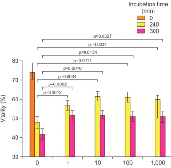
2. Sperm vitality
A total of 13 samples were evaluated for sperm vital- ity (Fig. 2; Supplementary Fig. 1). Since an increase in sperm motility was observed in 60 and 150 minutes, vitality would not be decreased at these time points, sperm vitality was instead analyzed at 3 time points (0, 240, and 300 minutes). A reduction in vitality was
observed as compared to the control group without ad- dition of RA. The addition of RA to the sperm samples caused sperm to retain their vitality.
3. Oxidation-reduction potential
A total of 8 samples were evaluated for ORP before and after exposure to RA. Samples were analyzed at 3 time points (0, 240, and 300 minutes) after the ob- served increase in sperm motility was observed in 60 and 150 minutes (Fig. 3; Supplementary Fig. 2). A non- significant trend (p=0.3125 and p=0.1953) of increase in ORP levels compared to the control was observed in all study groups after 240 and 300 minutes of incubation, respectively.
Motility(%)
Time (min) 80
70
60
50
CCF 2017
C
0 300
0 1 10 100 1,000
Concentration of ritalinic acid (ng/mL)
p=0.0059
p=0.0098
p=0.0020
60 150
p=0.0078
p=0.0742
240
Fig. 1. The impact of ritalinic acid on sperm motility.
Vitality(%)
Incubation time (min)
80
70
40
30
CCF 2017
C
0 1,000
0 240 300
Concentration of ritalinic acid (ng/mL)
1 10 100
60
50
p=0.0134 p=0.0017 p=0.0010 p=0.0034 p=0.0002 p=0.0012
p=0.0327 p=0.0034
Fig. 2. The impact of ritalinic acid on sperm vitality.
Oxidation-reductionpotential (mV/10/mL)6
280
230
180
0 1,000
Incubation time (min)
0 240 300
Concentration of ritalinic acid (ng/mL)
1 10 100
p=0.3282 p=0.0650 p=0.2786 p=0.5737
Fig. 3. The impact of ritalinic acid on oxidation reduction potential.
DISCUSSION
MPH in its various administration forms is widely used for treatment of ADHD in the general population, including those patients at reproductive age. Increas- ing age of the treated patients and increased rates of MPH abuse have been reported as well [16]. The direct impact of MPH on human sperm and the resulting im- pact on male reproduction is still unknown. This study is the first indicating that MPH metabolite, RA, has a temporary effect on sperm function including increased vitality, motility and possibly increased reactive oxygen species (ROS) levels as compared to controls.
Results of the current study clearly show that MPH main metabolite, RA, was able to increase human sperm motility, while sperm kinematic parameters, such as LIN, ALH, and VCL were not affected. In addi- tion, vitality was maintained over time at significantly higher levels as compared to the control. It is also im- portant to note that despite increased oxidative stress (OS) levels as measured as ORP after 240 and 300 min- utes, respectively, these increases were not significant.
Moreover, the impact of RA on sperm functions was comparable in all studied RA concentrations including the subclinical dose of 1 ng/mL, and the abuse equiva- lent doses of 100 and 1,000 ng/mL. However, due to the fact that the actual RA concentrations in semen have never been reported before, concentrations used in this study were only based on plasma levels and has to be considered as a limitation of our study.
The exact mechanism of action of MPH in ADHD treatment is still debatable. It is hypothesized that MPH acts as a catecholamine reuptake inhibitor im- pacting mainly dopamine and norepinephrine levels.
MPH blocks the dopamine transporter in the central nervous system, thus causing an increased dopamine concentration in the synaptic cleft [17]. In the male reproductive system, high concentrations of catechol- amines including dopamine [18] and the presence of dopamine receptors in the sperm cell [19] including a2- and b-adrenergic receptors and the dopamine type 2 receptor in testis, as well as spermatogenic cells and sperm cells have been described [20]. Catecholamines were reported to impact sperm cell motility [21]. While evaluating the impact of dopamine on sperm motil- ity, Urra et al [19] exposed human sperm to differ- ent dopamine concentrations. An inhibition of total sperm motility and progressive motility was observed
after incubation for 1, 3, and 6 hours at a dopamine concentration of 1 mM. Nevertheless, exposing sperm to dopamine in the absence and presence of a specific dopamine transporter inhibitor, the dopamine-induced inhibition of total sperm motility was partially re- versed. Thus, the temporary increase in sperm motil- ity observed in the current study corresponding to the MPH half-life may be explained by the inhibition of the dopamine uptake inhibition.
The observed increased motility cannot be attributed to decreased OS levels as measured by ORP since these slightly, but not significantly, increased. At this stage, it can also only be speculated whether this increased ROS activity is due to increased metabolic activity of the spermatozoa or simply caused by the elimination of protective antioxidants present in the seminal plasma.
Excessive ROS production that impairs the fine redox balance between oxidants and anti-oxidants (enzymatic and non-enzymatic) yield OS. In turn, OS has a well- established deteriorating effect on sperm function including reduced sperm motility, decreased ability of spermatozoa to adhere to the oocyte [22], to undergo capacitation [23] as well as OS-induced DNA damage, mainly DNA fragmentation [24]. Previous studies re- ported that a large percentage of male factor infertility is due to OS-mediated sperm damage [25,26]. In addi- tion, several extrinsic factors such as smoking [27,28] or varicocele [29] may cause OS.
On the other hand, naturally generated low ROS levels are major role players in male reproductive physiologic processes such as spermatogenesis and the triggering of sperm capacitation. Ultimately, the molecular processes involved in sperm binding to the zona pellucida are also mediated by hydrogen peroxide.
Likewise, ROS in low levels are important for proper sperm motility [24]. ROS, in general, are essential for proper spermatogenic mechanisms to occur, and hy- drogen peroxide, in particular, stimulates acrosomal reaction, tyrosine phosphorylation and hyperactivation in the male germ cell. The latter, in turn is a direct expression of changes taking place in the sperm kine- matic parameters VCL (>150 µm/s), LIN (<50%), and ALH (>7 µm). Considering that these parameters did not change during the incubation with MPH, however, one can assume that MPH did not stimulate intrinsic ROS production which, in turn, would trigger capaci- tation, sperm hyperactivation and acrosome reaction.
On the other hand, sperm exposed to dopamine and
norepinephrine after facilitating capacitation with heparin were reported to have a dose-dependent effect on the acrosome integrity [19,20] implying that sperm would not be able to fertilize the oocyte by the time it will reach it as capacitation and/or the acrosome reac- tion are prematurely triggered.
A potential application of the observed impact of RA (or any other dopamine reuptake inhibitor) on sperm cells may be as a motility enhancer. This would be of particular interest in patients with asthenozoospermia or during various fertility treatment procedures such as intra-uterine insemination (IUI). IUI is often a first line treatment in patients with adequate sperm num- bers since it is minimally invasive and far less expen- sive when compared to in-vitro fertilization. It is used when there is only mildly reduced sperm count, motil- ity or morphology as well as in cases of unexplained in- fertility. In this procedure, the sperm avoids the hostile vaginal environment and the need to pass through the uterine cervix. However, the transport to the fallopian tube and more importantly fertilization of the egg re- mains. Hence, an increased sperm motility observed af- ter the use of RA may be used to increase motility and therefore potentially increase the effectiveness of IUI.
However, before using RA during fertility treatments, its impact on sperm capacitation, acrosome reaction and sperm DNA must be determined. These processes are major role players in the fertilizing competence of sperm and are essential to enable the sperm to pen- etrate the oocyte and cause fertilization [30].
The current study has some limitations. The first is the small study sample of the evaluated parameters.
Another limitation is the fact that only selected sperm parameters including vitality, total motility, VCL, ALH, LIN, and ORP were measured. However, the fact that despite the small sample significant differences were observed between the study groups reflects the substantial effect of RA on sperm function parameters.
CONCLUSIONS
In conclusion, RA significantly increased sperm mo- tility and maintained vitality over a long incubation period. This may allow its in vitro use to improve sperm motility in cases of patients with asthenozoospermia.
However, it is unknown what effects RA has on other sperm functional characteristics such as: capacitation, acrosome reaction, intrinsic ROS production and sperm
nuclear DNA fragmentation. Further investigation is required prior to considering its clinical use.
ACKNOWLEDGEMENTS
The study was supported by funds from the Ameri- can Center for Reproductive Medicine. Andrology lab technologists helped with scheduling of subjects. Fig- ures were drawn by graphic artists from the Cleveland Clinic Arts department.
Special thanks to: Reva Prabha Peer, Ibrahim Al- tamimi, Mohamed Iesar Mohamed, Muna Abuayash, Abdullah Alzaaqi, Fares Bamajbuor.
Disclosure
The authors have no potential conflicts of interest to disclose.
Authors Contribution
Conceptualization: AH. Formal analysis: RH. Funding acquisi- tion: AA. Investigation: AH. Methodology: AH, LS. Supervision:
AA. Validation: RH, AA. Writing–original draft: AH. Writing–
review & editing: RH, LS, AA.
Supplementary Materials
Supplementary materials can be found via https://doi.
org/10.5534/wjmh.180127.
REFERENCES
1. Chai G, Governale L, McMahon AW, Trinidad JP, Staffa J, Murphy D. Trends of outpatient prescription drug utilization in US children, 2002-2010. Pediatrics 2012;130:23-31.
2. Renoux C, Shin JY, Dell’Aniello S, Fergusson E, Suissa S.
Prescribing trends of attention-deficit hyperactivity disorder (ADHD) medications in UK primary care, 1995-2015. Br J Clin Pharmacol 2016;82:858-68.
3. Moffitt TE, Houts R, Asherson P, Belsky DW, Corcoran DL, Hammerle M, et al. Is adult ADHD a childhood-onset neuro- developmental disorder? Evidence from a four-decade longi- tudinal cohort study. Am J Psychiatry 2015;172:967-77.
4. Jenson D, Yang K, Acevedo-Rodriguez A, Levine A, Brous- sard JI, Tang J, et al. Dopamine and norepinephrine receptors participate in methylphenidate enhancement of in vivo hippo- campal synaptic plasticity. Neuropharmacology 2015;90:23-32.
5. Kodama T, Kojima T, Honda Y, Hosokawa T, Tsutsui KI,
Watanabe M. Oral administration of methylphenidate (ritalin) affects dopamine release differentially between the prefrontal cortex and striatum: a microdialysis study in the monkey. J Neurosci 2017;37:2387-94.
6. Volkow ND, Wang GJ, Smith L, Fowler JS, Telang F, Logan J, et al. Recovery of dopamine transporters with methamphet- amine detoxification is not linked to changes in dopamine release. Neuroimage 2015;121:20-8.
7. Modi NB, Wang B, Noveck RJ, Gupta SK. Dose-proportional and stereospecific pharmacokinetics of methylphenidate delivered using an osmotic, controlled-release oral delivery system. J Clin Pharmacol 2000;40:1141-9.
8. Wargin W, Patrick K, Kilts C, Gualtieri CT, Ellington K, Mueller RA, et al. Pharmacokinetics of methylphenidate in man, rat and monkey. J Pharmacol Exp Ther 1983;226:382-6.
9. Adjei A, Teuscher NS, Kupper RJ, Chang WW, Greenhill L, Newcorn JH, et al. Single-dose pharmacokinetics of methyl- phenidate extended-release multiple layer beads administered as intact capsule or sprinkles versus methylphenidate imme- diate-release tablets (Ritalin(®)) in healthy adult volunteers. J Child Adolesc Psychopharmacol 2014;24:570-8.
10. Beckman DA, Schneider M, Youreneff M, Tse FL. Juvenile toxicity assessment of d,l-methylphenidate in rats. Birth De- fects Res B Dev Reprod Toxicol 2008;83:48-67.
11. Fazelipour S, Jahromy MH, Tootian Z, Kiaei SB, Sheibani MT, Talaee N. The effect of chronic administration of methylphe- nidate on morphometric parameters of testes and fertility in male mice. J Reprod Infertil 2012;13:232-6.
12. Nudmamud-Thanoi S, Thanoi S. Methamphetamine induces abnormal sperm morphology, low sperm concentration and apoptosis in the testis of male rats. Andrologia 2011;43:278-82.
13. Cansu A, Ekinci O, Ekinci O, Serdaroglu A, Erdogan D, Coskun ZK, et al. Methylphenidate has dose-dependent nega- tive effects on rat spermatogenesis: decreased round sperma- tids and testicular weight and increased p53 expression and apoptosis. Hum Exp Toxicol 2011;30:1592-600.
14. World Health Organization. WHO laboratory manual for the examination and processing of human semen. 5th ed. Ge- neva: World Health Organization; 2010.
15. Claassens OE, Wehr JB, Harrison KL. Optimizing sensitivity of the human sperm motility assay for embryo toxicity test- ing. Hum Reprod 2000;15:1586-91.
16. Cairns R, Daniels B, Wood DA, Brett J. ADHD medication overdose and misuse: the NSW Poisons Information Centre experience, 2004-2014. Med J Aust 2016;204:154.
17. Gronier B. In vivo electrophysiological effects of methylphe- nidate in the prefrontal cortex: involvement of dopamine D1 and alpha 2 adrenergic receptors. Eur Neuropsychopharmacol
2011;21:192-204.
18. Fait G, Vered Y, Yogev L, Gamzu R, Lessing JB, Paz G, et al.
High levels of catecholamines in human semen: a preliminary study. Andrologia 2001;33:347-50.
19. Urra JA, Villaroel-Espíndola F, Covarrubias AA, Rodríguez- Gil JE, Ramírez-Reveco A, Concha II. Presence and function of dopamine transporter (DAT) in stallion sperm: dopamine modulates sperm motility and acrosomal integrity. PLoS One 2014;9:e112834.
20. Ramírez AR, Castro MA, Angulo C, Ramió L, Rivera MM, Torres M, et al. The presence and function of dopamine type 2 receptors in boar sperm: a possible role for dopamine in vi- ability, capacitation, and modulation of sperm motility. Biol Reprod 2009;80:753-61.
21. Bavister BD, Chen AF, Fu PC. Catecholamine requirement for hamster sperm motility in vitro. J Reprod Fertil. J Reprod Fertil 1979;56:507-13.
22. Bromfield EG, Aitken RJ, Anderson AL, McLaughlin EA, Nixon B. The impact of oxidative stress on chaperone-mediat- ed human sperm-egg interaction. Hum Reprod 2015;30:2597- 613.
23. Morielli T, O’Flaherty C. Oxidative stress impairs function and increases redox protein modifications in human sperma- tozoa. Reproduction 2015;149:113-23.
24. Agarwal A, Durairajanayagam D, Halabi J, Peng J, Vazquez- Levin M. Proteomics, oxidative stress and male infertility.
Reprod Biomed Online 2014;29:32-58.
25. Shekarriz M, Thomas AJ Jr, Agarwal A. Incidence and level of seminal reactive oxygen species in normal men. Urology 1995;45:103-7.
26. Tremellen K. Oxidative stress and male infertility: a clinical perspective. Hum Reprod Update 2008;14:243-58.
27. Sharma R, Harlev A, Agarwal A, Esteves SC. Cigarette smok- ing and semen quality: a new meta-analysis examining the ef- fect of the 2010 World Health Organization laboratory meth- ods for the examination of human semen. Eur Urol 2016;70:
635-45.
28. Harlev A, Agarwal A, Gunes SO, Shetty A, du Plessis SS.
Smoking and male infertility: an evidence-based review.
World J Mens Health 2015;33:143-60.
29. Agarwal A, Sharma R, Harlev A, Esteves SC. Effect of vari- cocele on semen characteristics according to the new 2010 World Health Organization criteria: a systematic review and meta-analysis. Asian J Androl 2016;18:163-70.
30. Stival C, Puga Molina Ldel C, Paudel B, Buffone MG, Visconti PE, Krapf D. Sperm capacitation and acrosome reaction in mammalian sperm. Adv Anat Embryol Cell Biol 2016;220:93- 106.