INTRODUCTION
Erectile dysfunction (ED) is one of the most common
problems among men worldwide. The global prevalence of ED is predicted to increase rapidly due to population aging [1-3]. ED is frequently associated with cardio-
Received: Jul 5, 2019 Revised: Nov 1, 2019 Accepted: Dec 10, 2019 Published online Jan 9, 2020 Correspondence to: Dae Chul Jung https://orcid.org/0000-0001-5769-5083
Department of Radiology, Yonsei Severance Hospital, Yonsei University College of Medicine, 50-1 Yonsei-ro, Seodaemun-gu, Seoul 03722, Korea.
Tel: +82-2-2228-7400, Fax: +82-2-2227-8337, E-mail: [email protected] Copyright © 2021 Korean Society for Sexual Medicine and Andrology
Stiffness of the Central Corpus Cavernosum on Shear-Wave Elastography Is Inversely Correlated with the Penile Rigidity Score in Patients with Erectile Dysfunction
Joo Yong Lee1 , Dae Chul Jung2 , Seungsoo Lee2 , Nam Gyu Kang2,3 , Young Taik Oh2 , Kyunghwa Han2
1Department of Urology, Yonsei Severance Hospital, Urological Science Institute, Yonsei University College of Medicine, 2Department of Radiology, Yonsei Severance Hospital, Research Institute of Radiological Science, Yonsei University College of Medicine, Seoul, 3Korea &
Armed Forces Capital Hospital, Seongnam, Korea
Purpose:
Purpose: To perform real-time quantitative measurements of penile rigidity for patients with erectile dysfunction (ED) using shear-wave elastography (SWE).
Materials and Methods:
Materials and Methods: A total of 92 patients with clinically diagnosed ED filled out an abridged five-item version of the In- ternational Index of Erectile Function (IIEF-5) questionnaire and underwent SWE as well as penile color Doppler ultrasound (CDUS) after intracavernosal injection for penile erection. Elasticity measurements were repeated on two sites of the corpus cavernosum (central and peripheral elasticity of corpus cavernosum [ECC]) and the glans penis during the erection phase.
Correlations between penile elasticity and rigidity scores or IIEF-5 were evaluated statistically. Penile elasticity was also com- pared with the ED types based on CDUS.
Results:
Results: The mean age of all patients was 53.5±13.4 years, and the mean IIEF-5 score was 9.78±5.01. The rigidity score and central ECC value demonstrated a significant correlation (r=-0.272; 95% confidence interval: -0.464 to -0.056; p=0.015).
The IIEF-5 score was not significantly correlated with penile elasticity. Vascular ED patients showed significantly higher cen- tral ECC values than nonvascular ED patients (p<0.001). At a cut-off value of 8.05 kPa, the central ECC had a specificity of 41.5%, a sensitivity of 84.6%, and an area under the ROC curve of 0.720 with a standard error of 0.059 (p=0.019) for pre- dicting vascular ED.
Conclusions:
Conclusions: Quantitatively measuring Young’s modulus of the corpus cavernosum using SWE could be an objective tech- nique for assessing penile erectile rigidity and the vascular subtype in patients with ED.
Keywords:
Keywords: Elastography; Erectile dysfunction; Penis; Ultrasonography
This is an Open Access article distributed under the terms of the Creative Commons Attribution Non-Commercial License (http://creativecommons.org/licenses/by-nc/4.0) which permits unrestricted non-commercial use, distribution, and reproduction in any medium, provided the original work is properly cited.
pISSN: 2287-4208 / eISSN: 2287-4690 World J Mens Health 2021 Jan 39(1): 123-130 https://doi.org/10.5534/wjmh.190094
vascular diseases [4]. The epidemiological correlation between vasculogenic ED and cardiovascular risk has already been confirmed in previous studies [5-8]. Penile erection rigidity is an important index in the diagnosis and treatment monitoring of ED patients because a penile erection needs to reach and maintain sufficient rigidity for completing sexual intercourse. Special equipment for measuring penile erection rigidity in clinical setting is limited due to disadvantages associ- ated with availability, cost, and discomfort for both the patient and operator [9-12]. Therefore, patients are stratified initially and then monitored after treatment according to self-reported scores from the visual ana- logue scale (VAS) of penile rigidity as well as from an abridged five-item version of the International Index of Erectile Function (IIEF-5) questionnaire, which is considered a valid diagnostic tool. However, since these scoring systems have limitations in that they are based on subjectively self-reported data from patients in questionnaires, a more easily performed and objective diagnostic process for evaluating penile rigidity in ED patients that can supplement physical examinations and the patient’s history is needed [13].
Penile ultrasonography (USG) is a high-performing noninvasive imaging modality. USG reveals the inter- nal anatomy and gross pathological changes in real time. Additionally, temporal changes in penile blood flow, as observed in vasculogenic ED, can be analyzed with color Doppler ultrasound (CDUS) [14]. A recently developed technique, namely, shear-wave elastography (SWE), has been widely used for measuring the stiff- ness of target soft tissues and organs [15]. Such elastog- raphy can be used to calculate Young’s modulus of the living tissue, which is directly related to its stiffness.
SWE has been used to examine several organs and pathologies, yielding effective outcomes for breast, thy- roid, prostate, and liver diseases [16,17]. SWE is thought to be a promising candidate for the noninvasive evalu- ation of the stiffness of cavernosal tissues, including penile rigidity. To the best of our knowledge, no study has reported the effectiveness of SWE compared with CDUS for predicting penile rigidity in patients with ED or for assessing ED. Thus, the objective of this study was to perform real-time quantitative measure- ments of penile rigidity for ED patients using penile SWE.
MATERIALS AND METHODS
1. Ethics statement
The present study protocol was reviewed and ap- proved by the Institutional Review Board (IRB) of Yonsei University Health System, Yonsei Severance Hospital (Reg. No. 4-2018-1048). The IRB waived the re- quirement of informed consent.
2. Patient selection
Between January 2014 and March 2018, we reviewed the clinical data of consecutive patients with ED as their primary complaint at a Severance Hospital of the urology department. These subjects met the following inclusion criteria: (a) patients with ED were at least 18 years old; (b) patients were invited to complete the IIEF-5 at the time of their first assessment of ED [18];
and (c) patients underwent penile CDUS and SWE after intracavernosal injection to induce penile erec- tion (Fig. 1). The severity of ED was interpreted using published criteria [19]. Based on the Korean version of
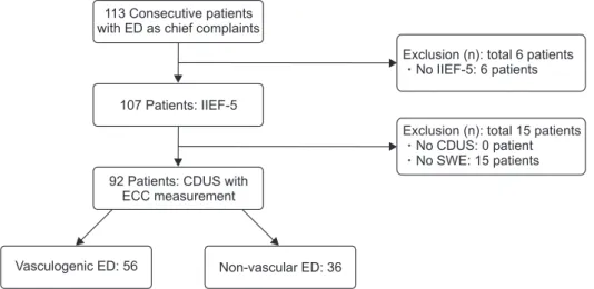
113 Consecutive patients with ED as chief complaints
107 Patients: IIEF-5
92 Patients: CDUS with ECC measurement
Exclusion (n): total 6 patient No IIEF-5: 6 patients
s
Exclusion (n): total 15 patient No CDUS: 0 patient No SWE: 15 patients
s
Vasculogenic ED: 56 Non-vascular ED: 36
Fig. 1. Flow diagram showing patient selection. ED: erectile dysfunction, IIEF- 5: abridged five-item version of the Inter national Index of Erectile Function, CDUS: color Doppler ultrasound, SWE:
shear-wave elastography, ECC: elasticity of corpus cavernosum.
the IIEF-5 [20], we stratified patients into the following four groups: normal, more than 18 points; mild, 14 to 17 points; moderate, 10 to 13 points; and severe, less than 9 points.
3. Ultrasonography technique
Penile USG was performed by a radiologist with 15 years of experience with a high-frequency linear trans- ducer (12–15 MHz) of an Aixplorer ultrasound system (SuperSonic Imagine SA, Aix-en-Provence, France) for all patients. Penile scans were performed on the ven- tral surface using longitudinal and transverse views for patients in the supine position. Grayscale sonogra- phy and CDUS were performed for evaluating anatom- ical structure of the penis and penile vessels in flaccid state. A mixture of agents such as papaverine, phentol- amine, and prostaglandin E1 (STANDRO®; Shinpoong Pharm. Co., Seoul, Korea) was injected into the lateral aspect of the mid-shaft of the penis after evaluating the flaccid state. After grayscale sonographic evalua- tions with a similar method to that described for the flaccid state, we switched to SWE mode.
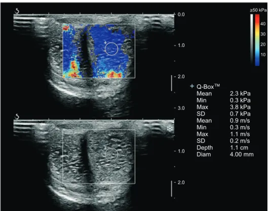
Elasticity measurements were repeated in the trans- verse plane from the mid-shaft and glans of penis every 5 minutes from 5 minutes beginning at 5 min- utes after injection. The first measurement was made
from the cavernosal artery-centered circular region of interest (ROI) with a 4.0-mm diameter, which was placed less than 2 cm in depth on each side of the cor- pus cavernosum (CC), namely, the central elasticity of corpus cavernosum (ECC) (Fig. 2). In addition, repeated measurements were performed with the same sized ROI at the peripheral portion of the CC near the tu- nica albuginea, namely, the peripheral ECC. Finally, we measured Young’s modulus by SWE at the glans penis (elasticity of glans penis on SWE, EG). At least three valid measurements were obtained for each ROI position on both sides of the cavernosum and glans.
All measurements were performed repeatedly at 10 to 20 minutes after intracavernosal injection. These measurements were recorded in kilopascals (kPa). The mean value of these measurements was used for sta- tistical analysis. Minimal compression was given to obtain the grayscale. SWE images were obtained with the goal of achieving identical images for all patients.
In addition, CDUS was simultaneously conducted. Af- ter intracavernosal injection, spectral sampling of the cavernosal artery was performed at the penile root under guidance of a colored Doppler signal. The peak systolic velocity and end diastolic velocity of the cav- ernosal artery were measured with angle correction.
Previously published CDUS criteria for diagnosing
0.0
1.0
2.0
3.0
1.0
2.0
Q-Box Mean Min Max SD Mean Min Max SD Depth Diam
2.3 kPa 0.3 kPa 3.8 kPa 0.7 kPa 0.9 m/s 0.3 m/s 1.1 m/s 0.2 m/s 1.1 cm 4.00 mm
40 30 20 10
>50 kPa
Fig. 2. Shear-wave elastography of a 56-year-old man with a penile rigidity score of 75. 4.0-mm2 circular region of in- terest (ROI) was placed in the left central corpus cavernosum. The mean elasticity of the ROI was 2.3 kPa. Min: minimum, Max: maximum, SD: standard deviation, Diam: diameter.
vascular ED after intracavernosal injection were used (Supplement) [21]. Measurements were not completed if the patient was unable to tolerate the study procedure due to illness or refusal to take the injection. After the CDUS exam, patients reported a VAS (rigidity score, 0–100) to gauge penile rigidity after intracavernosal injection. Patients were able to check the scale with in- formed reference for each score range. The tool asked them to consider the question, “How would you rate the hardness of your erection? (full range, 0–100)” and firstly, they selected one range of the following options (reference) and then, select a specific number within the selected range: 1–10: Penis does not enlarge; 10–50:
Penis is hard, but not hard enough for penetration;
50–70: Penis is hard enough for penetration, but not completely hard; 70–100: Penis is completely hard and sufficiently rigid. Subjective measurement of erection rigidity can be achieved by the VAS.
4. Statistical analysis
Data are presented as the mean±standard deviation or the median (25% quantile–75% quantile). Normal- ity tests for continuous variables were performed using the Shapiro–Wilk normality test. ECC measure- ments (kPa) were compared between measurement sites (central ECC, peripheral ECC, and EG) using the Wilcoxon signed-rank test with continuity correction.
Correlations of ECC measurements (kPa) with rigid- ity scores (0–100) or the IIEF-5 scores were analyzed using Pearson’s product-moment correlation according to measurement site. ECC measurements (kPa) were also compared between ED types based on CDUS by performing the Mann–Whitney U-test or one-way analysis of variance with Tukey’s post hoc adjustment.
The sensitivity, specificity, and positive and negative predictive values for predicting the patients with low rigidity scores were calculated by dichotomizing the results. The area under the receiver operator charac- teristic (ROC) curve (AUC) and the corresponding con- fidence intervals were estimated. Optimal cut-off value was selected using Youden index: the value correspond- ing with the maximum of the Youden’s index. The p- values <0.05 (two tailed) were considered statistically significant. Statistical analyses were performed using R software ver. 3.4.3 (R Foundation for Statistical Com- puting, Vienna, Austria).
RESULTS
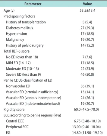
A total of 92 patients with ED who met our inclu- sion criteria were finally included in this study. A flow diagram of the patient selection process is shown in Fig. 1. The demographic characteristics of the patients enrolled are summarized in Table 1. The mean IIEF- 5 score of the total patient group was 9.78±5.01, which falls into a moderate to severe degree of ED. ECC mea- surements on SWE are summarized in Table 1 accord- ing to the penis measurement site. The central ECC value was significantly lower than the peripheral ECC value on the paired test (p<0.001, Wilcoxon signed-rank test).
1. Elasticity of corpus cavernosum vs. rigidity score
There was a significant correlation between the
Table 1. Demographic characteristics of 92 patients who underwent penile CDUS with shear-wave elastography
Parameter Value
Age (y) 53.5±13.4
Predisposing factors
History of transplantation 5 (5.4)
Diabetes mellitus 27 (29.3)
Hypertension 17 (18.5)
Malignancy 19 (20.7)
History of pelvic surgery 14 (15.2) Total IIEF-5 score
No ED (over than 18) 7 (7.6)
Mild ED (14–17) 17 (18.5)
Moderate ED (10–13) 22 (23.9)
Severe ED (less than 9) 46 (50.0) Penile CDUS classification of ED
Nonvascular ED 36 (39.1)
Vascular ED (arterial insufficiency) 13 (14.1) Vascular ED (venous incompetence) 24 (26.1) Vascular ED (indeterminate/mixed) 19 (20.7)
Rigidity score 60.0 (47.5–70.0)
ECC according to penile regions (kPa)
Central ECC 6.75 (5.48–10.19)
Peripheral ECC 13.00 (9.40–18.04)
EG 14.80 (11.90–19.10)
Values are presented as mean±standard deviation, number (%), or median (interquartile range).
CDUS: color Doppler ultrasound, IIEF-5: abridged five-item version of the International Index of Erectile Function, ED: erectile dysfunction, ECC: elasticity of corpus cavernosum on shear-wave elastography, EG:
elasticity of glans penis on shear-wave elastography.
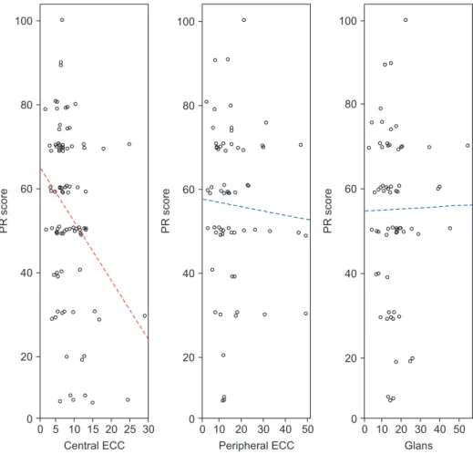
rigidity score and mean central ECC value (r=-0.285;
95% confidence interval [CI]: -0.4662 to -0.0799; p<0.01;
t=-2.7526, degree of freedom=86). In contrast, the pe- ripheral ECC and EG showed no significant correlation (Fig. 3). After the patients with rigidity scores of 50 were dichotomized, ROC analysis showed that at an ECC cut-off value of 8.6 kPa, the sensitivity, specificity, positive predictive value, and negative predictive value were 48.9%, 83.0%, 73.3%, and 62.9%, respectively. The AUC was 0.629 with a standard error of 0.059 (95% CI:
0.522–0.728, p=0.0187).
2. Elasticity of corpus cavernosum vs.
International Index of Erectile Function 5
Although the IIEF-5 scores did not show a significant correlation with the elasticity value in all three mea- surements, weak inverse correlations were found in the central ECC (p=0.219; r=-0.132), peripheral ECC (p=0.191;
r=-0.165), and EG values (p=0.209; r=-0.153) (Fig. 4).
3. Elasticity of corpus cavernosum comparison between types of erectile dysfunction
Vascular ED patients showed a significantly higher central ECC value than nonvascular ED patients (W=595.5, p<0.001 on Wilcoxon rank sum test with con- tinuity correction) (Fig. 5). ROC analysis showed that at an ECC cut-off value of 8.05 kPa, the sensitivity, speci- ficity, positive predictive value, and negative predictive value for predicting vascular ED were 44.4%, 83.7%, 2.99, and 0.61, respectively. The AUC was 0.720 with a standard error of 0.059 (p=0.019). There were also sig- nificant differences in the central ECC values between arteriogenic, venogenic, indeterminate and nonvascular ED (p=0.026 in the Kruskal Wallis one-way ANOVA with Tukey’s post hoc adjustment). The central ECC value was significantly higher for the arterial insuf- ficiency type of vascular ED than for nonvascular ED or the other types of vascular ED (p=0.011). Neither the peripheral ECC nor EG showed a significant difference between the types of ED. Venogenic or mixed-type ED showed no significant difference in the ECC value.
100
80
60
40
20
30
PRscore
25 20 15 10 5
Central ECC
100
80
60
40
20
50
PRscore
30 20 10
Peripheral ECC
100
80
60
40
20
50
PRscore
30 20 10
Glans
40 40
0 0
0 0
0 0
Fig. 3. Correlation between the elasticity of corpus cavernosum (ECC) and penile rigidity scores (PR score) according to penile regions. (A) Central ECC, (B) periph- eral ECC, and (C) glans (elasticity of glans penis) measurements.
DISCUSSION
During the erection phase, there is a significant dif- ference in the internal smooth muscle density between the central CC and peripheral CC (near the tunica
membrane) on grayscale USG. In this study, central CC measurements of Young’s modulus on SWE showed a significant correlation with changes in penile rigid- ity, while the peripheral CC and glans measurements did not show any significant differences. Turkay et al [22] performed stiffness measurements of the CC using SWE in ED patients with IIEF scores of 17 or lower and healthy controls that did not have ED. They found that there were statistically significant differences in mean SWE measurements of the two groups. However, their results could not reflect penile rigidity at erec- tion because they performed measurements only in the flaccid state. Cui et al [23] performed SWE measure- ments in the erectile state and reported that cavernosal SWE values at the erectile state are significantly lower than those in the flaccid state in ED patients. These results suggest that SWE can be used to evaluate ED.
Based on these studies, we aimed to find a quantitative method to predict penile rigidity by measuring SWE in ED patients. The findings of Cui et al [23] and Zheng et al [12] also showed that penile shear wave velocity values significantly decrease as tumescence progresses
Nonvascular ED 30
25 20 15 10 5
Vascular ED
CentralECC(kPa)
Types of ED
Fig. 5. Comparison of penile elasticity measurements (central elas- ticity of corpus cavernosum [central ECC]) according to the erectile dysfunction (ED) subtype. Box-plot shows a significant difference be- tween vascular ED and nonvascular ED, p<0.05 by Wilcoxon signed- rank test.
20
15
10
5
30
IIEF-5(total)
25 20 15 10 5
Central ECC
20
15
10
5
50
IIEF-5(total)
30 20 10
Peripheral ECC
20
15
10
5
50
IIEF-5(total)
30 20 10
Glans
40 40
0
0 0
0 0
0
Fig. 4. Correlation between the elasticity of corpus cavernosum (ECC) mean value and IIEF (total) score according to penile regions. (A) Central ECC, (B) peripheral ECC, and (C) glans (elasticity of glans pe- nis) measurements. IIEF-5: abridged five- item version of the International Index of Erectile Function.
because the amount of smooth muscle trabeculae with- in the ROI gradually decreases until it reaches a full erection phase. In the current study, stiffness measure- ments were performed in the center of the caverno- sum, which is known to have web-like configurations of many smooth muscle structures and various sizes of arterioles and capillaries. In the central CC area, there were significant inverse correlations between penile rigidity and Young’s modulus of the cavernosum (kPa) during erection. Most ED patients showed an increased smooth muscle density in the peripheral CC compared with the central CC during erection. Additionally, the glans penis showed higher echogenicity than the central CC. It is likely that SWE could not reflect the intracavernosal pressure change effectively in these areas due to the long distance from the cavernosal arteries as the main blood supply. Stiffness measure- ments for the peripheral CC and glans penis showed no significant relationship with any clinical parameters regarding ED.
Since the 1990s, CDUS has become an essential tool for identifying and classifying the organic causes of ED. Although its importance has decreased due to the introduction of effective oral medication (phospho- diesterase 5 inhibitor, PDE5-I), its certain roles have remained for evaluation of nonresponders of PDE5-I or patients who have undergone pelvic surgeries for pros- tate or rectal cancer. In addition, CDUS can be initially assessed to confirm vascular ED [21]. CDUS, however, fails to characterize ED patients who present with poor rigidity despite normal CDUS findings, known as a type of nonvascular ED. In our study, there were significant differences in central ECC measurements at erection among ED types, such as venogenic, arterio- genic, indeterminate and nonvascular ED. Additionally, vascular ED showed a higher central ECC value than nonvascular ED. The reason why arteriogenic ED mea- sured more relevant than venogenic ED in our results as follows; Our study cohort had more vasculogenic ED patients than non-vasculogenic type. Probably, that was caused by the enrollment of more patients who underwent pelvic injury or pelvic surgeries (33 pa- tients, 25.8%). This can be a source of bias against the major causes of ED in the general population. After pelvic surgeries, the cavernoso-occlusive mechanism is lost with subsequent venogenic ED when a high pro- portion of trabecular smooth muscle is affected [24].
Unfortunately, SWE measurements showed no sig-
nificant associations with the IIEF total or firmness subdomain. However, the current study suggests that SWE measurement can complement the subjective shortcoming of a visual analog scale for penile rigid- ity. It can be used to obtain a more detailed description of a patient’s condition when it is used in combination with IIEF questionnaires. This study has some limita- tions. First, its retrospective design and relatively small size of patients could lead to selection bias. The absence of a healthy control group might have also affected the performance of SWE parameters. A randomized con- trolled study with a larger patient group and an objec- tive rigidity measurement is needed in the future.
CONCLUSIONS
Quantitatively measuring Young’s modulus of CC using SWE could be an objective technique to assess the penile rigidity of ED patients and to predict vascu- lar ED. Patients with high ECC could have a high pos- sibility of ED and vascular ED patients showed higher ECC value than non-vascular ED. Thus, our results suggest that the central cavernosal stiffness during erection could be a quantitative clinical imaging index for penile rigidity and vascular ED in ED patients.
ACKNOWLEDGEMENTS
This research was supported by a grant (NRF- 2019R1A2C2004746) of the National Research Foun- dation of Korea funded by the Korean Government (MEST). It was also supported by a faculty research grant of Yonsei University College of Medicine for 2018 (6-2018-0066).
Conflict of Interest
The authors have nothing to disclose.
Author Contribution
Conceptualization: JYL, DCJ, SL, NGK, YTO, KH. Data cu- ration: JYL, DCJ, SL, NGK. Formal analysis: JYL, DCJ, KH.
Funding acquisition: DCJ. Investigation: JYL, DCJ, SL, NGK, YTO. Methodology: JYL, DCJ, SL, NGK, KH. Project adminis- tration: DCJ. Resources: JYL, DCJ. Software: JYL, DCJ. Super- vision: DCJ, YTO. Validation: JYL, DCJ, KH. Visualization: SL, NGK. Writing – original draft: JYL, DCJ. Writing – review &
editing: JYL, DCJ.
Data Sharing Statement
The data analyzed for this study have been deposited in HARVARD Dataverse and are available at https://doi.
org/10.7910/DVN/RFM11L.
Supplementary Material
Supplementary material can be found via https://doi.
org/10.5534/wjmh.190094.
REFERENCES
1. Jønler M, Moon T, Brannan W, Stone NN, Heisey D, Bruske- witz RC. The effect of age, ethnicity and geographical location on impotence and quality of life. Br J Urol 1995;75:651-5.
2. Laumann EO, Paik A, Rosen RC. Sexual dysfunction in the United States: prevalence and predictors. JAMA 1999;281:
537-44.
3. Ayta IA, McKinlay JB, Krane RJ. The likely worldwide in- crease in erectile dysfunction between 1995 and 2025 and some possible policy consequences. BJU Int 1999;84:50-6.
4. Thompson IM, Tangen CM, Goodman PJ, Probstfield JL, Moinpour CM, Coltman CA. Erectile dysfunction and subse- quent cardiovascular disease. JAMA 2005;294:2996-3002.
5. Guo W, Liao C, Zou Y, Li F, Li T, Zhou Q, et al. Erectile dys- function and risk of clinical cardiovascular events: a meta- analysis of seven cohort studies. J Sex Med 2010;7:2805-16.
6. Dong JY, Zhang YH, Qin LQ. Erectile dysfunction and risk of cardiovascular disease: meta-analysis of prospective cohort studies. J Am Coll Cardiol 2011;58:1378-85.
7. Nguyen HMT, Gabrielson AT, Hellstrom WJG. Erectile dys- function in young men: a review of the prevalence and risk factors. Sex Med Rev 2017;5:508-20.
8. Vlachopoulos CV, Terentes-Printzios DG, Ioakeimidis NK, Aznaouridis KA, Stefanadis CI. Prediction of cardiovascular events and all-cause mortality with erectile dysfunction: a systematic review and meta-analysis of cohort studies. Circ Cardiovasc Qual Outcomes 2013;6:99-109.
9. Eardley I, Ellis P, Boolell M, Wulff M. Onset and duration of action of sildenafil for the treatment of erectile dysfunction.
Br J Clin Pharmacol 2002;53 Suppl 1:61-5S.
10. Yuan YM, Zhou S, Zhang K. [Methods for evaluation of pe- nile erection hardness]. Zhonghua Nan Ke Xue 2010;16:642- 5. Chinese.
11. Allen RP, Smolev JK, Engel RM, Brendler CB. Comparison of
RigiScan and formal nocturnal penile tumescence testing in the evaluation of erectile rigidity. J Urol 1993;149:1265-8.
12. Zheng X, Ji P, Mao H, Wu J. Evaluation of penile erection rigidity in healthy men using virtual touch tissue quantifica- tion. Radiol Oncol 2012;46:114-8.
13. Rosen RC, Cappelleri JC, Smith MD, Lipsky J, Peña BM. De- velopment and evaluation of an abridged, 5-item version of the International Index of Erectile Function (IIEF-5) as a di- agnostic tool for erectile dysfunction. Int J Impot Res 1999;11:
319-26.
14. Huang ST, Hsieh ML. Different hemodynamic responses by color Doppler ultrasonography studies between sildenafil non-responders and responders. Asian J Androl 2007;9:129- 33.
15. Richards G, Goldenberg E, Pek H, Gilbert BR. Penile sono- elastography for the localization of a non-palpable, non-sono- graphically visualized lesion in a patient with penile curvature from Peyronie’s disease. J Sex Med 2014;11:516-20.
16. Nowicki A, Dobruch-Sobczak K. Introduction to ultrasound elastography. J Ultrason 2016;16:113-24.
17. Cosgrove D, Piscaglia F, Bamber J, Bojunga J, Correas JM, Gilja OH, et al; EFSUMB. EFSUMB guidelines and recom- mendations on the clinical use of ultrasound elastography.
Part 2: clinical applications. Ultraschall Med 2013;34:238-53.
18. Rosen RC, Riley A, Wagner G, Osterloh IH, Kirkpatrick J, Mishra A. The international index of erectile function (IIEF):
a multidimensional scale for assessment of erectile dysfunc- tion. Urology 1997;49:822-30.
19. Cappelleri JC, Rosen RC, Smith MD, Mishra A, Osterloh IH.
Diagnostic evaluation of the erectile function domain of the International Index of Erectile Function. Urology 1999;54:
346-51.
20. Ahn TY, Lee DS, Kang WC, Hong JH, Kim YS. Validation of an abridged Korean version of the International Index of Erectile Function (IIEF-5) as a diagnostic tool for erectile dysfunction. Korean J Urol 2001;42:535-40.
21. Jung DC, Park SY, Lee JY. Penile Doppler ultrasonography revisited. Ultrasonography 2018;37:16-24.
22. Turkay R, Inci E, Yenice MG, Tugcu V. Shear wave elastogra- phy: Can it be a new radiologic approach for the diagnosis of erectile dysfunction? Ultrasound 2017;25:150-5.
23. Cui A, Xu L, Mu J, Tong M, Peng C, Wu T. The role of shear wave elastography on evaluation of the rigidity changes of corpus cavernosum penis in venogenic erectile dysfunction.
Eur J Radiol 2018;103:1-5.
24. Gontero P, Fontana F, Kocjancic E, Frea B, Tizzani A. Male and female sexual dysfunction (SD) after radical pelvic uro- logical surgery. ScientificWorldJournal 2006;6:2302-14.
![Fig. 5. Comparison of penile elasticity measurements (central elas- elas-ticity of corpus cavernosum [central ECC]) according to the erectile dysfunction (ED) subtype](https://thumb-ap.123doks.com/thumbv2/123dokinfo/5408854.220803/6.892.77.649.92.868/comparison-elasticity-measurements-central-cavernosum-according-erectile-dysfunction.webp)