THE KOREAN JOURNAL OF HEMATOLOGY O R I G I N A L A R T I C L E
Prognostic significance of the FLT3 ITD mutation in patients with normal-karyotype acute myeloid leukemia in relapse
Sang Hyuk Park
1, Hyun-Sook Chi
1, Sook-Kyung Min
1, Young-Uk Cho
2, Seongsoo Jang
1, Chan-Jeoung Park
1, Jung-Hee Lee
3, Je-Hwan Lee
3, Kyoo-Hyung Lee
3, Ho-Joon Im
4, Jong-Jin Seo
41Department of Laboratory Medicine, University of Ulsan College of Medicine and Asan Medical Center, 2Department of Laboratory Medicine, Eulji University School of Medicine, Eulji General Hospital, Departments of 3Internal Medicine and 4Pediatrics, University of Ulsan College of Medicine and Asan Medical Center, Seoul, Korea
p-ISSN 1738-7949 / e-ISSN 2092-9129 DOI: 10.5045/kjh.2011.46.2.88 Korean J Hematol 2011;46:88-95.
Received on March 10, 2011 Revised on April 11, 2011 Accepted on May 17, 2011
Background
Fms-like tyrosine kinase 3 internal tandem duplication (FLT3 ITD) mutation is related to poor prognosis in normal-karyotype acute myeloid leukemia (AML). However, the prog- nostic significance of the mutation at relapse has not been adequately investigated. We investigated the prognostic significance of the FLT3 ITD mutation at relapse in normal-kar- yotype AML patients.
Methods
We analyzed 69 normal-karyotype AML patients, in whom paired bone marrow samples taken at initial diagnosis and subsequent relapse were analyzed for the FLT3 ITD mutation at the Asan Medical Center between 1995 and 2009.
Results
Forty patients showed a persistent wild-type genotype, 11 showed the FLT3 ITD mutation at diagnosis and relapse, and 9 lost and another 9 acquired the mutation at relapse. The mutation status at relapse affected the overall survival (OS), with the mutation group showing shorter OS and survival after relapse than the wild-type group did (P<0.001 and P<0.001, respectively), despite having received more frequent stem cell trans- plantation after relapse than the wild-type group did. However, no difference was detected in the OS and survival after relapse with regard to the mutation status at diagnosis.
Conclusion
The patients with FLT3 ITD mutation at relapse showed poorer prognoses than those with- out the mutation. However, mutation status at diagnosis did not affect the outcome. These results suggest that, in normal-karyotype AML patients with relapse, the prognostic sig- nificance of FLT3 ITD mutation at relapse is greater than that of the mutation status at diagnosis.
Key Words AML, Prognosis, FLT3 ITD, Relapse, Normal karyotype
Correspondence to Hyun-Sook Chi, M.D., Ph.D.
Department of Laboratory Medicine, Asan Medical Center and University of Ulsan College of Medicine, 86, Asanbyungwon- gil, Songpa-gu, Seoul 138-736, Korea Tel: +82-2-3010-4502
Fax: +82-2-478-0884 E-mail: hschi@amc.seoul.kr
Ⓒ 2011 Korean Society of Hematology
INTRODUCTION
The Fms-like tyrosine kinase 3 (FLT3) gene encodes a class III receptor tyrosine kinase that plays important roles in cellular proliferation and differentiation [1-8]. To date, 2 types of FLT3 mutations have been found to induce auto- phosphorylation through ligand-independent FLT3 dimeri- zation, leading to uncontrolled hematologic progenitor cell proliferation and malignancy. One such mutation is an in- ternal tandem duplication (ITD) of the region between exon
14 and 15 encoding the juxtamembrane domain; the other, in exon 20, is a point mutation in aspartic acid residue 835 (D835) within the activation loop of the second tyrosine kinase domain [9, 10]. The FLT3 ITD mutation is found in approximately 30% of newly diagnosed acute myeloid leukemia (AML) patients, and an association of the mutation with poor prognostic indicators, such as leukocytosis, higher marrow blast counts, shorter overall survival (OS) and event-free survival (EFS), and higher relapse rates, has been firmly established in those with newly diagnosed AML, espe- cially in the intermediate cytogenetic risk group [11-16].
The FLT3 D835 mutation occurs less frequently than the FLT3 ITD mutation does, and the prognostic significance of this mutation in newly diagnosed AML patients is less clear, with previous studies reporting conflicting outcomes [17-19]. In addition to the FLT3 ITD mutation, mutations in the nucleophosmin1 (NPM1) gene are the most frequent genetic alterations, being found in approximately 50% of patients with normal-karyotype AML, and known to be asso- ciated with better response to induction therapy and a more favorable outcome in the absence of FLT3 ITD mutations [20].
Thus far, the prognostic impact of the FLT3 ITD mutation at relapse has not been adequately evaluated in patients with normal-karyotype AML. Therefore, we analyzed FLT3 ITD mutation status at diagnosis and relapse in patients with normal-karyotype AML relapse, and investigated the prog- nostic significance of FLT3 ITD mutation at relapse.
MATERIALS AND METHODS
1. Selection of patients and assessment of clinical variables Paired bone marrow samples collected at diagnosis and relapse from 69 patients newly diagnosed with normal-kar- yotype AML between 1995 and 2009 at Asan Medical Center were analyzed for FLT3 ITD mutation status. Data for clinical variables, including CBC, blast counts in peripheral blood (PB) and bone marrow (BM), karyotype at initial diagnosis and relapse, and implementation of stem cell transplantation (SCT) were obtained from all patients. The dates of initial diagnosis, first complete remission (CR), first relapse, SCT performance, and death were established by performing a retrospective review of the patients’ medical records. CR was defined as the presence of less than 5% blasts with more than 20% cellularity in a standardized BM aspirate after the first or second course of induction chemotherapy.
Relapse was defined as more than 5% leukemic blasts in BM aspirates in patients who had previously achieved CR state. OS was defined as the time from diagnosis to death or last follow-up, and survival after relapse was defined as the time from first relapse to death or last follow-up.
2. Cytogenetic analysis
Cytogenetic analyses were performed on at least 20 meta- phases using paired diagnosis and relapse BM samples.
According to the classification system proposed by the US Southwest Oncology Group (SWOG), cytogenetic abnormal- ities at relapse were grouped into 4 categories [21]: (1) good- prognosis: inv(16)/t(16;16)/del(16q) or t(8;21) lacking del(9q) and complex karyotypes; (2) intermediate-prognosis: normal karyotype, +8, +6, Y, and del(12p); (3) poor-prognosis: 5/del (5q), 7/del(7q), abn(3q), t(6;9) and complex karyotypes; (4) unknown-prognosis: all the other above-mentioned abnor- malities.
3. Patient treatment
All patients received standard induction chemotherapy
with cytarabine and daunorubicin. This regimen included continuous intravenous infusion of 200 mg/m2/day (100 mg/
m2/day for patients aged >60 years) of cytarabine on days 1-7 and 45 mg/m2/day of daunorubicin on days 1-3. Patients who failed to achieve CR, but attained partial remission (PR) received a second identical cycle of induction chemo- therapy. At relapse, the patients received the same regimen of induction chemotherapy used following the initial diagnosis. Depending on patient age and availability of a suitable donor, the patients received autologous or allogeneic SCT on the first CR status or later. Patients with FLT3 ITD mutation at initial diagnosis were preferentially invited to receive autologous or allogeneic SCT at the first CR status.
However, there was no difference in the induction chemo- therapy regimen according to FLT3 ITD mutation status at initial diagnosis or relapse.
4. Detection of FLT3 ITD and NPM1 mutations
FLT3 ITD and NPM1 mutations were identified using PCR-capillary electrophoresis methods. Genomic DNA was extracted from BM mononuclear cells using the Qiagen DNA purification kit (Qiagen, Hilden, Germany) and stored at -20oC. Primer sets were designed to detect the FLT3 ITD and NPM1 4 base pair insertion mutations. Additionally, a specific set of HBG primers was used to ascertain DNA integrity and validate the precision of PCR amplification.
The sequences of the primers specific to FLT3, NPM1, and HBG were as follows. FLT3 primers: forward, 5'-FAM-AGCA ATT TAG GTA TGA AAG CCA GCTA-3'; reverse, 5'-CTT TCA GCA TTT TGA CGG CAA CC-3'. NPM1 primers: for- ward, 5'-GTT TCT TTT TTT TTT TTT CCA GGC TAT TCA AG-3', reverse, 5'-HEX-CAC GGT AGG GAA AGT TCT CAC TCT GC-3'. HBG primers: forward, 5'-HEX-CCA GAA GAG CCA AGG ACA GGT ACG-3', reverse, 5'-AGA TCC CCA AAG GAC TCA AAG AAC C-3'. The 20 μL multiplex PCR reaction solution consisted of 200 ng of genomic DNA, 20 pmol of each primer, 25 mmol/L MgCl2, 2.5 mmol/L dNTP, 2 μL of 10× PCR buffer, 0.2 μL of Qiagen HotTaq DNA polymerase and water. The PCR conditions were as follows: the mixture was heated to 95oC for 15 min for initial denaturation, and then 35 cycles of 95oC for 20 sec, 60oC for 40 sec, and 72oC for 40 sec were performed, followed by a final extension phase of 72oC for 30 min. The PCR products were then diluted 1:30 and analyzed by capillary electrophoresis using ABI 310 genetic analyzer (Applied Biosystems Inc., Foster city, CA, USA) and GeneScan Analysis software (version 1.2, Applied Biosystems Inc., Foster City, CA, USA). The data is represented as a ratio of mutant cells:wild-type cells. The lowest detection limit of this proto- col was 5%, which had been determined by previous serial dilution experiments [22]. For the specimens, in which NPM1 mutation was detected, additional direct sequencing was per- formed to confirm the mutation subtype.
5. Analysis of clinical variables related to FLT3 ITD paired mu- tation status at diagnosis and relapse
An analysis of clinical variables related to FLT3 ITD muta-
tion status at diagnosis and relapse was performed. Results were grouped into 4 categories: (1) persistent wild type:
NN (negative at diagnosis and relapse); (2) acquired mutation:
NP (negative at diagnosis and positive at relapse); (3) loss of mutation: PN (positive at diagnosis and negative at relapse);
and (4) persistent mutation: PP (positive at diagnosis and relapse). The clinical variables described above were com- pared individually between NN and PN, NN and NP, NN and PP, and PN and PP groups. Two additional comparisons were made: between patients positive for FLT3 ITD mutation at relapse (NP+PP) and those negative for FLT3 ITD mutation at relapse (NN+PN), and between patients positive for FLT3 ITD mutation at diagnosis (PN+PP) and those negative for FLT3 ITD mutation at diagnosis (NN+NP). Additionally, we compared the prognosis in patients without FLT3 ITD muta- tion stratifying according to NPM1 mutation status at diag- nosis or relapse to identify the prognostic impact of NPM1 mutations. We also compared the prognosis of patients on the basis of FLT3 ITD mutant:wild-type ratio in patients with the mutation at diagnosis or relapse in order to evaluate the effect of the amount of mutation on the prognosis.
6. Multivariate analysis of OS and survival after relapse with respect to variables, including FLT3 ITD mutation status at diagnosis and relapse
Multivariate analysis of OS and survival after relapse with respect to FLT3 ITD mutation status at diagnosis and relapse was performed. For the analysis at diagnosis, WBC counts and blast percentage in PB and BM, which were found to be significantly different among FLT3 ITD mutation groups in univariate analysis, were used to adjust the relevant out- comes. For the analysis at relapse, duration of CR and SCT performance rates after relapse, which showed significant differences among FLT3 ITD mutation groups in univariate analysis, were used to adjust the dependent measures.
7. Statistical analysis
Pearson’s chi-squared or Fisher’s exact tests (for small numbers) were used to compare categorical variables. Mann- Whitney U test was used to compare continuous variables.
Estimations of OS and survival after relapse were performed using Kaplan-Meier methods and compared using log-rank test. Multivariate analyses of OS and survival after relapse were performed using Cox’s proportional hazards model.
For all analyses, tests were two-tailed, and P-values≤0.05 were considered statistically significant. All calculations were performed using SPSS 13.0.1 for Windows (SPSS Inc, Chicago, IL, USA).
RESULTS
1. Overall patient characteristics
All patients achieved first CR after a median interval of 32.0 days and relapsed within a median of 7.6 months from the first CR. A second CR was achieved in 26 patients (37.7%), of whom 8 (30.8%) relapsed again after the second round
of induction chemotherapy. Fifty-five patients (79.7%) died after relapse with a median interval of 3.8 months. The FLT3 ITD mutation was detected in 20 patients (29.0%) at diagnosis and relapse, with median mutant:wild-type ratio as 0.3 (range, 0.05-4.36) and 0.3 (range, 0.05-9.94) at diagnosis and relapse, respectively. The NPM1 mutation was detected in 19 (27.5%) and 13 (18.8%) patients at diagnosis and relapse, with median mutant:wild-type ratio as 0.55 (range, 0.16-0.86) and 0.55 (range, 0.07-0.75) at diagnosis and relapse, respec- tively. Among the 19 patients who showed NPM1 mutation at diagnosis, 16 (84.2%) had NPM1 type-A mutation. Among the 13 patients who showed NPM1 mutation at relapse, 10 (76.9%) had NPM1 type-A mutation. The remaining 3 patients had NPM1 type-D mutation at diagnosis and relapse.
Cases showing a change of NPM1 mutation type from diag- nosis to relapse were not found.
2. Comparison of clinical variables and prognosis between patients with different paired FLT3 ITD mutation status Tables 1 and 2 show comparisons of the clinical variables and prognosis between patients with different FLT3 ITD mutation status at diagnosis and relapse. SCT performance rates at the first CR ranged from 25.0% to 44.4%, which is variable but this variation was not driven by FLT3 ITD mutation status at diagnosis and relapse. However, SCT per- formance rates after the first relapse were significantly different. Patients with acquired or persistent FLT3 ITD mutation at relapse more frequently underwent SCT than those with persistent negative status did (55.6% or 63.6%
vs. 20.0%; P=0.043, 0.009, respectively). There was no prog- nostic impact of being in the poor cytogenetic risk group at relapse (OS: 20.6 vs. 22.0 months, P=0.950; duration of CR: 7.5 vs. 8.9 months, P=0.669; survival after relapse: 10.2 vs. 14.3 months, P=0.214).
The PP group showed a significantly shorter median dura- tion of CR (6.9 months) than the NN group (10.3 months;
P=0.045); the PP group also showed a significantly higher median WBC count (44.1×10⁹/L vs. 8.8×10⁹/L; P=0.046) than the NN group, but these variables did not differ between the PP and PN groups. The PN group showed significantly higher median WBC counts (66.1×10⁹/L vs. 8.8×10⁹/L;
P=0.011) and blast percentages in PB (85.0% vs. 38.5%, P=0.006) and in BM (87.0% vs. 60.2%, P=0.001) than the NN group. The median OS was significantly shorter in the NP (9.2 months; P=0.002) and the PP (12.1 months; P=0.003) groups than in the NN group (19.5 months). The median survival after relapse was also significantly shorter in the NP (5.3 months; P=0.017) and the PP (4.9 months; P=0.024) groups than in the NN group (10.3 months). The median OS and survival after relapse in the PP group was significantly shorter than that in the PN group (45.5 months and 43.5 months; P=0.025 and 0.028, respectively).
3. Comparison of clinical variables and prognosis between groups with different FLT3 ITD mutation status at diagnosis and relapse
Table 3 shows 2 additional comparisons of clinical variables
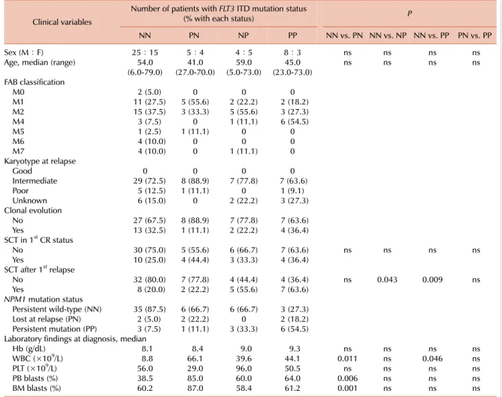
Table 1. Comparison of clinical variables between patients with different paired FLT3 ITD mutation status.
Clinical variables
Number of patients with FLT3 ITD mutation status
(% with each status) P
NN PN NP PP NN vs. PN NN vs. NP NN vs. PP PN vs. PP
Sex (M:F) 25:15 5:4 4:5 8:3 ns ns ns ns
Age, median (range) 54.0 41.0 59.0 45.0 ns ns ns ns
(6.0-79.0) (27.0-70.0) (5.0-73.0) (23.0-73.0) FAB classification
M0 2 (5.0) 0 0 0
M1 11 (27.5) 5 (55.6) 2 (22.2) 2 (18.2)
M2 15 (37.5) 3 (33.3) 5 (55.6) 3 (27.3)
M4 3 (7.5) 0 1 (11.1) 6 (54.5)
M5 1 (2.5) 1 (11.1) 0 0
M6 4 (10.0) 0 0 0
M7 4 (10.0) 0 1 (11.1) 0
Karyotype at relapse
Good 0 0 0 0
Intermediate 29 (72.5) 8 (88.9) 7 (77.8) 7 (63.6)
Poor 5 (12.5) 1 (11.1) 0 1 (9.1)
Unknown 6 (15.0) 0 2 (22.2) 3 (27.3)
Clonal evolution
No 27 (67.5) 8 (88.9) 7 (77.8) 7 (63.6)
Yes 13 (32.5) 1 (11.1) 2 (22.2) 4 (36.4)
SCT in 1st CR status
No 30 (75.0) 5 (55.6) 6 (66.7) 7 (63.6) ns ns ns ns
Yes 10 (25.0) 4 (44.4) 3 (33.3) 4 (36.4)
SCT after 1st relapse
No 32 (80.0) 7 (77.8) 4 (44.4) 4 (36.4) ns 0.043 0.009 ns
Yes 8 (20.0) 2 (22.2) 5 (55.6) 7 (63.6)
NPM1 mutation status
Persistent wild-type (NN) 35 (87.5) 6 (66.7) 6 (66.7) 3 (27.3) Lost at relapse (PN) 2 (5.0) 2 (22.2) 0 2 (18.2) Persistent mutation (PP) 3 (7.5) 1 (11.1) 3 (33.3) 6 (54.5) Laboratory findings at diagnosis, median
Hb (g/dL) 8.1 8.4 9.0 9.3 ns ns ns ns
WBC (×109/L) 8.8 66.1 39.6 44.1 0.011 ns 0.046 ns
PLT (×109/L) 56.0 29.0 96.0 50.5 ns ns ns ns
PB blasts (%) 38.5 85.0 60.0 64.0 0.006 ns ns ns
BM blasts (%) 60.2 87.0 58.4 61.2 0.001 ns ns ns
Abbreviations: FAB, French-American-British classification; CR, complete remission; FLT3 ITD, fms-like tyrosine kinase3 internal tandem duplication; NN, negative at diagnosis and relapse; PN, positive at diagnosis and negative at relapse; NP, negative at diagnosis and positive at relapse; PP, positive at diagnosis and relapse; ns, not significant; Hb, hemoglobin; WBC, white blood cell; PLT, platelet; PB, peripheral blood;
BM, bone marrow; SCT, stem cell transplantation; NPM1, nucleophosmin.
Table 2. Comparison of prognosis between patients with different paired FLT3 ITD mutation status.
Prognostic variables FLT3 ITD mutation status P
NN PN NP PP NN vs. PN NN vs. NP NN vs. PP PN vs. PP
Duration of CR, median (months) 10.3 6.6 5.4 6.9 ns ns 0.045 ns
Overall survival, median (months) 19.5 45.5 9.2 12.1 ns 0.002 0.003 0.025
Survival after relapse, median (months) 10.3 43.5 5.3 4.9 ns 0.017 0.024 0.028
Abbreviations: CR, complete remission; FLT3 ITD, fms-like tyrosine kinase3 internal tandem duplication; NN, negative at diagnosis and relapse;
PN, positive at diagnosis and negative at relapse; NP, negative at diagnosis and positive at relapse; PP, positive at diagnosis and relapse; ns, not significant.
between groups with different FLT3 ITD mutation status at diagnosis and relapse. A comparison between the NN+NP group, which comprised patients with negative FLT3 ITD mutation status at diagnosis, and the PN+PP group, which
included patients with positive FLT3 ITD mutation status at diagnosis, showed that the median WBC count at diagnosis was significantly higher in the PN+PP group (44.1×10⁹/L) than in the NN+NP group (12.4×10⁹/L; P=0.007). Moreover,
Table 3. Comparison of clinical variables and prognosis between groups with different FLT3 ITD mutation status at diagnosis and relapse.
Clinical variables
FLT3 ITD status (% with each status) P
NN+NP (Negative
at diagnosis) PN+PP (Positive
at diagnosis) NN+PN (Negative
at relapse) NP+PP (Positive
at relapse) NN+NP vs.
PN+PP NN+PN vs.
NP+PP
Sex (M:F) 29:20 13:7 30:19 12:8 ns ns
Age, median (range) 56.5 41.0 53.0 55.0 ns ns
(5.0-79.0) (5.0-79.0) (6.0-79.0) (5.0-79.0) SCT in 1st CR status
No 36 (73.5) 12 (66.7) 35 (71.4) 13 (65.0) ns ns
Yes 13 (26.5) 8 (33.3) 14 (28.6) 7 (35.0)
SCT after 1st relapse
No 36 (73.5) 11 (55.0) 39 (79.6) 8 (33.3) ns <0.001
Yes 13 (26.5) 9 (45.0) 10 (20.4) 12 (66.7)
Laboratory findings at diagnosis, median
Hb (g/dL) 8.3 8.6 8.1 8.9 ns ns
WBC (×109/L) 12.4 44.1 17.1 39.5 0.007 ns
PLT (×109/L) 58.0 41.0 51.0 69.0 ns ns
PB blasts (%) 47.0 68.0 55.0 60.0 0.006 ns
BM blasts (%) 61.3 87.0 64.6 64.2 0.005 ns
Duration of CR median (months) 8.6 6.9 10.0 6.8 ns 0.034
Overall survival, median (months) 17.6 13.7 23.1 12.0 ns <0.001
Survival after relapse, median (months) 6.9 6.4 11.8 5.5 ns <0.001
Abbreviations: CR, complete remission; Hb, hemoglobin; WBC, white blood cell; PLT, platelet; PB, peripheral blood; BM, bone marrow; SCT, stem cell transplantation; FLT3 ITD, fms-like tyrosine kinase3 internal tandem duplication; NN, negative at diagnosis and relapse; PN, positive at diagnosis and negative at relapse; NP, negative at diagnosis and positive at relapse; PP, positive at diagnosis and relapse; ns, not significant.
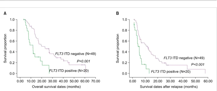
Fig. 1. Comparison of overall survival (A) and survival after relapse (B) between patients positive and negative for FLT3 ITD mutation at diagnosis.
the PN+PP group showed significantly higher blast percent- age in PB (68.0% vs. 47.0%; P=0.006) and in BM (87.0%
vs. 61.3%, P=0.005) than the NN+NP group did. However, there was no difference in the duration of CR, there was no difference in OS and survival after relapse between these groups (Fig. 1). Another comparison was performed between the NN+PN group, which included patients with negative FLT3 ITD mutation status at relapse, and the NP+PP group, which included patients with positive FLT3 ITD mutation status at relapse, revealed that the median duration of CR was significantly shorter in the NP+PP group (6.8 months) than in the NN+PN group (10.0 months; P=0.034). In partic-
ular, the median OS was significantly shorter in the NP+PP group (12.0 months) than in the NN+PN group (23.1 months;
P<0.001), as was survival after relapse (5.5 vs. 11.8 months;
P<0.001), despite the fact that SCT was performed sig- nificantly more frequently in the NP+PP group than in the NN+PN group after the first relapse (66.7% vs. 20.4%; P
<0.001) (Fig. 2).
4. Comparison of prognosis in patients without FLT3 ITD mu- tation stratified by NPM1 genotype status at diagnosis and relapse
Patients with NPM1 mutation at diagnosis showed a trend
Table 4. Multivariate analysis of OS and survival after relapse with respect to FLT3 ITD mutation status at diagnosis and relapse.
Clinical variables Overall survival Survival after relapse
HR (95% CI) P HR (95% CI) P
FLT3 ITD mutation at relapse 2.486 (1.203-5.138) 0.014a) 2.042 (1.062-4.045) 0.039a) (compared with negative at relapse)
FLT3 ITD mutation at diagnosis 1.084 (0.528-2.144) nsb) 1.147 (0.534-2.447) nsb)
(compared with negative at diagnosis)
a)P-value was adjusted for WBC count, duration of CR, blast percentage in PB and BM at diagnosis, and SCT performance rates after 1st relapse,
b)P-value was adjusted for WBC count and blast percentage in PB and BM at diagnosis.
Abbreviations: HR, hazard ratio; FLT3 ITD, fms-like tyrosine kinase3 internal tandem duplication; ns, not significant; CI, confidence interval.
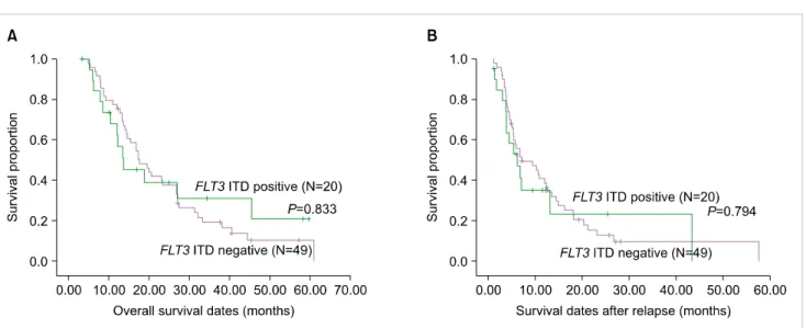
Fig. 2. Comparison of overall survival (A) and survival after relapse (B) between patients positive and negative for FLT3 ITD mutation at relapse.
towards a better outcome, such as longer OS (24.6 vs. 17.3 months; P=0.173) and duration of CR (10.0 vs. 7.5 months;
P=0.558) than those without NPM1 mutation at diagnosis.
However, these differences were not statistically significant.
Patients with NPM1 mutation at relapse also showed no differences in OS (15.4 vs. 23.1 months; P=0.609) and dura- tion of CR (8.6 vs. 10.3 months; P=0.657) compared with those without NPM1 mutation at relapse.
5. Comparison of prognosis in patients with FLT3 ITD muta- tion at diagnosis or relapse according to the amount of FLT3 ITD mutants
The high/low cut-off ratio was set at 0.66, as described in a previous study [23]. Six patients with a high mutant:wild ratio (≥0.66) at diagnosis showed trends towards shorter OS (9.3 vs. 22.5 months; P=0.370) and duration of CR (4.1 vs. 6.6 months; P=0.682) than 14 patients with a low ratio (<0.66) at diagnosis, suggesting possible poor prognosis, but the results were not statistically significant. Eight patients with high mutant:wild ratios (≥0.66) at relapse also showed similar OS (9.2 vs. 10.1 months; P=0.734) and duration of CR (4.1 vs. 5.4 months; P=0.701) as shown by 12 patients with a low ratio (<0.66) at relapse.
6. Multivariate analysis of OS and survival after relapse re- lated to FLT3 ITD mutation status at diagnosis and relapse Table 4 shows the results of multivariate analysis of OS and survival after relapse related to FLT3 ITD mutation status at diagnosis and relapse. FLT3 ITD mutation at relapse, irre- spective of the initial mutation status, showed a statistically significant association with a poor prognosis on OS (HR, 2.486; P=0.014) and survival after relapse (HR, 2.042; P= 0.039). FLT3 ITD mutation at diagnosis did not show statisti- cally significant prognostic impact on OS or survival after relapse.
DISCUSSION
In this study, the FLT3 ITD mutation was present in 20 (29.0%) patients each at diagnosis and relapse, similar to the findings of a recent study [22]. Between diagnosis and relapse, the FLT3 ITD mutation status changed in 18 patients (26.1%). This figure is quite high relative to the recently reported value of 17.5%, and reflects the heterogeneity of FLT3 ITD mutation populations [24].
Patients with persistent FLT3 ITD mutation showed higher initial WBC counts than those with persistent wild-type
status. Patients with loss of the FLT3 ITD mutation at relapse showed higher initial WBC count, blast percentage in PB and BM than those with persistent wild-type status, con- sistent with the previous findings [11-16]. Patients who ac- quired the FLT3 ITD mutation at relapse showed significantly shorter OS and survival after relapse than those with persis- tent wild-type status, and similar differences were observed between patients who acquired the FLT3 ITD mutation at relapse and patients with persistent FLT3 ITD mutation.
Additionally, patients with persistent FLT3 ITD mutation showed a significantly shorter duration of CR than those with persistent wild-type status. These findings were most pronounced in patients with FLT3 ITD mutation at relapse, compared to those with mutation-negative status at relapse, despite SCT being significantly more frequently performed after the first relapse in patients with FLT3 ITD mutation.
Collectively, the results suggest that FLT3 ITD mutation at relapse is a poor prognostic indicator, and are consistent with recent data that reported a shorter time-to-relapse in patients with FLT3 ITD mutation at relapse compared to those without the mutation [24]. On the other hand, FLT3 ITD mutation status at diagnosis influenced WBC count and blast percentage in PB and BM at diagnosis, but prognostic differences according to mutation status at diagnosis were not statistically significant. These differences between the prognostic potential of mutation status at diagnosis and re- lapse may indicate that the prognostic impact of FLT3 ITD mutation status at relapse is more pronounced than the muta- tion status at diagnosis in normal-karyotype AML patients with relapse.
Multivariate analysis revealed the FLT3 ITD mutation at relapse to be an independent indicator of poor prognosis for both OS and survival after relapse. Otherwise, the pres- ence of FLT3 ITD mutation at diagnosis did not show any prognostic effect. These results support our speculation that the impact of FLT3 ITD mutation status at relapse is of greater prognostic value than the mutation status at diagnosis in normal-karyotype AML patients with relapse. Thus far, FLT3 ITD mutation status at diagnosis has been firmly estab- lished as a prognostic indicator for normal-karyotype AML [25, 26]. Moreover, the ratio of mutant:wild-type cells has been suggested to be a possible prognostic indicator by a previous study, which showed significantly lower 5-year relapse-free survival rates (57% vs. 79%, P=0.048) in patients with a high mutant:wild-type ratio (≥0.66) than in those with a low ratio. However, in our study, not only did the FLT3 ITD mutation status at diagnosis not predict outcome but there was also no evidence of a prognostic impact of the relative amount of the mutant cells. One possible ex- planation for the discordant results is the different nature of the patient group included in the analysis. Previous studies have focused on FLT3 ITD mutation status at initial diagnosis;
therefore, all patients could be included into analysis regard- less of their status at relapse. However, in our study, only patients who had undergone a relapse could be enrolled.
Because the clinical outcome between relapsed and non- relapsed patients is quite different, this heterogeneity might
influence the clinical outcome.
Our study had some limitations. First, several molecular aberrations which have potential prognostic impacts in nor- mal karyotype AML, such as CCAAT-enhancer binding pro- tein-α (CEBPA) and Wilm’s tumor 1 (WT1), were not analyzed. These factors may have influenced our results.
Second, the number of patients was relatively small, which may lead to false positives or negatives and thus would have affected our conclusions. Third, although we found that a prognostic impact of NPM1 mutation was not evident at either diagnosis or relapse in patients in the absence of FLT3 ITD mutation, this analysis suffered from low statistical pow- er due to the limited number of patients. For this reason, we could not perform more categorized analysis based on the different mutation status combination of FLT3 ITD and NPM1, which has been widely used in recent studies.
Therefore, further studies with larger patient populations will be required for more accurate analysis.
In conclusion, patients with acquired or persistent FLT3 ITD mutation at relapse showed significantly shorter OS and survival after relapse than those with persistent wild- type status. These statistical differences were most pro- nounced in patients with FLT3 ITD mutation at relapse, when compared to those with mutation-negative status at this time point. However, the mutation status at diagnosis did not affect the outcome. Collectively, these results suggest that the impact of FLT3 ITD mutation status at relapse is expected to be of greater prognostic value than the mutation status at diagnosis in normal karyotype-AML patients with relapse.
REFERENCES
1. Gilliland DG, Griffin JD. The roles of FLT3 in hematopoiesis and leukemia. Blood 2002;100:1532-42.
2. Brasel K, Escobar S, Anderberg R, de Vries P, Gruss HJ, Lyman SD.
Expression of the flt3 receptor and its ligand on hematopoietic cells. Leukemia 1995;9:1212-8.
3. Drexler HG. Expression of FLT3 receptor and response to FLT3 ligand by leukemic cells. Leukemia 1996;10:588-99.
4. Turner AM, Lin NL, Issarachai S, Lyman SD, Broudy VC. FLT3 receptor expression on the surface of normal and malignant hu- man hematopoietic cells. Blood 1996;88:3383-90.
5. Birg F, Courcoul M, Rosnet O, et al. Expression of the FMS/
KIT-like gene FLT3 in human acute leukemias of the myeloid and lymphoid lineages. Blood 1992;80:2584-93.
6. Carow CE, Levenstein M, Kaufmann SH, et al. Expression of the hematopoietic growth factor receptor FLT3 (STK-1/Flk2) in hu- man leukemias. Blood 1996;87:1089-96.
7. Stacchini A, Fubini L, Severino A, Sanavio F, Aglietta M, Piacibello W. Expression of type III receptor tyrosine kinases FLT3 and KIT and responses to their ligands by acute myeloid leu- kemia blasts. Leukemia 1996;10:1584-91.
8. Levis M, Small D. FLT3: ITDoes matter in leukemia. Leukemia 2003;17:1738-52.
9. Hayakawa F, Towatari M, Kiyoi H, et al. Tandem-duplicated Flt3
constitutively activates STAT5 and MAP kinase and introduces autonomous cell growth in IL-3-dependent cell lines. Oncogene 2000;19:624-31.
10. Yamamoto Y, Kiyoi H, Nakano Y, et al. Activating mutation of D835 within the activation loop of FLT3 in human hematologic malignancies. Blood 2001;97:2434-9.
11. Abu-Duhier FM, Goodeve AC, Wilson GA, et al. FLT3 internal tandem duplication mutations in adult acute myeloid leukaemia define a high-risk group. Br J Haematol 2000;111:190-5.
12. Fröhling S, Schlenk RF, Breitruck J, et al. Prognostic significance of activating FLT3 mutations in younger adults (16 to 60 years) with acute myeloid leukemia and normal cytogenetics: a study of the AML Study Group Ulm. Blood 2002;100:4372-80.
13. Kottaridis PD, Gale RE, Frew ME, et al. The presence of a FLT3 internal tandem duplication in patients with acute myeloid leuke- mia (AML) adds important prognostic information to cytogenetic risk group and response to the first cycle of chemotherapy: analysis of 854 patients from the United Kingdom Medical Research Council AML 10 and 12 trials. Blood 2001;98:1752-9.
14. Kiyoi H, Naoe T, Nakano Y, et al. Prognostic implication of FLT3 and N-RAS gene mutations in acute myeloid leukemia. Blood 1999;93:3074-80.
15. Kottaridis PD, Gale RE, Linch DC. Flt3 mutations and leukaemia.
Br J Haematol 2003;122:523-38.
16. Thiede C, Steudel C, Mohr B, et al. Analysis of FLT3-activating mutations in 979 patients with acute myelogenous leukemia: asso- ciation with FAB subtypes and identification of subgroups with poor prognosis. Blood 2002;99:4326-35.
17. Mead AJ, Linch DC, Hills RK, Wheatley K, Burnett AK, Gale RE.
FLT3 tyrosine kinase domain mutations are biologically distinct from and have a significantly more favorable prognosis than FLT3 internal tandem duplications in patients with acute myeloid leukemia. Blood 2007;110:1262-70.
18. Whitman SP, Ruppert AS, Radmacher MD, et al. FLT3 D835/I836
mutations are associated with poor disease-free survival and a dis- tinct gene-expression signature among younger adults with de no- vo cytogenetically normal acute myeloid leukemia lacking FLT3 internal tandem duplications. Blood 2008;111:1552-9.
19. Mead AJ, Gale RE, Hills RK, et al. Conflicting data on the prog- nostic significance of FLT3/TKD mutations in acute myeloid leu- kemia might be related to the incidence of biallelic disease. Blood 2008;112:444-5.
20. Rau R, Brown P. Nucleophosmin (NPM1) mutations in adult and childhood acute myeloid leukaemia: towards definition of a new leukaemia entity. Hematol Oncol 2009;27:171-81.
21. Ferrara F, Palmieri S, Leoni F. Clinically useful prognostic factors in acute myeloid leukemia. Crit Rev Oncol Hematol 2008;66:
181-93.
22. Huang Q, Chen W, Gaal KK, Slovak ML, Stein A, Weiss LM. A rap- id, one step assay for simultaneous detection of FLT3/ITD and NPM1 mutations in AML with normal cytogenetics. Br J Haema- tol 2008;142:489-92.
23. Chillón MC, Santamaría C, García-Sanz R, et al. Long FLT3 in- ternal tandem duplications and reduced PML-RARalpha ex- pression at diagnosis characterize a high-risk subgroup of acute promyelocytic leukemia patients. Haematologica 2010;95:745- 51.
24. Cloos J, Goemans BF, Hess CJ, et al. Stability and prognostic influ- ence of FLT3 mutations in paired initial and relapsed AML samples. Leukemia 2006;20:1217-20.
25. Kim YK, Kim HN, Lee SR, et al. Prognostic significance of nucleo- phosmin mutations and FLT3 internal tandem duplication in adult patients with cytogenetically normal acute myeloid leukemia. Korean J Hematol 2010;45:36-45.
26. Chang SH, Lee NY, Kim DH, Sohn SK, Suh JS. FLT3 Gene Mutations as a Prognostic Factor for Acute Myeloid Leukemia.
Korean J Lab Med 2006;26:233-40.