D I A B E T E S & M E T A B O L I S M J O U R N A L
This is an Open Access article distributed under the terms of the Creative Commons At- tribution Non-Commercial License (http://creativecommons.org/licenses/by-nc/3.0/) which permits unrestricted non-commercial use, distribution, and reproduction in any medium, provided the original work is properly cited.
Role of Peroxisome Proliferator-Activated Receptor α in Diabetic Nephropathy
Sungjin Chung, Cheol Whee Park
Division of Nephrology, Department of Internal Medicine, The Catholic University of Korea College of Medicine, Seoul, Korea
With a developing worldwide epidemic of diabetes mellitus, the renal complications associated with diabetes have become a seri- ous health concern. Primary therapy for treating diabetic nephropathy is a multifactorial process. Peroxisome proliferator-acti- vated receptor alpha (PPARα) agonists have been used primarily in clinical practice for the treatment of dyslipidemia and insulin resistance. Given that PPARα expression and regulation of metabolic pathways are involved in oxidative stress, inflammation, blood pressure regulation, and the renin-angiotensin aldosterone system, PPARα likely influences the development and patho- genesis of diabetic nephropathy via indirect effects on glucose and lipid homeostasis and also by direct action on the kidneys.
These findings suggest that PPARα may become an important therapeutic target for treating diabetic renal complications.
Keywords: Diabetes mellitus; Kidney; PPAR alpha
Corresponding author: Cheol Whee Park,
Division of Nephrology, Seoul St. Mary’s Hospital, The Catholic University of Korea College of Medicine, 505 Banpo-dong, Seocho-gu, Seoul 137-701, Korea
PEROXISOME PROLIFERATOR-ACTIVATED RECEPTORS
Peroxisome proliferator-activated receptors (PPAR) are ligand- activated transcription factors that are members of the nuclear hormone receptor superfamily [1,2]. There are three PPAR isoforms of the distinct genes commonly designated as PPARα (NR1C1), PPARγ (NR1C3) and PPARβ/δ (NR1C2) (or simply δ) [1]. The identification and designation of this PPAR subfam- ily of nuclear receptors in the 1990s was the result of over 25 cumulative years of work with peroxisome proliferators [2].
The PPARs heterodimerize with another nuclear receptor, the 9-cis-retinoic acid receptor (RXR), to form a complex that in- teracts with specific DNA-response elements within the pro- moter regions of the target genes. This heterodimer complex is activated by appropriate ligand binding, so it can therefore re- cruit transcription coactivators and oversee the transcription of genes involved in the regulation of lipid and carbohydrate
metabolism [3].
Tissue expressions differ based on the PPAR subtype [4,5].
PPARα is highly expressed in the liver, renal cortex, intestinal mucosa, and heart, which are all organs that possess high mi- tochondrial and β-oxidation activity. Lower expression of PPARα is also observed in several other tissues. Similar tissue expression profiles of PPARα have been found in rodents and humans [4]. PPARα is abundantly expressed in the proximal tubules and the medullary thick ascending limbs, and to a less- er extent, in the glomerular mesangial cells [6,7]. Given the high level of expression in the renal proximal tubules, PPARα has been implicated in the metabolic control of the kidney in main- taining a sustained balance of energy production and expendi- ture.
PPARs basically function as sensors for fatty acid deriva- tives and control important metabolic pathways involved in lipid and energy metabolism. PPARs also play an important role in various pathophysiologic conditions, such as immuni- pISSN 2233-6079 · eISSN 2233-6087
ty, inflammation, apoptosis, and cell differentiation [8]. Each member of the PPAR subfamily has additional actions [9,10].
PPARα plays an additional role in lipoprotein synthesis, in- flammatory responses and the fatty acid oxidation system [2].
In general, PPARα functions as catabolic regulators of energy [2,11].
Numerous studies in experimental and clinical models have shown the beneficial effects of PPARs in improving organ function in some diseases [12,13]. The PPARα agonists, such as fenofibrate and clofibrate, are traditionally proven lipid- lowering drugs [13]. Despite their serendipitous discovery and clinical use in the treatment of dyslipidemia, the actual phar- macological profile of activity of the PPARα agonists is a new discovery [14]. In addition to their involvement in lipid and li- poprotein metabolism, recent evidence supports the theory that PPARα critically regulates inflammation and vascular function in the kidney, which has led to renewed interest in PPARα as a renal protective option.
ROLE OF PPARα IN NORMAL PHYSIOLOGY
The PPARα gene in the human, which spans ~93.2 kb, is lo- cated on chromosome 22q12-q13.1 and, encodes a protein of 468 amino acids. The PPARα gene that encodes mRNA is de- rived from 8 exons with a 5′ untranslated region encoded by exons 1, 2, and part of exon 3 [15]. The remainder of exon 3 and exons 4-8 are known to contribute to the coding region of PPARα. The last 232 bp of exon 8 contribute to the 3′-untrans- lated region. In the mouse, the PPARα gene is located on chro- mosome 15E2, and it encodes a protein of 468 amino acids [2].
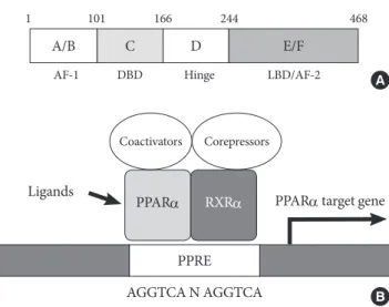
The encoded protein shares functional domains with other nuclear hormone receptors. As with other nuclear receptors and PPARs, PPARα contains four major functional domains, including the N-terminal ligand-independent transactivation domain (A/B domain), the DNA binding domain (DBD or C domain), the co-factor docking domain (D domain), and the C-terminal E/F domain (including the ligand binding domain [LBD] and the ligand-dependent transactivation domain (AF- 2 domain)) (Fig. 1) [13]. The A/B domain contains an activa- tion function-1 (AF-1) region, which has a low level of basal transactivation activity and functions independently of ligand- binding. In humans, DBD encompasses amino acids 101-166, which contain two very highly conserved zinc finger motifs and architectural elements that are capable of sequence-specif- ic binding to DNA [15]. D domain, a flexible hinge domain,
connects the DBD and LBD. This hinge region binds co-re- pressor proteins, with the characteristic LXXXIXXXL repres- sor motif, to the receptor in its quiescent, unliganded state [2].
LBD in the human PPARα protein, which contains an AF-2 region composed of two α-helices flanking one four-stranded β-sheet, extends from amino acids 280 to 468 [15]. The AF-2 domain is repressed until ligand-binding occurs. Following li- gand-binding, the AF-2 domain undergoes a conformational shift, which allows the formation of hydrogen bonds between Tyr-314 and Tyr-464 as well as the formation of a charge clamp between Glu-462 and Lys-292. This conformational change in the protein allows interaction of the receptor with the LXXLL (L, leucine; X, any amino acid) motifs located in co-activator proteins [16]. In a similar fashion as other nuclear receptors, PPARα undergoes conformational adjustment upon binding to a ligand to achieve the co-regulator exchanges and activa- tion of the target genes [2,16].
PPARα, like the other two PPAR isoforms, is localized to the nucleus, which is characteristic of the type II nuclear re- ceptor family [2]. PPARα regulates many target genes, and the
A/B
1 101 166 244 468
AF-1 DBD Hinge LBD/AF-2
C D E/F
PPARα target gene PPRE
Ligands
AGGTCA N AGGTCA
PPARα RXRα
Coactivators Corepressors
A
B
Fig. 1. Structure and molecular mechanism of action of per- oxisome proliferator-activated receptor alpha (PPARα). (A) PPARα has four functional domains: the N-terminal ligand- independent transactivation domain (A/B domain); DNA binding domain (DBD or C domain), including an activation function-1 (AF-1); co-factor docking domain (D domain); and C-terminal E/F domain including a ligand binding domain (LBD) and an activation function-2 (AF-2). (B) The PPARα and retinoid X receptor-α (RXRα) heterodimer, which can re- cruit diverse coactivators and corepressors that modulate the transcriptional activity of PPARα, binds to PPAR-response el- ements (PPRE) to activate target gene transcription.
expression of the PPARα gene is also affected by other tran- scription factors [17]. According to an experimental study, PPARα is regulated by various physiological conditions such as stress, hormones, glucocorticoids, insulin, and leptin [18].
Its expression additionally appears to be related to aging [19].
PPARα is also regulated at the transcriptional level by nuclear receptors such as hepatocyte nuclear factor 4 (HNF4) and the orphan receptor, known as chicken ovalbumin upstream pro- moter-transcription factor II (COUP-TFII). HNF4 positively affects PPARα expression via a direct repeat 1 (DR1) element, which is composed of the consensus sequence AGG(A/T)CA separated with a single nucleotide spacing between two repeats [17]. The DR1 element in the human PPARα promoter is an- tagonized by COUP-TFII. PPARα also appears to modulate its own expression [20]; transcript levels are induced during mac- rophage differentiation by high glucose levels, and PPARα is regulated by the ubiquitin proteasomal degradation system [21,22].
PPARα functions as an obligate heterodimer with another nuclear receptor: retinoid X receptor (RXR; NR2B) [2]. The PPARα/RXR heterodimers bind to a specific DNA sequence element called a peroxisome proliferator response element (PPRE), which is located in the promoter region of the target genes [23,24]. PPRE consists of a direct repeat of hexametric core recognition elements spaced by 1 bp (DR1, 5′AGGT- CANAGGTCA-3′) located in the promoter regions of target genes [13]. The upstream extended hexamer of DR1 interacts with PPARs, whereas the downstream hexamer is known to interact with RXR of the PPAR/RXR heterodimer [15]. Inter- estingly, PPARγ binds more strongly than the other isoforms, while conservation of the 5′-flanking extension is more im- portant for the binding of PPARα and PPARβ/δ [25]. After activation of the PPARα/RXR heterodimer at the PPRE, the PPARα/RXR complex recruits diverse nuclear receptor co-fac- tors that modulate transcriptional activity of the PPAR and RXR receptor heterodimer [13]. As with other nuclear recep- tors, transcriptional activation of PPAR genes involves the participation of many transcription co-regulators, and PPARs interact with co-activators, such as steroid receptor co-activa- tor-1 (SRC-1), or co-repressors, such as the nuclear co-repres- sor (N-CoR) and the silencing mediator for retinoid and thy- roid hormone receptors (SMRT) [26,27]. PPARα-interacting co-activators and co-repressors augment or repress, respectively, the PPARα transactivation activity [28].
PPAR activities are regulated by postranslational modifica-
tion such as phosphorylation. This phosphorylation of PPARα is mediated by insulin and stress. Stress stimuli cause an in- crease in PPARα phosphorylation in rat neonatal cardiac my- ocytes via the p38 mitogen-activated protein kinase (MAPK) pathway [29]. PPARα is also reported to be phosphorylated by protein kinase C (PKC). Inhibition of PKC activity impairs li- gand-activated PPARα transactivation activity but enhances PPARα transexpression activity, which suggests that the PKC signaling pathway may act as a molecular switch for the trans- activation and transexpression properties of PPARα and also that PKC phosphorylation may play a role in statin-mediated anti-inflammatory effects [30,31].
PPARα basically regulates all three fatty acid oxidation sys- tems, mitochondrial and peroxisomal β-oxidative processing and microsomal ω-oxidation, which indicates that PPARα functions mostly as a catabolic regulator of energy expendi- ture [32]. In this regard, activation of PPARα by pharmaco- logical intervention proved useful in combating diet-induced obesity-associated complications [33,34]. PPARα also has hy- polipidemic effects, which shows that PPARα ligands reduce VLDL production and enhance the catabolism of triglyceride (TG)-rich particles. This process indirectly decreases small dense LDL particles, enhancing the formation of HDL parti- cles and hepatic elimination of excess cholesterol [35].
PPARα IN PATHOLOGICAL CONDITIONS
In association with their critical role as a primary sensor and regulator of lipid metabolism, PPARα agonists have been re- ported to decrease inflammation. According to numerous ex- perimental studies, PPARα appears to influence both acute and chronic inflammatory disorders involving neutrophils and macrophages. Since some preference for specific fatty acids by each PPAR has been demonstrated, fatty acids and their deriv- atives (including 8(S)-hydroxyeicosatetraenoic acid, lipoxy- genase metabolite leukotriene B4 [LTB4], and the arachido- nate mono-oxygenase metabolite epoxyeicosatrienoic acids) have been shown to activate PPARα [13]. Among them, LTB4 is a powerful chemotactic inflammatory eicosanoid that in- duces transcription of genes of the β- and ω-oxidation path- ways that neutralize and degrade LTB4 itself to regulate the in- flammatory response [2,35]. Without PPARα regulation, the LTB4-induced inflammatory response tends to continue. Fur- thermore, experimental agents containing LTB4 (or its precur- sor arachidonic acid) when applied to the ears of PPARα
knockout and wild-type mice showed that the inflammatory response was significantly prolonged in PPARα-null mice compared to the wild-type controls, suggesting that PPARα affects the duration of the inflammatory response, possibly by limiting cytokine expression and also by inducing genes that metabolize LTB4 [35]. In addition, PPARα activation may re- sult in reduced leukocyte adhesion to activated endothelial cells of the arterial lumen and subsequent inhibition of the formation of macrophage foam cells by regulating the expres- sion of genes involved in reverse cholesterol transport and re- active oxygen species (ROS) output [36]. PPARα agonists seem to inhibit lipopolysaccharide activation of peritoneal macrophages, indicating a role in inflammation that is inde- pendent of macrophage polarization [37]. Fibrate treatment also has been reported to reduce atherosclerosis in apoE-defi- cient mice and in human ApoAI transgenic apoE-deficient mice [38]. Therefore, activation of PPARα may be beneficial in ameliorating the formation and progression of atherosclerotic plaques by minimizing lipoprotein oxidative modifications.
According to several experimental studies, PPARα ligands influence the levels of pro-inflammatory cytokines, such as in- terleukin (IL)-1, IL-6, tumor necrosis factor-α (TNF-α), cy- clooxygenase-2, and inducible nitric oxide synthase (iNOS).
They regulate these cytokines by inhibiting the translocation of the p65 subunit of nuclear factor κB (NF-κB), increasing IκB (the inhibitor of NF-κB) and decreasing phosphorylation of the c-jun subunit of AP-1 [37,39]. PPARα agonists appear to inhibit TNF-α-induced vascular cell adhesion molecule (VCAM)-1 expression in endothelial cells by suppressing tran- scriptional activity of NF-κB [37]. Considering that PPARα is expressed in both vascular endothelial cells and smooth mus- cle cells, PPARα can therefore be expected to be involved in vascular pathologic processes. PPARα activation has been re- ported to inhibit vascular smooth muscle cell (VSMC) prolif- eration by suppressing telomerase activity through the p16/
retinoblastoma/E2F transcriptional pathway. In addition, PPARα agonists have been shown to inhibit inflammation in VSMCs; conversely, PPARα deficiency leads to more profound inflammation in these cells [37]. PPARα has the ability to in- hibit vascular inflammation, oxidative stress, and cell growth and migration, as evidenced by experimental results that showed that PPARα blocked NF-κB, transforming growth factor (TGF)-β/Smad and MAPK pathways [40]. Additionally, several other studies have demonstrated a similar, important role for PPARα in modulating inflammation in vascular endo-
thelial cells, cartilage and bone tissue, kidney, adipose tissue, and the central nervous system [2].
PPARα has also been implicated in blood pressure regula- tion. Experimental studies demonstrated that PPARα agonists reduced angiotensin II-induced hypertension in rats, probably by improving endothelial cell function [41]. A PPARα agonist has also been reported to decrease blood pressure in a deoxy- corticosterone acetate (DOCA)-salt-induced hypertensive mouse model by increasing renal 20-hydroxyeicosatetraenoic acid production and therefore decreasing sodium retention [37]. In addition, PPARα has been found to co-localize with arachidonic CYP450 4A enzymes in the renal proximal tu- bule; knockout cyp4A14 mice exhibited androgen-dependent hypertension [42], which suggested that regulation of CYP4A by PPARα may play a role in sodium homeostasis and blood pressure regulation. Interestingly, PPARα also appears to be associated with tissue renin-angiotensin system (RAS) activity.
Our recent study showed that PPARα content in the kidney was negatively correlated with activation of intrarenal RAS [33].
Fenofibrate treatment in spontaneously hypertensive rats fed with a high-fat diet attenuated weight gain, fat mass and insu- lin resistance, whereas angiotensin receptor blockers or anti- oxidant treatment did not improve metabolic parameters [33, 43]. Besides the recovery of the diet-induced decrease in intra- renal PPARα expression and the increase in lipid accumula- tion with administration of PPARα agonists, the most impor- tant finding was that PPARα abolished intrarenal RAS activa- tion and oxidative stress while also providing protection against increased blood pressure and renal injury [33].
Based on these observations, we surmised that PPARα li- gands exerted potential organ-protective effects in modulating inflammatory processes, atherosclerosis, blood pressure, and the RAS.
PPARα AGONIST IN DIABETIC NEPHROPATHY
Hyperglycemia, endothelial dysfunction, lipotoxicity, and dys- lipidemia are common denominators of the pathological mechanism that gives rise to diabetic renal complications in both type 1 and type 2 diabetes mellitus [44]. Increasing evi- dence suggests that PPARα may play critical regulatory roles in a variety of biologic events, such as lipid metabolism, ener- gy homeostasis, insulin sensitivity, inflammation and blood pressure regulation; a therapeutic potential for PPARα ago-
nists in renal complications of diabetes has also been suggest- ed. PPARα has been implicated in the pathogenesis of obesity and insulin resistance, which are among the diagnostic criteria for the metabolic syndrome seen in up to 75% of patients with type 2 diabetes [13].
In vivo experiments
Activation of PPARα reduces weight gain in animals, and a high-fat diet in PPARα-null mice leads to a more dramatic in- crease in body weight [33,45]. Treatment with PPARα agonists also improves insulin resistance and glycemic control in db/db mice and OLETF rats and prevents the development of diabe- tes in obese OLETF rats [46,47].
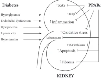
In addition to systemic effects, multiple intrarenal mecha- nisms have been implicated in the beneficial effect of PPARα ligands (Fig. 2). An experimental study with a type 2 diabetic model showed that PPARα activation by fenofibrate improved insulin sensitivity, glucose control, and diabetic nephropathy, as evidenced by urinary albumin excretion and attenuated glomerular mesangial expansion [48]. Such beneficial effects on renal outcome with fibrates may result from anti-inflam-
matory, anti-atherosclerotic, antihypertensive, and anti-RAS actions as noted above. In an experimental study to investigate the role of PPARα in type 1 diabetic nephropathy, more severe structural changes (such as glomerulosclerosis and mesangial area expansion) as well as an effect on albuminuria were noted in diabetic PPARα-knockout mice; these changes were associ- ated with an increase in the profibrotic, pro-inflammatory and pro-apoptotic pathways implicated in renal extracellular ma- trix accumulation [49]. In PPARα deficiency, the glomerular lesions exhibited increased type IV collagen and TGF-β ex- pression in diabetic kidney disease, suggesting that the activa- tion of PPARα ligands effectively prevents the glomerular ma- trix expansion that accompanies apoptosis and inflammatory cell infiltration in the glomerulus [49].
In our study, which investigated the effect of the glucagon- like peptide-1 analog exendin-4 on the progression of type 2 diabetic nephropathy, we observed significantly increased PPARα expression in a dose-dependent manner in exendin-4- treated db/db kidneys in mice compared with that seen in con- trol db/db kidneys [50]. This increase in PPARα expression was accompanied by reduced glomerular immunostaining for F4/80 and caspase-3 as well as for TGF-β1. Furthermore, exen- din-4 treatment decreased 24-hour urinary 8-hydroxy-deoxy- guanosine concentration, which was consistent with the re- duction in oxidative DNA damage and oxidative stress. These findings suggest that TGF-β1 expression mediated by oxida- tive stress may be suppressible by PPARα activation [50]. In- terestingly, there is evidence that starved PPARα-null mice develop increased albuminuria and exhibit albumin accumu- lation in the proximal tubules, which indicates that PPARα ac- tivity may facilitate albumin reabsorption and degradation in this nephron segment [51]. This mechanism may contribute to the beneficial effect of PPARα agonists on albuminuria in type 2 diabetic nephropathy.
In vitro experiments
In mesangial cells, PPARα agonists reduce the production of TGF-β and extracellular matrix. The TGF-β signaling pathway may be one possible mechanism that relates to the effect of PPARα agonists on the mesangial matrix production. One study showed that clofibrate directly inhibits oxidant stress–in- duced TGF-β1 expression in these cells, indicating that PPARα agonists block the TGF-β signaling pathway, thereby attenuat- ing glomerular matrix production [52]. PPRE3X luciferase re- porter analysis demonstrated that the fenofibrate significantly
↑RAS PPARα
Diabetes
KIDNEY
↑TGFβ
↑Albuminuria
VEGF imbalance
↑Inflammation
Hyperglycemia Endothelial dysfunction Dyslipidemia Lipotoxicity Hypertension
↑Oxidative stress
↑Apoptosis
↑Fibrosis
Fig. 2. Protective actions of peroxisome proliferator-activated receptor alpha (PPARα) agonists for the improvement of dia- betic nephropathy. In diabetic patients, symptoms such as hy- perglycemia, dyslipidemia, endothelial dysfunction, lipotoxic- ity, and high blood pressure all can contribute to renal compli- cations of diabetes systemically or locally via increased inflam- mation, activation of the renin-angiotensin system (RAS), en- hanced oxidative stress, increased apoptosis and vasculopathy, which can be attenuated by the activation of PPARα. TGF, transforming growth factor; VEGF, vascular endothelial growth factor.
increased luciferase activity in mesangial cells, which is con- sistent with the existence of endogenous PPARα activity in these cells [48,49]. This finding suggests that increased PPARα activity in the tubule may exert anti-inflammatory and anti-fi- brotic effects via paracrine action resulting from increased PPRE activity in the glomeruli [49].
Importantly, we need to pay attention to the role PPARα in vascular biology. PPARα ligands appear to modulate renal en- dothelial cell proliferation and migration, probably through their ability to interfere with the vascular endothelial growth factor (VEGF)-mediated signaling pathway. VEGF is crucial for maintaining the function and integrity of the endothelium [44]. In the kidney, the VEGF receptors (VEGFR) are expressed in the endothelium of the glomeruli, the peritubular capillar- ies and, to a lesser extent, the mesangial and tubular cells [53].
In the glomerular endothelial cells, VEGF-A stimulates the VEGFR-2/Akt axis to regulate endothelial NOS (eNOS) phos- phorylation. eNOS is activated by the phosphorylation of ser- ine (Ser1177) of the protein kinase Akt/PKB and is also known to regulate glomerular hemodynamics by generation of nitric oxide (NO) [53,54]. Several type 1 or type 2 animal models have shown that VEGF stimulates renal pathological progres- sions, as demonstrated by glomerular hyperfiltration and hy- pertrophy and urinary albumin excretion [44]. By contrast, excessively low levels of VEGF are associated with renal dete- rioration in the type 2 diabetic model [44,53], suggesting that a too-low VEGF level can be just as damaging as when the VEGF level is too high [53]. Interestingly, PPARα agonists have been shown to inhibit endothelial VEGFR-2 expression, and fenofibrate treatment induces a significant reduction of VEGF levels in serum [53,55]. Although the data regarding the effects of administration with PPARα agonists on VEGF changes in diabetic kidneys are lacking, angiogenic modula- tion and endothelial cell stabilization by PPARα agonists might be responsible for the potential renoprotective effects in dia- betic nephropathy models. Recently, we found that dual VEG- FR1 and VEGFR2 inhibition in db/db mice aggravated diabet- ic peripheral neuropathy, including a decrease in nerve con- duction velocity and an increase in the tactile threshold of the sciatic nerve associated with vascular rarefaction resulting from endothelial cell apoptosis, which completely recovered to levels of non-diabetic db/m mice by fenofibrate (unpublished data).
As renal lipotoxicity can lead to chronic kidney disease, an overload of free fatty acid-bound albumin in the proximal epi- thelial cells induced tubular cell injury resulting from not only
a decrease in the lipolytic enzymes but also increases in lipid accumulation and oxidative stress [56]. Fenofibrate, by con- trast, inhibited palmitate-induced expressions of both mono- cyte chemoattractant protein-1 and PAI-1 and oxidative stress in the proximal tubular cells, which were associated with the overexpression of lipolytic enzymes and enhancement of renal lipolysis. Therefore, we need more experimental evidences us- ing various types of renal cells including podocytes in order to investigate whether pharmacological activation of PPARα could be a therapeutically suitable strategy against glomerular and tubuleinterstitial lesions in diabetic nephropathy.
Clinical studies
Recent studies have shown a beneficial effect of fibrate treat- ment on type 2 diabetes and diabetic nephropathy [57-59]. In normotensive patients with non–insulin-dependent diabetes, effective treatment of dyslipidemia by a PPARα activator gem- fibrozil for one year was associated with significant stabiliza- tion of urine albumin excretion [58]. The Fenofibrate Inter- vention and Event Lowering in Diabetes (FIELD) study re- ported that fenofibrate treatment was associated with reduc- tions in cardiovascular disease outcomes [59]. This study in- cluded 9,795 patients with type 2 diabetes. The results demon- strated that fenofibrate treatment was associated with an 11%
reduction in total cardiovascular disease events [59]. Interest- ingly, the FIELD study showed that fenofibrate resulted in sig- nificantly more patients’ regressing or not progressing in their urinary albumin excretion. In the fenofibrate group, albumin- uria progressed in 9.5% of the patients compared to 11.0% of patients in the placebo group. Albuminuria regressed in 9.4%
of patients in the fenofibrate treatment group and in 8.2% in the placebo group participants [44,59]. The Action to Control Cardiovascular Risk in Diabetes (ACCORD) study also dem- onstrated that fibrate therapy with intensive glucose control could significantly reduce microalbuminuria (38.2% vs. 41.6%, P=0.01) and macroalbuminuria (10.5% vs. 12.3%, P=0.03) despite the marginal effects of fibrates in the management of dyslipidemia [60,61]. Unfortunately, most trials with PPARα agonists have been designed for cardiovascular disease, not re- nal disease, as a primary end point. The effect of fenofibrate on urinary protein excretion appears to be minor compared with the major effects on cardiovascular disease events [62]. An- other weakness is that most studies have been limited to peo- ple with type 2 diabetes. In the future, more large-scale, pro- spective, randomized trials will be necessary to evaluate the
efficacy of fibrates on renal outcomes in patients with type 1 or type 2 diabetes.
LIMITATIONS OF PPARα AGONISTS
Despite evidence of the beneficial effects of PPARα agonists in patients with diabetes, there are still many issues to be ad- dressed concerning their safety in clinical use. The most im- portant concern is due to the fact that fibrate treatment typi- cally results in increased serum levels of creatinine and cys- tatin C and might potentially decrease the estimated glomeru- lar filtration rate and creatinine clearance [44]. More care should be taken when prescribing fibrates to patients with mild-to-moderate renal insufficiency. Currently, it is recom- mended that fenofibrate dosages should be reduced by one third in chronic kidney disease (CKD) stage 2, by an addition- al one third in CKD stage 3 and 4, and avoided in CKD stage 5 [62]. Although the mechanisms of deterioration in renal func- tion with fibrates are not clear, renal hemodynamic changes in association with the reduction of cylcooxygenase 2 levels, an inhibitory action of fenofibrate on the excretion of creatinine by the kidneys, or an increase in the flow of creatinine from muscle might be an explanation [44,63].
In addition, the fact that PPARα is subject to tissue-specific regulation should be considered. Over-expression of PPARα in the heart results in increased fatty acid oxidation, elevated lipid droplets, and worsened cardiomyopathy, suggesting that cardiac PPARα activation may be harmful [54,64]. By contrast, an increase in PPARα expression in the diabetic kidney is evi- dent [49], indicating compensatory PPARα activation in re- sponse to the renal damage suffered. The proper balance be- tween overactivation and underactivation of PPARα may dif- fer in each type of affected organ or tissue. To maximize the therapeutic potential and minimize the harmful effects of PPARα, future investigations should examine the development of selective agents with tissue-, organ- or disease-specific effects and targeted gene-selective activities. This will require devel- opment of more selective PPARα modulators.
CONCLUSION
Recent studies have suggested beneficial roles for PPARα and PPARα target genes as therapeutic targets in the treatment of disorders involving inflammation, atherosclerosis, oxidative stress, angiogenesis, and RAS. PPARα agonists have tradition-
ally been used to lower circulating fatty acids and TG. Current- ly, considerable evidence suggests that PPARα is involved in the pathogenesis of diabetic nephropathy and contributes to the extrametabolic control of renal function. Although the mechanisms of the beneficial effects of fibrates in the kidneys are still under investigation, PPARα would be a promising therapeutic target in the management of diabetes and diabetic nephropathy.
CONFLICTS OF INTEREST
No potential conflict of interest relevant to this article was re- ported.
ACKNOWLEDGMENTS
This research was supported by a grant of the Korean Health Technology R&D Project, Minister of Health and Welfare, Re- public of Korea, (to C. W. Park; A111055) and also by Seoul St.
Mary’s Hospital Clinical Medicine Research Program year of 2010 through The Catholic University of Korea.
REFERENCES
1. Cheng CF, Chen HH, Lin H. Role of PPARα and its agonist in renal diseases. PPAR Res 2010;2010:345098.
2. Pyper SR, Viswakarma N, Yu S, Reddy JK. PPARalpha: energy combustion, hypolipidemia, inflammation and cancer. Nucl Recept Signal 2010;8:e002.
3. Berger JP, Akiyama TE, Meinke PT. PPARs: therapeutic targets for metabolic disease. Trends Pharmacol Sci 2005;26:244-51.
4. Bookout AL, Jeong Y, Downes M, Yu RT, Evans RM, Mangels- dorf DJ. Anatomical profiling of nuclear receptor expression reveals a hierarchical transcriptional network. Cell 2006;126:
789-99.
5. Guan Y, Zhang Y, Schneider A, Davis L, Breyer RM, Breyer MD. Peroxisome proliferator-activated receptor-gamma activ- ity is associated with renal microvasculature. Am J Physiol Re- nal Physiol 2001;281:F1036-46.
6. Guan Y, Zhang Y, Davis L, Breyer MD. Expression of peroxi- some proliferator-activated receptors in urinary tract of rabbits and humans. Am J Physiol 1997;273(6 Pt 2):F1013-22.
7. Ruan XZ, Moorhead JF, Fernando R, Wheeler DC, Powis SH, Varghese Z. PPAR agonists protect mesangial cells from inter- leukin 1beta-induced intracellular lipid accumulation by acti-
vating the ABCA1 cholesterol efflux pathway. J Am Soc Nephrol 2003;14:593-600.
8. Maeda T, Kishioka S. PPAR and pain. Int Rev Neurobiol 2009;
85:165-77.
9. Kliewer SA, Xu HE, Lambert MH, Willson TM. Peroxisome proliferator-activated receptors: from genes to physiology. Re- cent Prog Horm Res 2001;56:239-63.
10. Yu S, Matsusue K, Kashireddy P, Cao WQ, Yeldandi V, Yeldan- di AV, Rao MS, Gonzalez FJ, Reddy JK. Adipocyte-specific gene expression and adipogenic steatosis in the mouse liver due to peroxisome proliferator-activated receptor gamma1 (PPARga- mma1) overexpression. J Biol Chem 2003;278:498-505.
11. Ruan X, Zheng F, Guan Y. PPARs and the kidney in metabolic syndrome. Am J Physiol Renal Physiol 2008;294:F1032-47.
12. Letavernier E, Perez J, Joye E, Bellocq A, Fouqueray B, Hay- mann JP, Heudes D, Wahli W, Desvergne B, Baud L. Peroxisome proliferator-activated receptor beta/delta exerts a strong pro- tection from ischemic acute renal failure. J Am Soc Nephrol 2005;16:2395-402.
13. Guan Y. Peroxisome proliferator-activated receptor family and its relationship to renal complications of the metabolic syn- drome. J Am Soc Nephrol 2004;15:2801-15.
14. Fruchart JC. Peroxisome proliferator-activated receptor-alpha (PPARalpha): at the crossroads of obesity, diabetes and cardio- vascular disease. Atherosclerosis 2009;205:1-8.
15. Desvergne B, Wahli W. Peroxisome proliferator-activated re- ceptors: nuclear control of metabolism. Endocr Rev 1999;20:
649-88.
16. Xu HE, Lambert MH, Montana VG, Plunket KD, Moore LB, Collins JL, Oplinger JA, Kliewer SA, Gampe RT Jr, McKee DD, Moore JT, Willson TM. Structural determinants of ligand binding selectivity between the peroxisome proliferator-acti- vated receptors. Proc Natl Acad Sci U S A 2001;98:13919-24.
17. Pineda Torra I, Jamshidi Y, Flavell DM, Fruchart JC, Staels B.
Characterization of the human PPARalpha promoter: identifi- cation of a functional nuclear receptor response element. Mol Endocrinol 2002;16:1013-28.
18. Inoue J, Satoh S, Kita M, Nakahara M, Hachimura S, Miyata M, Nishimaki-Mogami T, Sato R. PPARalpha gene expression is up-regulated by LXR and PXR activators in the small intestine.
Biochem Biophys Res Commun 2008;371:675-8.
19. Poynter ME, Daynes RA. Peroxisome proliferator-activated receptor alpha activation modulates cellular redox status, re- presses nuclear factor-kappaB signaling, and reduces inflam- matory cytokine production in aging. J Biol Chem 1998;273:
32833-41.
20. Corton JC, Anderson SP, Stauber A. Central role of peroxisome proliferator-activated receptors in the actions of peroxisome proliferators. Annu Rev Pharmacol Toxicol 2000;40:491-518.
21. Rigamonti E, Chinetti-Gbaguidi G, Staels B. Regulation of macrophage functions by PPAR-alpha, PPAR-gamma, and LXRs in mice and men. Arterioscler Thromb Vasc Biol 2008;
28:1050-9.
22. Genini D, Carbone GM, Catapano CV. Multiple interactions between peroxisome proliferators-activated receptors and the ubiquitin-proteasome system and implications for cancer pathogenesis. PPAR Res 2008;2008:195065.
23. Chandra V, Huang P, Hamuro Y, Raghuram S, Wang Y, Burris TP, Rastinejad F. Structure of the intact PPAR-gamma-RXR- nuclear receptor complex on DNA. Nature 2008;456:350-6.
24. van der Meer DL, Degenhardt T, Vaisanen S, de Groot PJ, Hei- naniemi M, de Vries SC, Muller M, Carlberg C, Kersten S.
Profiling of promoter occupancy by PPARalpha in human hepatoma cells via ChIP-chip analysis. Nucleic Acids Res 2010;
38:2839-50.
25. Juge-Aubry C, Pernin A, Favez T, Burger AG, Wahli W, Meier CA, Desvergne B. DNA binding properties of peroxisome proliferator-activated receptor subtypes on various natural peroxisome proliferator response elements. Importance of the 5’-flanking region. J Biol Chem 1997;272:25252-9.
26. Zhu Y, Qi C, Calandra C, Rao MS, Reddy JK. Cloning and identification of mouse steroid receptor coactivator-1 (mSRC- 1), as a coactivator of peroxisome proliferator-activated recep- tor gamma. Gene Expr 1996;6:185-95.
27. Dowell P, Ishmael JE, Avram D, Peterson VJ, Nevrivy DJ, Leid M. Identification of nuclear receptor corepressor as a peroxi- some proliferator-activated receptor alpha interacting protein.
J Biol Chem 1999;274:15901-7.
28. Feige JN, Gelman L, Michalik L, Desvergne B, Wahli W. From molecular action to physiological outputs: peroxisome prolif- erator-activated receptors are nuclear receptors at the cross- roads of key cellular functions. Prog Lipid Res 2006;45:120-59.
29. Barger PM, Browning AC, Garner AN, Kelly DP. p38 mitogen- activated protein kinase activates peroxisome proliferator-acti- vated receptor alpha: a potential role in the cardiac metabolic stress response. J Biol Chem 2001;276:44495-501.
30. Burns KA, Vanden Heuvel JP. Modulation of PPAR activity via phosphorylation. Biochim Biophys Acta 2007;1771:952-60.
31. Blanquart C, Mansouri R, Paumelle R, Fruchart JC, Staels B, Glineur C. The protein kinase C signaling pathway regulates a
molecular switch between transactivation and transrepression activity of the peroxisome proliferator-activated receptor alpha.
Mol Endocrinol 2004;18:1906-18.
32. Ricote M, Valledor AF, Glass CK. Decoding transcriptional programs regulated by PPARs and LXRs in the macrophage:
effects on lipid homeostasis, inflammation, and atherosclero- sis. Arterioscler Thromb Vasc Biol 2004;24:230-9.
33. Shin SJ, Lim JH, Chung S, Youn DY, Chung HW, Kim HW, Lee JH, Chang YS, Park CW. Peroxisome proliferator-activated re- ceptor-alpha activator fenofibrate prevents high-fat diet-in- duced renal lipotoxicity in spontaneously hypertensive rats.
Hypertens Res 2009;32:835-45.
34. Zhao X. Prevention of local lipotoxicity: a new renoprotective mechanism of peroxisome proliferator-activated receptor-al- pha activation in hypertension and obesity? Hypertens Res 2009;32:821-3.
35. Devchand PR, Keller H, Peters JM, Vazquez M, Gonzalez FJ, Wahli W. The PPARalpha-leukotriene B4 pathway to inflam- mation control. Nature 1996;384:39-43.
36. Zandbergen F, Plutzky J. PPARalpha in atherosclerosis and in- flammation. Biochim Biophys Acta 2007;1771:972-82.
37. Duan SZ, Usher MG, Mortensen RM. PPARs: the vasculature, inflammation and hypertension. Curr Opin Nephrol Hyper- tens 2009;18:128-33.
38. Duez H, Chao YS, Hernandez M, Torpier G, Poulain P, Mundt S, Mallat Z, Teissier E, Burton CA, Tedgui A, Fruchart JC, Fievet C, Wright SD, Staels B. Reduction of atherosclerosis by the peroxisome proliferator-activated receptor alpha agonist fenofibrate in mice. J Biol Chem 2002;277:48051-7.
39. Ramanan S, Kooshki M, Zhao W, Hsu FC, Robbins ME. PPA- Ralpha ligands inhibit radiation-induced microglial inflam- matory responses by negatively regulating NF-kappaB and AP-1 pathways. Free Radic Biol Med 2008;45:1695-704.
40. Kintscher U, Lyon C, Wakino S, Bruemmer D, Feng X, Goetze S, Graf K, Moustakas A, Staels B, Fleck E, Hsueh WA, Law RE.
PPARalpha inhibits TGF-beta-induced beta5 integrin tran- scription in vascular smooth muscle cells by interacting with Smad4. Circ Res 2002;91:e35-44.
41. Diep QN, Amiri F, Touyz RM, Cohn JS, Endemann D, Neves MF, Schiffrin EL. PPARalpha activator effects on Ang II-in- duced vascular oxidative stress and inflammation. Hyperten- sion 2002;40:866-71.
42. Holla VR, Adas F, Imig JD, Zhao X, Price E Jr, Olsen N, Kovacs WJ, Magnuson MA, Keeney DS, Breyer MD, Falck JR, Water- man MR, Capdevila JH. Alterations in the regulation of an-
drogen-sensitive Cyp 4a monooxygenases cause hypertension.
Proc Natl Acad Sci U S A 2001;98:5211-6.
43. Chung S, Park CW, Shin SJ, Lim JH, Chung HW, Youn DY, Kim HW, Kim BS, Lee JH, Kim GH, Chang YS. Tempol or candesartan prevents high-fat diet-induced hypertension and renal damage in spontaneously hypertensive rats. Nephrol Dial Transplant 2010;25:389-99.
44. Hiukka A, Maranghi M, Matikainen N, Taskinen MR. PPA- Ralpha: an emerging therapeutic target in diabetic microvas- cular damage. Nat Rev Endocrinol 2010;6:454-63.
45. Kim BH, Won YS, Kim EY, Yoon M, Nam KT, Oh GT, Kim DY. Phenotype of peroxisome proliferator-activated receptor- alpha(PPARalpha)deficient mice on mixed background fed high fat diet. J Vet Sci 2003;4:239-44.
46. Aasum E, Belke DD, Severson DL, Riemersma RA, Cooper M, Andreassen M, Larsen TS. Cardiac function and metabolism in type 2 diabetic mice after treatment with BM 17.0744, a novel PPAR-alpha activator. Am J Physiol Heart Circ Physiol 2002;283:H949-57.
47. Koh EH, Kim MS, Park JY, Kim HS, Youn JY, Park HS, Youn JH, Lee KU. Peroxisome proliferator-activated receptor (PPAR)- alpha activation prevents diabetes in OLETF rats: comparison with PPAR-gamma activation. Diabetes 2003;52:2331-7.
48. Park CW, Zhang Y, Zhang X, Wu J, Chen L, Cha DR, Su D, Hwang MT, Fan X, Davis L, Striker G, Zheng F, Breyer M, Guan Y. PPARalpha agonist fenofibrate improves diabetic ne- phropathy in db/db mice. Kidney Int 2006;69:1511-7.
49. Park CW, Kim HW, Ko SH, Chung HW, Lim SW, Yang CW, Chang YS, Sugawara A, Guan Y, Breyer MD. Accelerated dia- betic nephropathy in mice lacking the peroxisome prolifera- tor-activated receptor alpha. Diabetes 2006;55:885-93.
50. Park CW, Kim HW, Ko SH, Lim JH, Ryu GR, Chung HW, Han SW, Shin SJ, Bang BK, Breyer MD, Chang YS. Long-term treat- ment of glucagon-like peptide-1 analog exendin-4 ameliorates diabetic nephropathy through improving metabolic anomalies in db/db mice. J Am Soc Nephrol 2007;18:1227-38.
51. Kamijo Y, Hora K, Tanaka N, Usuda N, Kiyosawa K, Nakajima T, Gonzalez FJ, Aoyama T. Identification of functions of per- oxisome proliferator-activated receptor alpha in proximal tu- bules. J Am Soc Nephrol 2002;13:1691-702.
52. Wilmer WA, Dixon CL, Hebert C, Lu L, Rovin BH. PPAR-al- pha ligands inhibit H2O2-mediated activation of transforming growth factor-beta1 in human mesangial cells. Antioxid Redox Signal 2002;4:877-84.
53. Kim HW, Lim JH, Kim MY, Chung S, Shin SJ, Chung HW,
Choi BS, Kim YS, Chang YS, Park CW. Long-term blockade of vascular endothelial growth factor receptor-2 aggravates the diabetic renal dysfunction associated with inactivation of the Akt/eNOS-NO axis. Nephrol Dial Transplant 2011;26:1173-88.
54. Park CW, Kim HW, Lim JH, Yoo KD, Chung S, Shin SJ, Chung HW, Lee SJ, Chae CB, Kim YS, Chang YS. Vascular endothelial growth factor inhibition by dRK6 causes endothelial apopto- sis, fibrosis, and inflammation in the heart via the Akt/eNOS axis in db/db mice. Diabetes 2009;58:2666-76.
55. Grau R, Diaz-Munoz MD, Cacheiro-Llaguno C, Fresno M, Iniguez MA. Role of peroxisome proliferator-activated recep- tor alpha in the control of cyclooxygenase 2 and vascular en- dothelial growth factor: involvement in tumor growth. PPAR Res 2008;2008:352437.
56. Tanaka Y, Kume S, Araki S, Isshiki K, Chin-Kanasaki M, Saka- guchi M, Sugimoto T, Koya D, Heneda M, Kashiwagi A, Mae- gawa H, Uzu T. Fenofibrate, a PPARα agonist, has renoprotec- tive effects in mice by enhancing renal lipolysis. Kidney Int 2011;79:871-82.
57. Fried LF, Orchard TJ, Kasiske BL. Effect of lipid reduction on the progression of renal disease: a meta-analysis. Kidney Int 2001;59:260-9.
58. Smulders YM, van Eeden AE, Stehouwer CD, Weijers RN, Slaats EH, Silberbusch J. Can reduction in hypertriglyceridae- mia slow progression of microalbuminuria in patients with non-insulin-dependent diabetes mellitus? Eur J Clin Invest 1997;27:997-1002.
59. Keech A, Simes RJ, Barter P, Best J, Scott R, Taskinen MR, Forder P, Pillai A, Davis T, Glasziou P, Drury P, Kesaniemi YA,
Sullivan D, Hunt D, Colman P, d’Emden M, Whiting M, Ehnholm C, Laakso M; FIELD study investigators. Effects of long-term fenofibrate therapy on cardiovascular events in 9795 people with type 2 diabetes mellitus (the FIELD study): ran- domised controlled trial. Lancet 2005;366:1849-61.
60. Ismail-Beigi F, Craven T, Banerji MA, Basile J, Calles J, Cohen RM, Cuddihy R, Cushman WC, Genuth S, Grimm RH Jr, Hamilton BP, Hoogwerf B, Karl D, Katz L, Krikorian A, O’Connor P, Pop-Busui R, Schubart U, Simmons D, Taylor H, Thomas A, Weiss D, Hramiak I; ACCORD trial group. Effect of intensive treatment of hyperglycaemia on microvascular outcomes in type 2 diabetes: an analysis of the ACCORD ran- domised trial. Lancet 2010;376:419-30.
61. ACCORD Study Group, Ginsberg HN, Elam MB, Lovato LC, Crouse JR 3rd, Leiter LA, Linz P, Friedewald WT, Buse JB, Gerstein HC, Probstfield J, Grimm RH, Ismail-Beigi F, Bigger JT, Goff DC Jr, Cushman WC, Simons-Morton DG, Byington RP. Effects of combination lipid therapy in type 2 diabetes mel- litus. N Engl J Med 2010;362:1563-74.
62. Molitch ME. Management of dyslipidemias in patients with diabetes and chronic kidney disease. Clin J Am Soc Nephrol 2006;1:1090-9.
63. Valensi P, Picard S. Lipids, lipid-lowering therapy and diabetes complications. Diabetes Metab 2011;37:15-24.
64. Finck BN, Han X, Courtois M, Aimond F, Nerbonne JM, Ko- vacs A, Gross RW, Kelly DP. A critical role for PPARalpha-me- diated lipotoxicity in the pathogenesis of diabetic cardiomyop- athy: modulation by dietary fat content. Proc Natl Acad Sci U S A 2003;100:1226-31.