서 론
쇄골 간부 골절은 비교적 흔한 골절의 하나로 모든 성인 골절의 2.6%-10.0%를 차지한다.1,2) 과거로부터 많은 연구에서 비수술적 치료로 좋은 결과를 보고하여 왔지만,3,4) 최근 비수술적 치료 후 불유합으로 인한 재수술 및 쇄골 길이의 단축으로 인한 기능 저 하 및 생역학적인 변화가 보고되고 있으며 수술적 치료 후 빠른 일상 생활로의 복귀 및 기능 회복을 위하여 전위된 쇄골 간부 골
Copyright © 2017 by The Korean Orthopaedic Association
“This is an Open Access article distributed under the terms of the Creative Commons Attribution Non-Commercial License (http://creativecommons.org/licenses/by-nc/4.0/) which permits unrestricted non-commercial use, distribution, and reproduction in any medium, provided the original work is properly cited.”
The Journal of the Korean Orthopaedic Association Volume 52 Number 5 2017 Received October 26, 2016 Revised February 27, 2017 Accepted March 23, 2017 Correspondence to: Woong Kyo Jeong, M.D.
Department of Orthopaedic Surgery, Korea University College of Medicine, 73 Inchon-ro, Seongbuk-gu, Seoul 02841, Korea
TEL: +82-2-920-5925 FAX: +82-2-924-5271 E-mail: drshoulder@korea.ac.kr
쇄골 간부 분쇄 골절에서 최소 침습 금속판 고정술의 유용성: 고식적 수술법과 비교
강태욱 • 황현정 • 이동기 • 한승범 • 정웅교
고려대학교 의과대학 고려대학교 안암병원 정형외과학교실
The Usefulness of Minimally Invasive Plate Osteosynthesis to Manage Comminuted Mid-Clavicle Fracture:
A Comparison with Conventional Open Plating
Tae Wook Kang, M.D., Hyun Jung Hwang, M.D., Dong Ki Lee, M.D., Seung Bum Han, M.D., and Woong Kyo Jeong, M.D.
Department of Orthopaedic Surgery, Korea University Anam Hospital, Korea University College of Medicine, Seoul, Korea
Purpose: The purpose of this study was to compare the clinical and radiological outcomes of minimally invasive plate osteosynthesis (MIPO)
with those of conventional open plating (COP) for treating comminuted mid-clavicular fractures and to evaluate the usefulness of MIPO.Materials and Methods: Thirty-nine patients who underwent surgical treatment for mid-clavicular comminuted fractures were analyzed
retrospectively. Nineteen patients were treated with MIPO and twenty patients with COP. Radiological evaluation included time to union, fracture healing, and clavicular length difference measured as the proportional length difference with the unaffected side. Clinical assessment was performed using the simple shoulder test score, American Shoulder and Elbow Surgeons score, and quick disability of the arm, shoulder and hand score. Moreover, the mean operation and radiation times, as well as exposure were compared.Results: All clavicles achieved bone union. The mean time to union was 12.1 weeks in the MIPO group, and 14.6 weeks in the COP group
(p=0.587). There was no significant difference between the two groups regarding the functional and radiological outcomes at 2-year follow- up. A significantly shorter operation time was observed in the MIPO group than in the COP group (75.8 min vs. 106.9 min, p=0.002). More radiation time and exposure were identified in the MIPO group (52.8 s vs. 37.1 s, p=0.002; 209.4 mGy vs. 43.1 mGy, p=0.005).Conclusion: Both COP and MIPO were shown to be effective treatment options for mid-clavicular comminuted fractures. MIPO may be
a better alternative to COP due to shorter operation time and no need for a bone graft, although the functional and radiological outcomes were not significantly different. However, all surgeons should pay close attention to minimize radiation hazard.Key words: clavicle, fracture, minimally invasive plate osteosynthesis
절의 경우 수술적 치료의 필요성이 대두되고 있다.5-7) 이를 위해 금속판 및 나사못을 이용한 고정법, 골수강 내 고정법 등 다양한 수술 방법이 시행되고 있다.8,9) 골수강 내 고정법은 금속판을 이 용한 고정법보다 덜 침습적이라는 장점이 있지만 고정 강도가 약 하여 골절부의 변형이나 쇄골의 단축을 초래할 수 있다는 단점 이 보고되고 있다.10,11) 금속판 및 나사못을 이용한 고정법은 전통 적으로 널리 시행되고 있으며 우수한 치료 결과가 보고되고 있으
나,7,12) 골절을 정복하는 과정에서 주위 연부 조직의 과도한 손상
으로 인한 수술부위 감염, 불유합 및 이로 인한 내고정 실패 등의 합병증이 보고되고 있다.13,14)
장골 간부 분쇄 골절에 있어서 최소 침습적 금속판 고정술 (minimally invasive plate osteosynthesis, MIPO)을 이용한 치료가 널리 사용되며 매우 효과적으로 알려져 있다. 이 술식은 절개부 위가 작아 미용적인 면에서 더 선호되고, 골절부를 직접 노출하 지 않음으로써 골막 및 연부 조직의 박리가 적어 골절 치유에 도 움을 줄 수 있는 방법으로 알려져 있다.15,16) 최근 쇄골 간부 골절 의 치료로 MIPO의 방법과 만족스런 임상적 결과가 보고되고 있
으나,15-17) 고식적 수술법(conventional open plating, COP)과의 비
교 연구는 제한적이다. 이에 저자들은 전위된 쇄골 간부 분쇄 골 절에 있어서 COP와 MIPO의 수술 후 임상적, 영상의학적 결과를 비교하고 MIPO의 유용성을 평가하고자 하였다.
대상 및 방법
본 연구는 고려대학교 안암병원의 의학연구윤리심의위원회(승 인번호: AN13105-003)의 승인을 받아 진행되었다.
2006년 4월부터 2012년 12월까지 본원에 내원하여 쇄골 간부 분쇄 골절로 수술을 시행 받은 후 1년 이상 추시 관찰이 가능하였 던 환자를 대상으로 하였다. 본원에서는 2006년부터 쇄골 골절에 대하여 MIPO 술식을 적용하기 시작하였으며 14명의 환자의 임 상적, 방사선적 결과를 평가하였다(unpublished data). 14명의 환자 중 2예에서 재수술이 시행되었고, 재수술의 원인으로 1예는 외상 으로 인한 재골절, 다른 1예는 불유합이었다. 불유합이 발생한 1 예는 단순 골절로 횡형 골절이었고, 복합 골절이었던 12예에서는 모두 골유합을 얻었다. 이후 저자들은 쇄골의 분쇄 골절은 MIPO 방법을 사용하여 수술하였다. 본 연구는 MIPO 술식이 본격적으 로 시행된 2009년을 기준으로, 그 이전의 쇄골 분쇄 골절 환자에 서 시행된 COP군과 2009년 이후의 MIPO군을 비교하였다. 그 중 에서 병적 골절 및 개방성 골절, 원위 및 근위 1/3 부위의 골절, 전 신 마취하 수술 불가능한 환자, 반대측의 쇄골 골절력이 있는 환 자, 소아 환자는 제외하였다. 총 39예의 환자가 연구 대상으로 포 함되었으며, 그 중 COP를 시행한 환자는 20예(51.3%)였고, MIPO 를 시행한 환자는 19예(48.7%)였다.
성별은 남자가 32예(82.1%), 여자가 7예(17.9%)였고, 연령은 최
소 10세에서 최고 66세로 COP의 경우 평균 44.1세, MIPO의 경 우 평균 38.0세였다. 수상 원인은 실족이 16예(41.0%)로 가장 많 았고, 스포츠 손상이 12예(30.8%), 교통사고가 11예(28.2%)였다.
Robinson의 분류에 따르면 B1형 24예(61.5%), B2형 15예(38.5%) 였다(Table 1). 나이, 성별, 골편의 수는 두 군 간 통계적 차이는 없 었다.
1. 수술 방법
수술은 한 명의 정형외과 전문의가 시행하였으며, 전신 마취 혹 은 신경 차단 마취 후 해면 의자 위치로 환자를 위치시켰다.
1) 최소 침습적 금속판 고정술(minimally invasive plate osteosynthesis)
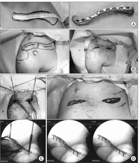
모든 환자에서 3.5 mm 잠김 압박 성형 금속판(locking compres- sion reconstruction plate; Depuy Synthes, Warsaw, IN, USA)을 사 용하였다. 수술 전날 건측의 쇄골 길이를 측정한 후 반대 방향의 비슷한 길이를 가진 사체 쇄골에 금속판을 대고 형태에 맞추어 성형을 하고 고압 멸균 소독하여 준비하였다(Fig. 1A). 골절부를 중심으로 쇄골의 상연을 따라 근위부와 원위부에 각각 2 cm의 피 부 절개를 시행하였다(Fig. 1B). 피부 절개 후 골절 부위의 연부 조 직 및 골막이 박리되지 않도록 조심하며 뭉툭한 kelly를 이용하
Table 1. Demographic Data between COP and MIPO Group
Variable COP group MIPO group
No. of patient 20 19
Age (yr) 44.1 38.0
Sex (male:female) 16:4 16:3 Involved side (right:left) 7:13 9:10
No. of fragment 3.45 3.37
Fracture site gap (mm) 15.58 21.33 Factors affecting union
Smoking 5 7
DM 0 1
Obesity (BMI>25 kg/m2) 6 3 Trauma mechanism
Slip down 9 7
Sports injury 2 10
Motor vehicle accident 9 2 Robinson classification
Simple, wedge (B1) 11 13 Multifragmentary (B2) 9 6
Values are presented as number only or mean only. COP, conventional open plating; MIPO, minimally invasive plate osteosynthesis; DM, diabetes mellitus; BMI, body mass index.
여 피하 조직과 골막 사이를 박리한 후 원위부 피부 절개를 통하 여 금속판을 삽입한 다음 쇄골의 전 상방에 위치시켰다. 금속판 삽입 후 금속판의 내측과 외측에 각각 하나의 drill sleeve를 미리 장착하고 drill을 삽입하며 손잡이로 사용하여 단축된 쇄골의 길 이를 회복시켰다. 방사선 투과 촬영기하에 bone hook, reduction forcep 등을 이용하여 골절부를 정복하였다(Fig. 1C). 쇄골의 전체 적인 모양 및 길이가 만족스러울 경우 내, 외측에 각각 한 개의 압 박 나사를 사용하여 고정하고, 1개 또는 2개의 잠김 나사를 추가 로 삽입하고 절개부위를 봉합하였다(Fig. 1D).
2) 고식적 수술법(conventional open plating)
골절부를 중심으로 쇄골의 상연을 따라 피부 절개을 한 뒤, 골절 부를 노출시켰다. 분쇄된 골편을 부착된 연부 조직과 골막의 손 상을 최소화하면서 정복하고 지연 나사를 사용하여 고정하였으
며, 이후 보호 금속판(protection plating)을 추가하였다. 지연 나사 를 삽입할 수 없을 정도로 골편이 작은 경우 골편을 고정하고 K- 강선을 이용하여 일시적으로 고정한 다음, 압박 금속판(compres- sion plating)으로 골편을 고정하여 골편의 고정을 도모하였고 K- 강선을 제거하였다. 전 예에서 cerclage wiring은 시행하지 않았다.
골결손이 발생한 경우는 자가골 이식을 시행하였다.
2. 수술 후 재활
수술 후 2주간 팔걸이를 착용시켰고 수술 직후 주관절과 수부의 능동 운동은 허용하였으며 환자가 수술 후 통증을 견딜 수 있을 때 견관절 운동을 시작하였다. 수술 후 1개월간 무거운 물체를 들 어올리는 활동을 제한하였으며 2개월 후부터 견관절 강화 운동 을 시작하였고 6개월 후부터 contact sports를 허용하였다.
A
B
C D
Figure 1. Operation steps of minimally invasive plate osteosynthesis for clavicle fracture. (A) After measuring the contralateral clavicle length, pre-bending of the plate was performed along the contour of the cadaveric bone. (B) Two small incisions (approximately 2 cm) were made at the medial and lateral sides of the fracture site. (C) Two drill-bits were inserted through the locking drill sleeves, located at the medial and lateral end of the plate, then displacement was reduced using the joystick technique. (D) After restoration of alignment and length, one cortical screw and two locking screws were inserted at each side of the fracture.
3. 임상적 평가
임상적 평가는 수술 후 1년 추시 시의 simple shoulder test (SST) score, American Shoulder and Elbow Surgeons (ASES) score, quick disability of the arm, shoulder and hand (Q-DASH) score를 이용하 여 비교하였고, 또한 두 군 간의 평균 수술 시간과 방사선 조사 시 간, 조사량(mGy)을 비교하였다. 수술 시간은 피부 절개 시작부터 봉합을 완료할 때까지로 정하였다.
4. 방사선적 평가
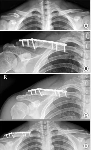
주기적으로(수술 후 2주, 1개월, 2개월, 4개월, 6개월, 1년) 촬영한 쇄골의 전후방 및 사면 단순 방사선 사진을 이용하여 골유합 여 부를 판정하였으며, 골절부의 상, 하면이 모두 연결되거나 골절 선이 사라지고 골소주가 골절부를 통과하는 때를 골유합 시기로 판정하였다(Fig. 2). 또한 최종 추시 시의 쇄골 길이를 건 측과 비 교하여 길이의 변화를 비교하였다. 쇄골의 길이는 쇄골의 전후방 방사선 사진에서 쇄골의 내측 단과 외측 단의 중심을 연결하는 직선으로 측정하였다.
5. 통계 방법
통계 분석은 SPSS ver. 16.0 (SPSS Inc., Chicago, IL, USA)을 사용 하여 Mann-Whitney U-test를 이용하였고, p값이 0.05 미만인 경 우를 통계적 유의성이 있는 것으로 판정하였다.
결 과
1. 임상적 결과
COP군은 23.1개월(12-60개월), MIPO군은 평균 18.4개월(12-39개 월) 추시가 가능하였다. COP군 20예 중 14예에서 지연 나사와 보 호 금속판(protection plate)을 사용하여 골편을 고정하였고, 2예에 서는 압박 금속판만을 사용하였으며, 4예에서 골이식을 시행하였 다. 최종 추시 시 측정한 SST score, ASES score, Q-DASH score는 양 군에서 모두 우수한 결과를 나타내었으며 유의한 차이를 보이 지 않았다. 수술 시간은 COP군에서 평균 106.9분으로 MIPO군의 평균 75.8분보다 오래 걸렸으며 이는 통계적으로 유의한 차이를 보였다(p=0.002). MIPO군에서 방사선 조사 시간과 조사량이 더 많았으며 이는 통계적으로 유의한 차이를 보였다(p=0.002, 0.005) (Table 2). 두 군 모두에서 수술 부위 감염, 불유합, 재골절, 내고정 실패 등의 합병증은 관찰되지 않았다.
2. 방사선적 결과
A
B
C
D
Figure 2. Serial plain radiographs of mid-claviclar fractre treated with minimally invasive plate osteosynthesis. (A) Preoperative radiograph shows that the right clavicle is shortened and displaced compared with the left clavicle. (B) Clavicle anteroposterior (AP) radiograph taken just after the operation shows a reduced and fixed dispaced fragment. (C) Clavicle AP radiograph taken 3 months after the operation shows bone union. (D) Clavicle AP radiograph taken 1 year after the operation shows restroation length and alignment of right clavicle.Table 2. Clinical Outcomes between COP and MIPO Group
Variable COP group MIPO group p-value
Operation time (min) 106.9 75.8 0.002 Radiation time (s) 37.1 52.8 0.002 Radiation exposure (mGy) 43.1 209.4 0.005 Simple shoulder test 10.2 11.3 0.521ASES score 92.5 94.3 0.325
Q-DASH score 5.0 4.8 0.322
Values are presented as mean only. COP, conventional open plating;
MIPO, minimally invasive plate osteosynthesis; ASES, American Shoulder and Elbow Surgeons; Q-DASH, quick disability of the arm, shoulder and hand.
COP군과 MIPO군의 모든 환자에서 만족스러운 골유합을 얻었 다. 골유합까지 걸린 시간은 COP군에서 14.6주, MIPO군에서 12.1 주로 COP군에서 더 오래 걸렸으나, 두 군 간에 유의한 차이는 보이지 않았다(p=0.587). 환측과 건측의 쇄골 길이 비를 비교하 였을 때에도 두 군 간에 유의한 차이를 보이지 않았다(p=0.477) (Table 3).
고 찰
MIPO는 골절부 주변의 연부 조직과 근막의 혈류를 보존하여 골 유합을 촉친하는 방법으로 하지 골절에서 많이 사용되고 있으며 상지 골절에서도 적용이 증가하고 있다.18-20) 최근 일부 저자들은 MIPO를 쇄골 간부 골절에 적용하였으며, 임상적, 영상의학적으 로 우수한 결과를 보고하였고,15-17,21) COP와도 비견할 만한 결과 가 보고되고 있다.22,23) Jiang과 Qu22)는 COP와 MIPO 각각 32예를 비교하였으며 두 군 간에 임상적 결과는 차이가 없었으다. COP에 서 10예의 피부 감각 저하의 부작용이 발생하였고, MIPO에서 미 용적으로 환자의 만족도가 높았다고 보고했다. Sohn 등23)은 COP 14예, MIPO 19예를 비교하였고 MIPO에서 수술 시간이 짧았으나 통계적으로 유의하지 않았으며 COP에서 4예의 피부 감각 저하를 보였다고 하였다. 임상적 결과는 두 군 간에 통계적 유의성이 없 음을 보고하였다.
본 연구에서도 전위된 쇄골 간부 골절에서 COP와 MIPO로 수 술을 시행 받은 양 군에서 임상적, 영상의학적으로 만족스러운 결과를 보였고 두 군 간에 통계적으로 유의한 차이를 보이지 않 았다. 하지만 MIPO가 COP보다 통계적으로 유의하게 수술 시간 이 적게 걸리는 것을 확인할 수 있었다. 이는 COP로 수술하는 경 우 분쇄, 전위된 골편을 해부학적으로 정복하고 고정하는 데 더 많은 시간이 소요되며 MIPO에서 피부 절개가 적어 봉합 시간이 적기 때문이라고 생각된다.
골절 수술 시 방사선 투과 촬영기가 이용되면서 수술자의 방 사선 노출에 대한 위험성이 제기되고 있고, 장기간의 방사선 노 출은 갑상선암, 백내장 등의 많은 부작용을 일으킬 수 있어 이 에 대한 주의가 필요하다고 알려져 있다.24) 과거 연구들에 따르 면 MIPO는 골절의 정렬을 맞추기 위해 더 많은 방사선 촬영이 불 가피하여 수술 중 방사선 피폭이 COP에 비해 많은 것이 가장 큰 단점 중의 하나로 알려져 있다. 하지만 이전 연구에서는 쇄골의
MIPO 수술 시 방사선 조사량에 대해서는 보고된 바 없다. 본 연 구에서는 MIPO 시 평균 약 52초간 방사선이 조사되었고, 이전 연 구에서 측정된 상완골 골절 MIPO의 68초,25) 경골 골절의 MIPO의 39.9초26) 사이에서 방사선이 조사되었다. 방사선 에너지를 나타내 는 단위인 Gy (Gray)는 1 kg의 물체가 방사선을 통해 1 J의 에너지 를 흡수했을 때의 흡수선량으로, Sv (Sievert)는 인체가 받는 방사 선 영향을 나타낸 수치이며, 감마, 베타선의 경우 Gy와 Sv는 동일 한 값을 갖는다. 국제 방사선 방어 위원회(The International Com- mission on Radiological Protection)에서는 정형외과 의사의 방사 선 노출 허용량을 제시하였고 이는 1년간 눈에 150 mSv, 갑상선 에 300 mSv이며 전신에는 20 mSv까지 허용되는 것으로 규정하고 있다.27) 우리 나라에서도
‘
진단용 방사선 발생 장치의 안전관리에 관한 규칙’
에 방사선 관례 종사자의 선량 한도는 연간 50 mSv 이 하 및 5년간 누적 선량은 100 mSv 이하여야 한다고 규정하고 있 다. 식품의약품안전청에서도‘
진단용 방사선 안전 관리 규정’
에 따라 3개월간 5 mSv 이하 또는 연간 20 mSv 미만이 되도록 개인 피폭선량을 관리하고 있다. 본 연구에서는 방사선 투과 촬영기 에서 직접 발생한 방사선 조사량을 측정하여 신체에 직접적으로 얼마나 많은 방사선이 피폭되는지는 알 수 없지만, COP에 비해 MIPO가 상당한 양의 방사선에 피폭될 가능성이 있다는 것을 알 수 있었다. 따라서 MIPO 시 방사선 노출량을 줄이기 위해서는 철 저한 방사선 차폐 가운 착용 및 방사선 투과 촬영기와의 적절한 거리를 유지하여야 하고 역방향 방사선 투과 장비의 사용을 고려하는 등28,29) 방사선 노출 피해를 줄이기 위해 노력하여야 한다.
본 연구는 다음과 같은 제한점이 있다. 첫째, 무작위 배정에 의 한 전향적 연구가 아니라 후향적인 연구로 통계적 결과를 일반 화하기에는 제한점이 있다. 하지만 연구 기간 중 환자 소실이 없 었고 골절 형태와 환자의 나이가 비슷하여 선택 편향을 최소화 할 수 있었다. 또한 일정한 기준에 따라 수술 후 평가하였기 때문 에, 비록 후향적 연구이지만 전향적 자료 수집을 할 수 있었다. 둘 째, 대상 환자 수가 적었다. 셋째, 쇄골 골절 수술 후 발생할 수 있 는 합병증 중의 하나인 감각 저하에 대한 조사를 하지 않았다. 넷 째, 분쇄 골절에만 적용을 하여 단순 골절에서는 다른 결과를 가 져올 수 있다는 점이다. 다섯째, 방사선 투과 장비의 방사선 조사 량만을 측정하여 수술자에 대한 직접적인 방사선 피폭량은 알 수 없다는 점이다.
Table 3. Radiological Outcomes between COP and MIPO Group
Variable COP group MIPO group p-value
Time to bone union (wk) 14.6 12.1 0.587
Length ratio (operation side/non-injured side) 1.05 1.01 0.477 Values are presented as mean only. COP, conventional open plating; MIPO, minimally invasive plate osteosynthesis.
결 론
쇄골 간부 분쇄 골절에서 MIPO는 고식적 금속판 고정술과 비교 하여 임상적, 영상의학적으로 모두 우수한 결과를 보였고 짧은 수술 시간과 골이식술이 필요없는 효과적인 치료법으로 추천될 수 있을 것으로 생각된다. 하지만 고식적 금속판 고정술에 비해 방사선 노출량이 많아 보호장구의 착용 등 이의 피해를 줄이기 위한 노력을 명심하여야 할 것으로 생각된다.
CONFLICTS OF INTEREST
The authors have nothing to disclose.
REFERENCES
1. Postacchini F, Gumina S, De Santis P, Albo F. Epidemiology of clavicle fractures. J Shoulder Elbow Surg. 2002;11:452-6.
2. Robinson CM. Fractures of the clavicle in the adult. Epidemi- ology and classification. J Bone Joint Surg Br. 1998;80:476-84.
3. Neer CS 2nd. Nonunion of the clavicle. J Am Med Assoc.
1960;172:1006-11.
4. Rowe CR. An atlas of anatomy and treatment of midclavicu- lar fractures. Clin Orthop Relat Res. 1968;58:29-42.
5. McKee RC, Whelan DB, Schemitsch EH, McKee MD. Opera- tive versus nonoperative care of displaced midshaft clavicular fractures: a meta-analysis of randomized clinical trials. J Bone Joint Surg Am. 2012;94:675-84.
6. Robinson CM, Goudie EB, Murray IR, et al. Open reduction and plate fixation versus nonoperative treatment for dis- placed midshaft clavicular fractures: a multicenter, random- ized, controlled trial. J Bone Joint Surg Am. 2013;95:1576-84.
7. Canadian Orthopaedic Trauma Society. Nonoperative treat- ment compared with plate fixation of displaced midshaft clavicular fractures. A multicenter, randomized clinical trial.
J Bone Joint Surg Am. 2007;89:1-10.
8. Ferran NA, Hodgson P, Vannet N, Williams R, Evans RO.
Locked intramedullary fixation vs plating for displaced and shortened mid-shaft clavicle fractures: a randomized clinical trial. J Shoulder Elbow Surg. 2010;19:783-9.
9. Nordqvist A, Petersson CJ, Redlund-Johnell I. Mid-clavicle fractures in adults: end result study after conservative treat- ment. J Orthop Trauma. 1998;12:572-6.
10. Frigg A, Rillmann P, Perren T, Gerber M, Ryf C. Intramedul- lary nailing of clavicular midshaft fractures with the titanium
elastic nail: problems and complications. Am J Sports Med.
2009;37:352-9.
11. Smekal V, Irenberger A, Struve P, Wambacher M, Krappinger D, Kralinger FS. Elastic stable intramedullary nailing versus nonoperative treatment of displaced midshaft clavicular fractures-a randomized, controlled, clinical trial. J Orthop Trauma. 2009;23:106-12.
12. Zlowodzki M, Zelle BA, Cole PA, Jeray K, McKee MD;
Evidence-Based Orthopaedic Trauma Working Group. Treat- ment of acute midshaft clavicle fractures: systematic review of 2144 fractures: on behalf of the evidence-based orthopae- dic trauma working group. J Orthop Trauma. 2005;19:504-7.
13. Liu PC, Hsieh CH, Chen JC, Lu CC, Chuo CY, Chien SH. In- fection after surgical reconstruction of a clavicle fracture us- ing a reconstruction plate: a report of seven cases. Kaohsiung J Med Sci. 2008;24:45-9.
14. Duncan SF, Sperling JW, Steinmann S. Infection after clavicle fractures. Clin Orthop Relat Res. 2005;439:74-8.
15. Jung GH, Park CM, Kim JD. Biologic fixation through bridge plating for comminuted shaft fracture of the clavicle: technical aspects and prospective clinical experience with a minimum of 12-month follow-up. Clin Orthop Surg. 2013;5:327-33.
16. Lee HJ, Oh CW, Oh JK, et al. Percutaneous plating for comminuted midshaft fractures of the clavicle: a surgical technique to aid the reduction with nail assistance. Injury.
2013;44:465-70.
17. Sohn HS, Kim BY, Shin SJ. A surgical technique for mini- mally invasive plate osteosynthesis of clavicular midshaft fractures. J Orthop Trauma. 2013;27:e92-6.
18. Kim JJ, Oh HK, Bae JY, Kim JW. Radiological assessment of the safe zone for medial minimally invasive plate osteosyn- thesis in the distal femur with computed tomography angiog- raphy. Injury. 2014;45:1964-9.
19. Apivatthakakul T, Arpornchayanon O, Bavornratanavech S. Minimally invasive plate osteosynthesis (MIPO) of the humeral shaft fracture. Is it possible? A cadaveric study and preliminary report. Injury. 2005;36:530-8.
20. Röderer G, Erhardt J, Graf M, Kinzl L, Gebhard F. Clinical results for minimally invasive locked plating of proximal hu- merus fractures. J Orthop Trauma. 2010;24:400-6.
21. Sohn HS, Shin SJ, Kim BY. Minimally invasive plate osteo- synthesis using anterior-inferior plating of clavicular mid- shaft fractures. Arch Orthop Trauma Surg. 2012;132:239-44.
22. Jiang H, Qu W. Operative treatment of clavicle midshaft frac-
tures using a locking compression plate: comparison between mini-invasive plate osteosynthesis (MIPPO) technique and conventional open reduction. Orthop Traumatol Surg Res.
2012;98:666-71.
23. Sohn HS, Kim WJ, Shon MS. Comparison between open plating versus minimally invasive plate osteosynthe- sis for acute displaced clavicular shaft fractures. Injury.
2015;46:1577-84.
24. Radhi AM, Masbah O, Shukur MH, Shahril Y, Taiman K.
Radiation exposure to operating theatre personnel during fluoroscopic-assisted orthopaedic surgery. Med J Malaysia.
2006;61 Suppl A:50-2.
25. Kim JW, Oh CW, Byun YS, Kim JJ, Park KC. A prospective randomized study of operative treatment for noncommi- nuted humeral shaft fractures: conventional open plating ver-
sus minimal invasive plate osteosynthesis. J Orthop Trauma.
2015;29:189-94.
26. Polat A, Kose O, Canbora K, Yanık S, Guler F. Intramedullary nailing versus minimally invasive plate osteosynthesis for dis- tal extra-articular tibial fractures: a prospective randomized clinical trial. J Orthop Sci. 2015;20:695-701.
27. 1990 Recommendations of the international commission on radiological protection. Ann ICRP. 1991;21:1-201.
28. Mehlman CT, DiPasquale TG. Radiation exposure to the or- thopaedic surgical team during fluoroscopy: "how far away is far enough?". J Orthop Trauma. 1997;11:392-8.
29. Tremains MR, Georgiadis GM, Dennis MJ. Radiation ex- posure with use of the inverted-c-arm technique in upper- extremity surgery. J Bone Joint Surg Am. 2001;83:674-8.
쇄골 간부 분쇄 골절에서 최소 침습 금속판 고정술의 유용성: 고식적 수술법과 비교
강태욱 • 황현정 • 이동기 • 한승범 • 정웅교
고려대학교 의과대학 고려대학교 안암병원 정형외과학교실
목적: 본 연구의 목적은 전위된 쇄골 간부 분쇄 골절에 있어서 고식적 수술법(conventional open plating, COP)과 최소 침습적 금속 판 고정술(minimally invasive plate osteosynthesis, MIPO) 사이의 임상적, 영상의학적 결과를 비교하고 MIPO의 유용성을 평가하고 자 하였다.
대상 및 방법: 쇄골 간부 골절로 수술을 시행한 39예 중 COP 20예, MIPO 19예를 대상으로 후향적으로 조사하였다. 영상의학적 평 가는 골유합 기간과 쇄골 길이를 건 측과 비교하여 길이의 변화를 비교하였고, 임상적 평가는 simple shoulder test score, American Shoulder and Elbow Surgeons score, quick disability of the arm, shoulder and hand score를 이용하여 비교하였다. 또한 두 군 간 의 평균 수술 시간과 방사선 조사 시간, 조사량을 비교하였다.
결과: 모든 환자에서 골유합을 얻었으며 골유합까지 걸린 시간은 MIPO에서 12.1주로 COP의 14.6주보다 짧았으나 유의한 차이는 보이지 않았다(p=0.587). 2년 추시 시 임상적, 영상의학적으로 만족스러운 결과를 보였고 두 군 간에 통계적으로 유의한 차이를 보이 지 않았다. MIPO의 경우 COP보다 통계적으로 유의하게 수술 시간이 적게 걸렸으며(75.8분 vs. 106.9분, p=0.002) 방사선 조사 시간 과 조사량이 많았다(52.8초 vs. 37.1초, p=0.002; 209.4 mGy vs. 43.1 mGy, p=0.005).
결론: 쇄골 간부 분쇄 골절에서 MIPO는 임상적, 영상의학적으로 모두 우수한 결과를 보였고 수술 시간이 짧으며 골이식술이 불필요 한 효과적인 치료법으로 판단된다. 하지만 방사선 피해를 줄이는 노력을 간과하지 말아야 한다.
색인단어: 쇄골, 골절, 최소 침습적 금속판 고정술
접수일 2016년 10월 26일 수정일 2017년 2월 27일 게재확정일 2017년 3월 23일 책임저자 정웅교
02841, 서울시 성북구 인촌로 73, 고려대학교 의과대학 정형외과학교실 TEL 02-920-5925, FAX 02-924-5271, E-mail drshoulder@korea.ac.kr
Copyright © 2017 by The Korean Orthopaedic Association
“This is an Open Access article distributed under the terms of the Creative Commons Attribution Non-Commercial License (http://creativecommons.org/licenses/by-nc/4.0/) which permits unrestricted non-commercial use, distribution, and reproduction in any medium, provided the original work is properly cited.”