78 Ann Dermatol (Seoul)
Received June 8, 2008, Accepted for publication August 31, 2008 Reprint request to: Seon-Wook Hwang, M.D., Department of Derma- tology, Busan Paik Hospital, College of Medicine, Inje University, 633-165, Gaegum-dong, Busanjin-gu, Busan 614-735, Korea. Tel: 82- 51-890-6135, Fax: 82-51-897-6391, E-mail: skindoctor@paran.com
Ann Dermatol (Seoul) Vol. 21, No. 1, 2009
CASE REPORT
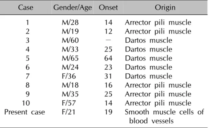
Table 1. The reported cases of acquired smooth muscle hamar- toma in the English medical literature
Case Gender/Age Onset Origin
1 2 3 4 5 6 7 8 9 10 Present case
M/28 M/19 M/60 M/33 M/65 M/24 F/36 M/18 M/35 F/57 F/21
14 12
− 25 64 23 31 16 25 14 19
Arrector pili muscle Arrector pili muscle Dartos muscle Dartos muscle Dartos muscle Dartos muscle Dartos muscle Arrector pili muscle Arrector pili muscle Arrector pili muscle Smooth muscle cells of
blood vessels
A Case of Acquired Smooth Muscle Hamartoma on the Sole
Deborah Lee, M.D., Sang-Hyun Kim, M.D., Soon-Kwon Hong, M.D., Ho-Suk Sung, M.D., Seon-Wook Hwang, M.D.
Department of Dermatology, Busan Paik Hospital, College of Medicine, Inje University, Busan, Korea
A smooth muscle hamartoma is a benign proliferation of smooth muscle bundles within the dermis. It arises from smooth muscle cells that are located in arrector pili muscles, dartos muscles, vascular smooth muscles, muscularis mammillae and the areolae. Acquired smooth muscle hamartoma (ASMH) is rare, with only 10 such cases having been reported in the English medical literature to date. Most of these cases of ASMH were shown to have originated from arrector pili and dartos muscles. Only one case was reported to have originated from vascular smooth muscle cells. A 21 year-old woman presented with a tender pigmented nodule, with numbness, on the sole of her foot, and this lesion had developed over the previous 18 months. The lesion showed no hyperpigmentation or hypertrichosis, and the biopsies demonstrated increased smooth muscle bundles in the dermis, and especially around the blood vessels. Moreover, the specimens stained positive with Masson trichrome stain and α-smooth-muscle actin antibodies, thus supporting our diagnosis of ASMH of the foot sole. Herein, we report on a rare case of ASMH on the foot sole, and this lesion originated from vascular smooth muscle cells. This type of case has not been previously described in the English medical literature.
(Ann Dermatol (Seoul) 21(1) 78∼80, 2009) -Keywords-
Acquired smooth muscle hamartoma, Sole, Vascular smooth muscle cell
INTRODUCTION
Smooth muscle hamartomas (SMH) are a result of benign proliferation of smooth muscle bundles in the dermis. SMH- associated smooth muscle cells originate from arrector pili-, dartos-, vulvar-, mammillary- and vascular wall- muscles1-3. SMH is characterized by slightly pigmented plaques that contain vellus hairs, and SMH is subdivided into two types: congenital smooth muscle hamartoma (CSMH) and acquired smooth muscle hamartoma (ASMH)1-3. ASMH is a very rare form of SMH that was first described by Wong and Solomon1 in 1985, with only such 10 cases having been reported in the English medical literature (Table 1). These lesions usually occur on the proximal extremities1,2, scrotum4-7, vulva8, shoulders2 and trunk9-11.
CASE REPORT
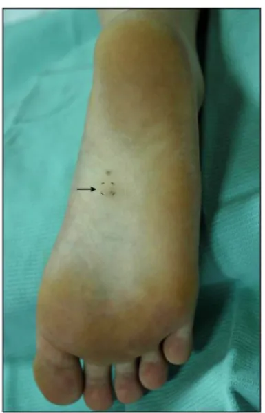
A 21 year-old women presented with an 18-month history of a slow-growing 0.5×0.5 cm nodule on the right sole (Fig. 1). She complained that the lesion was mildly tender
A Case of Acquired Smooth Muscle Hamartoma on the Sole
Vol. 21, No. 1, 2009 79 Fig. 1. A skin colored, 0.5×0.5 cm sized tender nodule on the
right sole.
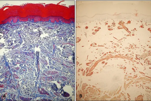
Fig. 2. An increased number of haphazardly oriented bundles of smooth muscle cells were observed in the dermis (A: H&E stain, ×40, B:
H&E stain, ×100).
and somewhat numb. Any evidence of trauma, allergic reaction, hyperpigmentation and hypertrichosis was not found and the Pseudo-Darier's sign could not be elicited.
Moreover, the results of the complete blood count, blood chemistry and urinalysis were all within the normal limits.
A skin biopsy specimen from the lesion revealed bundles of smooth muscle cells with cigar shaped nuclei, and these cells were arranged haphazardly throughout the dermis and especially around the blood vessels (Fig. 2).
Furthermore, the spindle cells stained positive with Masson trichrome stain and α-smooth-muscle actin antibodies.
The nodule was subsequently excised, and there has been no evidence of recurrence.
DISCUSSION
SMH is usually manifested at birth, but it can also appear later in life. CSMH is a congenital lesion that's principally composed of smooth muscle with or without vellus hair and/or epidermal hyperpigmentation. It is usually located on the trunk and extremities and it appears as variable sized papules, patches or plaques4. Transient piloerection or elevation of a lesion that's induced by rubbing is referred to as Pseudo-Darier's sign and this is the charac- teristic diagnostic clue for CSMH1-4. ASMH appears later in life as a slightly hyperpigmented or hypertrichotic patch or plaque with associated itching, pain or numbness.
These lesions are usually negative for Pseudo-Darier's sign7. The histopathologic hallmark of SMH is a local in- crease of large, randomly oriented bundles of smooth muscle cells with central, cigar-shaped nuclei. These smooth muscle bundles are arranged haphazardly around blood vessels in the dermis, and the smooth muscle bundles stain positive with Masson trichrome stain and α- smooth-muscle actin antibodies (Fig. 3).
The differential diagnosis for SMH includes leiomyomas, angioleiomyomas and Becker's nevus. In contrast to SMH, the smooth muscle bundles in leiomyomas are more tightly interlacing and poorly demarcated, and they can also be painful or tender. Angioleiomyomas contain thick-walled blood vessels with smooth muscle fibers deposited in concentric layers that merge with the mus- cular stroma. Hence, angioleiomyomas can be differen- tiated from SMH by the histological findings.
Becker's nevus often resembles SMH both clinically and histologically. Becker's nevus is a well-defined, uniformly hyperpigmented plaque with hypertrichosis. Histologi- cally, Becker's nevus exhibits an abnormal number of large, deeply seated terminal hair follicles that are some- times associated with hyperplastic pilar smooth muscles.
D Lee, et al
80 Ann Dermatol (Seoul)
Fig. 3. The haphazardly arranged smooth muscle bundles were mainly located around blood vessels (A:
Masson's trichrome stain, ×40, B: α- smooth-muscle actin antibody stain,
×40).
However, Becker's nevus can also present with a pro- liferation of dermal smooth muscle. Because of the overlapping histological features, some authors have speculated that Becker's nevus and SMH are two polar entities at either end of a continuous spectrum2,4,9. Since Wong and Solomon1 reported on the first case of ASMH in 1983, only 10 cases of ASMH have currently been reported in the English medical literature. ASMH is usually located on the skin of the forearm, chest, neck, scrotum, penis, major labium or shoulder. Five cases of ASMH had been reported to originate from the dartos muscle and 5 cases have been reported to originate from the arrector pili muscle (Table 1). In this case, we hypothesize that the hamartomatous proliferation of smooth muscle cells originated from blood vessel walls due to the haphazard arrangement of smooth muscle bundles around the dermal vasculature and the fact that arrector pili and dartos muscles are not normally present in foot soles. To the best of our knowledge, this is the first case of a smooth muscle hamartoma of the sole to be reported in the English medical literature.
REFERENCES
1. Wong RC, Solomon AR. Acquired dermal smooth-muscle
hamartoma. Cutis 1985;35:369-370.
2. ul Bari A, Rahman SB. Acquired smooth muscle hamartoma.
Indian J Dermatol Venereol Leprol 2006;72:178.
3. van Kooten EO, Hage JJ, Meinhardt W, Horenblas S, Mooi WJ. Acquired smooth-muscle hamartoma of the scrotum: a histological simulator? J Cutan Pathol 2004;31:388-392.
4. Quinn TR, Young RH. Smooth-muscle hamartoma of the tunica dartos of the scrotum: report of a case. J Cutan Pathol 1997;24:322-326.
5. Hsiao GH, Chen JS. Acquired genital smooth-muscle hamar- toma. A case report. Am J Dermatopathol 1995;17:67-70.
6. Semerci B, Ilbey O, Yurtseven O. Acquired penoscrotal smooth muscle hamartoma. J Urol 1997;158:885-886.
7. Oiso N, Fukai K, Ishii M, Hayashi T, Uda H, Imanishi M. A case of acquired smooth muscle hamartoma of the scrotum.
Clin Exp Dermatol 2005;30:523-524.
8. Kwon KS, Lee CW, Seo KH, Park JH, Oh CK, Jang HS, et al.
Acquired vulvar smooth muscle hamartoma: a case report and review of the literature. J Dermatol 2000;27:56-59.
9. Morales-Callaghan A, Vila JB, Cardenal EA, Bernier MA, Fraile HA, Miranda-Romero A. Acquired cutaneous smooth muscle hamartoma. J Eur Acad Dermatol Venereol 2005;
19:142-143.
10. Darling TN, Kamino H, Murray JC. Acquired cutaneous smooth muscle hamartoma. J Am Acad Dermatol 1993;
28:844-845.
11. Ryu HJ, Kim G, Song HJ, Oh CH. A case of acquired cutaneous smooth muscle hamartoma. Ann Dermatol 2002;
14:161-163.