ABSTRACT
Objective: To determine patterns among gynecologic oncologists in sentinel lymph node mapping (SLNM) for endometrial cancer (EC) and cervical cancer (CC).
Methods: A online survey assessing the practice of SLNM, including incidence, patterns of usage, and reasons for non-use was distributed to Society of Gynecologic Oncology candidate and full members in August 2017. Descriptive statistics and univariate analysis was performed.
Results: The 1,117 members were surveyed and 198 responses (17.7%) were received. Of the 70% (n=139) performing SLNM, the majority reported use for both CC and EC (64.0%) or EC alone (33.1%). In those using SLNM in EC, the majority (86.6%) performed SLNM in >50%
of cases for all patients (56.3%), International Federation of Gynecology and Obstetrics grade 1 (43.0%) and 2 (42.2%). Reported benefits of SLNM in EC were reduced surgical morbidity (89.6%), lymphedema (85.2%), and operative time (63.7%). Among those using SLNM for CC, the majority (73.1%) did so in >50% of cases. In EC, 77.2% and 21.3% reported that micro-metastatic disease (0.2–2.0 cm) and isolated tumor cells (ITCs) should be treated as node positive, respectively. In those not using SLNM for EC (n=64) and CC (n=105), concerns were regarding efficacy of SLNM and lack of training. When queried regarding training, 73.7% felt that SLNM would impact skill in full lymphadenectomy (LND).
Conclusion: The SLNM is utilized frequently among gynecologic oncologists for EC and CC staging. Common reasons for non-uptake include uncertainty of current data, lack of training and technology. Concerns exist regarding impact of SLNM in fellowship training of LND.
Keywords: Sentinel Lymph Node; Endometrial Cancer; Cervical Cancer; Lymphadenectomy
INTRODUCTION
Endometrial cancer (EC) is the most common gynecologic malignancy diagnosed in the United States [1]. The standard of care for EC is hysterectomy and bilateral salpingo-
oophorectomy via a minimally invasive surgical (MIS) approach [2]. Pelvic lymphadenectomy (LND) and/or para-aortic LND may be performed depending upon uterine factors, cancer stage and grade [3]. While 2 randomized control trials have studied the role of LND in EC and demonstrated no benefit in disease-specific or overall survival (OS), nodal information plays a crucial role in disease staging and treatment planning [4,5].
Original Article
Received: Aug 14, 2018 Revised: Nov 20, 2018 Accepted: Nov 21, 2018 Correspondence to Laura Moulton Chambers Division of Gynecologic Oncology,
Ob/Gyn & Women's Health Institute, Cleveland Clinic Foundation, 9500 Euclid Ave, Cleveland, OH 44195, USA.
E-mail: [email protected] Copyright © 2019. Asian Society of Gynecologic Oncology, Korean Society of Gynecologic Oncology
This is an Open Access article distributed under the terms of the Creative Commons Attribution Non-Commercial License (https://
creativecommons.org/licenses/by-nc/4.0/) which permits unrestricted non-commercial use, distribution, and reproduction in any medium, provided the original work is properly cited.
ORCID iDs
Laura Moulton Chambers
https://orcid.org/0000-0002-3773-7060 Roberto Vargas
https://orcid.org/0000-0003-4262-7824 Chad M. Michener
https://orcid.org/0000-0001-5170-6626 Conflict of Interest
No potential conflict of interest relevant to this article was reported.
Laura Moulton Chambers , Roberto Vargas , Chad M. Michener
Division of Gynecologic Oncology, Ob/Gyn & Women's Health Institute, Cleveland Clinic Foundation, Cleveland, OH, USA
Sentinel lymph node mapping in endometrial and cervical cancer:
a survey of practices and attitudes in
gynecologic oncologists
Author Contributions
Conceptualization: C.L.M., V.R., M.C.M.; Data curation: C.L.M.; Formal analysis: C.L.M.;
Investigation: C.L.M., V.R.; Methodology:
C.L.M., V.R., M.C.M.; Project administration:
V.R., M.C.M.; Resources: C.L.M., M.C.M.;
Software: C.L.M.; Supervision: V.R., M.C.M.;
Validation: C.L.M.; Visualization: C.L.M., V.R., M.C.M.; Writing - original draft: C.L.M., V.R., M.C.M.; Writing - review & editing: C.L.M., V.R., M.C.M.
There is growing evidence supporting the role of sentinel lymph node mapping (SLNM) in EC [6-12]. In the prospective multicenter study by Rossi et al. [11], specificity of greater than 97% was reported in patients with early stage EC undergoing SLNM. Similarly, data has increasingly demonstrated a role for SLNM in cervical cancer (CC) [13-16]. SLNM has become standard protocol at some institutions but despite growing evidence supporting use of SLNM and support of organizations including National Comprehensive Cancer Network (NCCN) and the Society of Gynecologic Oncology (SGO) consensus group, this technology has not become widespread in the United States [12,17-19]. Identification of the provider trends and attitudes and reasons for non-incorporation of SLNM may be helpful in guiding future studies and technologic advancements for MIS surgical staging in EC and CC.
The objective of this study is to identify trends in the utilization of SLNM for EC and CC among gynecologic oncologists and identify reasons for non-adoption of SLNM techniques among gynecologic oncologists.
MATERIALS AND METHODS
1. Study design
We designed an anonymous online questionnaire to assess member demographics, practice patterns, and attitudes regarding the use of SLNM in EC and CC. The study was approved by the Institutional Review Board at Cleveland Clinic Foundation, the Case Western Reserve University Comprehensive Cancer Center Protocol Review and Monitoring Committee (# 17- 1050) and by the SGO for the purpose of surveying candidate members and full members.
2. Survey creation and study variables
The survey consisted of 24 questions querying demographics, SLNM usage in practice, perceived benefits of SLNM, reasons for non-uptake of SLNM, attitudes regarding usage patterns, and interest in additional training. The first part of the survey queried demographic variables including years of practice, gender, practice setting, and location.
The second part of the survey assessed whether members were using SLNM for EC alone, CC alone, both EC and CC, or were not using SLNM in practice. If members reported use of SLNM in practice for EC, CC, or both, questions were asked regarding patterns of percentage of use, stage, and histologies in which SLNM was being used, surgical platform of use (MIS, multi-port laparoscopy, single port laparoscopy, robotic assisted laparoscopy; and laparotomy) and medium used for mapping (isosulfan blue, indocyanine green [ICG], technectium-99, and other). Questions were asked regarding use of full LND with alongside SLNM and personal mapping rate awareness and percentage, if known. For members who reported use of SLNM, questions were perceived benefits of SLNM in EC, CC, or both. In members not using SLNM for EC, CC, or both, they were asked for reasons for non-use (including concerns for efficacy of mapping, missing nodal positive disease and impact of ultra-staging, aid in training full LND, pathology department unsure how to process specimens, and lack of training during fellowship). Next, training practices for SLNM were assessed including where training was received, number of sentinel and full lymphadenectomies performed during fellowship and preparedness for SLNM after fellowship. Members were then queried whether they agreed, disagreed or were neutral regarding practice patterns in SLNM. Topics that were queried included perception of current data supporting SLNM in CC and EC, utility of current technology in SLNM, impact on fellowship training, whether they felt that micro-metastatic disease (0.2–2.0 mm) or
isolated tumor cells (ITCs) should be staged and treated as node positive. The complete survey is presented in Appendix.
3. Study participants
An E-mail invitation to participate in the anonymous study was distributed to all full and candidate gynecologic oncologist members of the SGO in August and September 2017. Fellows were excluded. Within the body of the E-mail, members were invited to participate in the confidential survey via the Research Electronic Data Capture (REDCap) website on a voluntary basis with an “opt-out” option [20]. No identifying data was collected, including the respondent's fellowship or current institution. Secure data storage was maintained with REDCap software (Vanderbilt, Nashville, TN, USA) [20]. The study was open for 4 weeks and members received an initial E-mail invitation, followed by up to 2 additional E-mails for members who did not complete the study which was automated in the REDCap program [20]. All members who were emailed the survey were included in the denominator of total surveys to calculate the response rate.
4. Statistical analysis
All returned surveys, including those with incomplete responses, were used for the final analysis. Descriptive statistics were performed to assess member demographics and for all survey responses. Percentage of response per question was divided by the denominator of answering responses per question. Respondent survey responses were analyzed with for multiple co-variates using Fisher's exact test and χ2 tests. Selected co-variates used to compare survey responses included years of practice, geographical location, gender, and training for SLNM. Two-tailed p-values <0.05 were considered significant. Statistical analysis was performed with JMP software (13.0; SAS Institute Inc., Cary, NC, USA).
RESULTS
1. Demographics
Of the 1,117 members who were surveyed, 198 responses (17.7%) were received. Table 1 displays the respondent demographics. Among those participating in the survey, there was a varied distribution of experiences within gynecologic oncology with over half (n=108;
54.5%) of respondents reporting more than 10 years of clinical practice. There was a broad geographic representation from across the United States among the responding gynecologic oncologists. The majority were in academic practice (n=103; 52.0%) or a combination of academic, and private practice (n=53; 26.8%). Among the 198 respondents, 70.2% (n=139) reported use of SLNM mapping for either EC, CC, or both in their practice.
2. Trends in SLNM for EC
Of the 139 of gynecologic oncologists using SLNM in their practice, 97.2% (n=135) endorsed use for EC. Table 2 displays the trends in usage in SLNM in EC. Robotic assisted laparoscopy was used as the most common surgical platform for SLNM in EC (n=109; 80.7%) and ICG was the most frequently used mapping dye (n=131; 97.0%).
The majority of respondents (n=94; 70.7%) utilized SLNM in over three-quarters of their surgical cases with 24.8% (n=33) using SLNM in all EC surgeries. When queried regarding usage of SLNM for EC, over half (56.3%; n=76) reported use for all patients regardless of histology without evidence of extra-uterine disease. Additionally, 43.0% (n=58) utilized SLNM for International Federation of Gynecology and Obstetrics (FIGO) 1 and 42.2%
(n=57) for FIGO2. Use of SLNM was less frequently reported for patients with FIGO3 (n=30;
22.2%), complex atypical hyperplasia (n=26; 19.3%), high-risk histologies including serous carcinoma, clear cell carcinoma, and carcinosarcoma (n=24; 17.8%).
Over half of respondents (n=85; 63%) reported concurrent use of SLNM and full systematic LND in EC staging when they first adopted the technique. The median number of reported concurrent cases was 25 (range, 2–250). Less than half of respondents (n=59; 43.7%) were aware of their individual mapping rate for SLNM. Median unilateral SLNM rate was 90%
(range, 0%–100%) and median bilateral SLNM rate was 85% (range, 50%–100%).
Among those performing SLNM for EC perceived benefits included reduced surgical morbidity (n=121; 89.6%), decreased lymphedema (n=115; 85.2%), lower blood loss (n=55;
40.7%), and increased speed vs. full LND (n=86; 63.7%). Additional reported benefits included faster recovery (n=28; 20.7%), improved detection of small volume metastatic disease (n=8; 5.9%), and reduced lymphocyst or lymphocele formation (n=3; 2.2%).
3. Trends in SLNM for cervical cancer
Of the 139 of gynecologic oncologists using SLNM in their practice, 67.0% (n=93) reported use for CC. Table 3 displays the trends in usage in SLNM in CC. Similar to EC, robotic assisted laparoscopy was used as the most common surgical platform for SLNM in CC (n=74;
80.0%) and ICG was the most frequently used mapping dye (n=86; 92.5%). The majority of respondents (n=57; 64.0%) utilized SLNM in over three-quarters of their surgical cases for CC. When queried regarding usage of SLNM for CC, the majority used the technology for stage IA2 (n=87; 93.5%) and stage IB1 (n=88; 95.0%) disease. Additionally, 50.5% (n=47) utilized SLNM for stage IA1 and 40.9% (n=38) for stage IB2 patients.
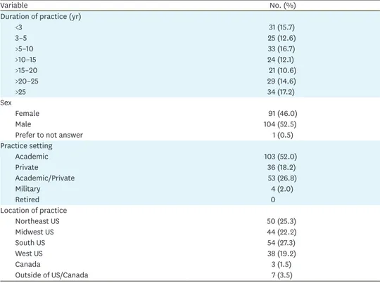
Table 1. Demographic information for survey respondents
Variable No. (%)
Duration of practice (yr)
<3 31 (15.7)
3–5 25 (12.6)
>5–10 33 (16.7)
>10–15 24 (12.1)
>15–20 21 (10.6)
>20–25 29 (14.6)
>25 34 (17.2)
Sex
Female 91 (46.0)
Male 104 (52.5)
Prefer to not answer 1 (0.5)
Practice setting
Academic 103 (52.0)
Private 36 (18.2)
Academic/Private 53 (26.8)
Military 4 (2.0)
Retired 0
Location of practice
Northeast US 50 (25.3)
Midwest US 44 (22.2)
South US 54 (27.3)
West US 38 (19.2)
Canada 3 (1.5)
Outside of US/Canada 7 (3.5)
US, United States.
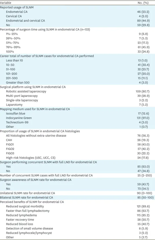
Table 2. Trends in usage in SLNM in endometrial cancer
Variable No. (%)
Reported usage of SLNM
Endometrial CA 46 (23.2)
Cervical CA 4 (2.0)
Endometrial and cervical CA 89 (44.9)
No 59 (29.8)
Percentage of surgeon time using SLNM in endometrial CA (n=133)
1%–25% 9 (6.8)
26%–50% 7 (5.3)
51%–75% 23 (17.3)
76%–99% 61 (45.9)
100% 33 (24.8)
Career total of number of SLNM cases for endometrial CA performed
Less than 10 13 (1.0)
10–50 41 (30.4)
51–100 32 (23.7)
101–200 27 (20.0)
201–500 15 (11.1)
Greater than 500 4 (3.0)
Surgical platform using SLNM in endometrial CA
Robotic assisted laparoscopy 109 (80.7)
Multi-port laparoscopy 39 (28.9)
Single-site laparoscopy 3 (2.2)
Laparotomy 7 (5.2)
Mapping medium used for SLNM in endometrial CA
Isosulfan blue 17 (12.6)
Indocyanine Green 131 (97.0)
Technectium-99 4 (3.0)
Other 1 (0.7)
Proportion of usage of SLNM in endometrial CA histologies
All histologies without extra-uterine disease 76 (56.3)
CAH 26 (19.3)
FIGO1 58 (43.0)
FIGO2 57 (42.2)
FIGO3 30 (22.2)
High-risk histologies (USC, UCC, CS) 24 (17.8)
Surgeon performing concurrent SLNM with full LND for endometrial CA
Yes 85 (63.0)
No 47 (34.8)
Number of concurrent SLNM cases with full LND for endometrial CA 25 (2–250) Surgeon awareness of SLNM rate for endometrial CA
Yes 59 (43.7)
No 73 (54.1)
Unilateral SLNM rate for endometrial CA 90 (0–100)
Bilateral SLNM rate for endometrial CA 85 (50–100)
Perceived benefits of SLNM for endometrial CA
Reduced surgical morbidity 121 (89.6)
Faster than full lymphadectomy 86 (63.7)
Reduced lymphedema 115 (85.2)
Faster recovery time 28 (20.7)
Reduced blood loss 55 (40.7)
Detection of small volume disease 8 (5.9)
Reduced lymphocele/lymphocyst 3 (2.2)
Other 5 (3.7)
The SLNM mapping rates and number of concurrent SLNM cases are reported as median +/− reported range.
CA, cancer; CAH, complex atypical hyperplasia; CS, carcinosarcoma; FIGO, International Federation of Gynecology and Obstetrics; LND, lymphadenectomy; SLNM, sentinel lymph node mapping; UCC, clear cell carcinoma; USC, uterine serous carcinoma.
Over three-quarters of respondents (n=72; 77.4%) reported concurrent use of SLNM and full systematic LND in CC staging when they first adopted the technique with a median number of 20 cases (range, 5–400). Less than one-third of respondents (n=25; 26.9%) were aware of their individual mapping rate for SLNM in CC, specifically. The median reported SLNM rate was 91.5% (range, 70%–100%).
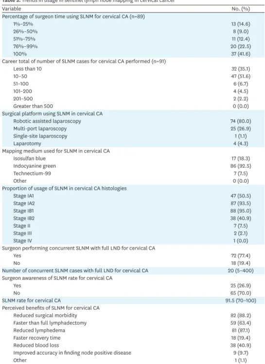
Table 3. Trends in usage in sentinel lymph node mapping in cervical cancer
Variable No. (%)
Percentage of surgeon time using SLNM for cervical CA (n=89)
1%–25% 13 (14.6)
26%–50% 8 (9.0)
51%–75% 11 (12.4)
76%–99% 20 (22.5)
100% 37 (41.6)
Career total of number of SLNM cases for cervical CA performed (n=91)
Less than 10 32 (35.1)
10–50 47 (51.6)
51–100 6 (6.7)
101–200 4 (4.5)
201–500 2 (2.2)
Greater than 500 0 (0.0)
Surgical platform using SLNM in cervical CA
Robotic assisted laparoscopy 74 (80.0)
Multi-port laparoscopy 25 (26.9)
Single-site laparoscopy 1 (1.1)
Laparotomy 4 (4.3)
Mapping medium used for SLNM in cervical CA
Isosulfan blue 17 (18.3)
Indocyanine green 86 (92.5)
Technectium-99 7 (7.5)
Other 0 (0.0)
Proportion of usage of SLNM in cervical CA histologies
Stage IA1 47 (50.5)
Stage IA2 87 (93.5)
Stage IB1 88 (95.0)
Stage IB2 38 (40.9)
Stage II 7 (7.5)
Stage III 2 (2.1)
Stage IV 1 (0.0)
Surgeon performing concurrent SLNM with full LND for cervical CA
Yes 72 (77.4)
No 18 (19.4)
Number of concurrent SLNM cases with full LND for cervical CA 20 (5–400) Surgeon awareness of SLNM rate for cervical CA
Yes 25 (26.9)
No 65 (70.0)
SLNM rate for cervical CA 91.5 (70–100)
Perceived benefits of SLNM for cervical CA
Reduced surgical morbidity 82 (88.2)
Faster than full lymphadectomy 59 (63.4)
Reduced lymphedema 81 (87.1)
Faster recovery time 18 (19.4)
Reduced blood loss 38 (40.9)
Improved accuracy in finding node positive disease 9 (9.7)
Other 1 (1.1)
The SLNM mapping rates and number of concurrent SLNM cases are reported as median +/− reported range.
CA, cancer; LND, lymphadenectomy; SLNM, sentinel lymph node mapping.
Among gynecologic oncologists utilizing SLNM for CC perceived benefits included reduced surgical morbidity (n=82; 88.2%), decreased lymphedema (n=81; 87.1%), lower blood loss (n=38; 40.9%), and increased speed vs. full LND (n=59; 63.4%). Additional reported benefits included faster recovery (n=18; 19.4%) and improved detection of small volume metastatic disease (n=9; 9.7%).
4. Reasons for non-uptake of SLNM
Of the 198 gynecologic oncologists who responded to the survey, 30% (n=59) did not use SLNM in their practice in the management of both EC and CC. When non-use was stratified by type of malignancy, 31.8% (n=63) reported non-use of SLNM for EC and 53.0% (n=105) reported non-use of SLNM for CC. Table 4 displays reported reasons for non-use of SLNM in EC and CC.
Reported reasons for non-use of SLNM for EC staging included uncertainty of data (n=26;
46.0%), lack of training (n=23; 36.5%), technology (n=22; 34.9%), issues with specimen processing for ultra-staging (n=11; 17.5%), and to aid with training of fellows in full LND (n=8; 12.7%). Concerns reported by non-users regarding SLNM included efficacy of mapping (n=20; 31.7%), implications of missing nodal positive disease (n=18; 28.6%), and uncertainty for how SLNM data will impact patient outcomes (n=20; 31.7%). Similarly for SLNM in CC, respondents reported uncertainty of data (n=62; 59.0%), lack of training (n=22; 21.0%), technology (n=19; 18.1%), and issues with specimen processing for ultra- staging (n=10; 9.5%) as reasons for non-uptake. Concerns reported by non-users regarding SLNM included efficacy of mapping (n=25; 23.8%), implications of missing nodal positive disease (n=34; 32.4%) and uncertainty for how SLNM data will impact patient outcomes (n=20; 19.0%).
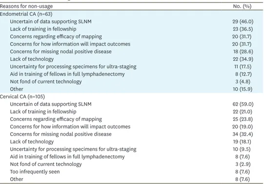
Table 4. Reasons for non-usage of SLNM in endometrial and cervical CA
Reasons for non-usage No. (%)
Endometrial CA (n=63)
Uncertain of data supporting SLNM 29 (46.0)
Lack of training in fellowship 23 (36.5)
Concerns regarding efficacy of mapping 20 (31.7)
Concerns for how information will impact outcomes 20 (31.7)
Concerns for missing nodal positive disease 18 (28.6)
Lack of technology 22 (34.9)
Uncertainty for processing specimens for ultra-staging 11 (17.5)
Aid in training of fellows in full lymphadenectomy 8 (12.7)
Not fond of current technology 3 (4.8)
Other 10 (15.9)
Cervical CA (n=105)
Uncertain of data supporting SLNM 62 (59.0)
Lack of training in fellowship 22 (21.0)
Concerns regarding efficacy of mapping 25 (23.8)
Concerns for how information will impact outcomes 20 (19.0)
Concerns for missing nodal positive disease 34 (32.4)
Lack of technology 19 (18.1)
Uncertainty for processing specimens for ultra-staging 10 (9.5)
Aid in training of fellows in full lymphadenectomy 8 (7.6)
Not fond of current technology 3 (2.9)
Too infrequently seen 8 (7.6)
Other 8 (7.6)
CA, cancer; SLNM, sentinel lymph node mapping.
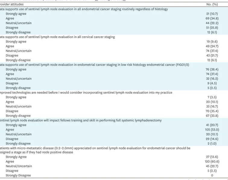
5. Provider attitudes toward SLNM
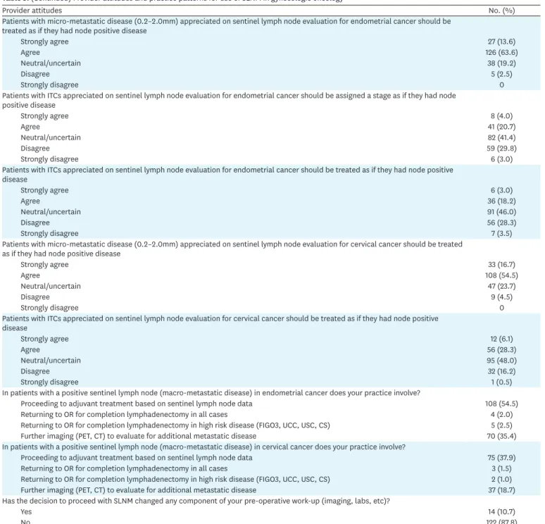
Provider Attitudes and Practices toward SLNM are displayed in Table 5. For EC, 45.5% (n=90) of respondents reported that data supported the use of SLNM in all EC cases regardless of histology, while 75.8% (n=150) felt that SLNM was supported in low-risk histologies (FIGO1/2) only. While the majority of respondents (n=154; 77.2%) agreed that micro- metastatic disease (0.2–2.0 cm) should be treated as node positive, only 21.3% (n=42) agreed for ITCs. When queried regarding practice patterns after positive lymph nodes were identified after SLNM, the majority reported proceeding direct to adjuvant treatment (n=108; 54.5%) or performing additional imaging to evaluate for additional metastatic disease (n=70; 35.4%).
When queried regarding SLNM in CC, approximately one-third of respondents both independently agreed (n=68; 34.4%) and disagreed (n=55; 27.8%) that data supported use.
Similar to EC, the majority of respondents (n=141; 71.2%) agreed that micro-metastatic disease (0.2–2.0 cm) should be treated as node positive, only 34.3% (n=68) agreed for ITCs. While the majority of gynecologic oncologists disagreed that improved technologies would increase
Table 5. Provider attitudes and practice patterns for use of SLNM in gynecologic oncology
Provider attitudes No. (%)
Data supports use of sentinel lymph node evaluation in all endometrial cancer staging routinely regardless of histology
Strongly agree 21 (10.7)
Agree 69 (34.8)
Neutral/uncertain 44 (22.2)
Disagree 51 (25.8)
Strongly disagree 12 (6.1)
Data supports use of sentinel lymph node evaluation in all cervical cancer staging
Strongly agree 19 (9.6)
Agree 49 (24.7)
Neutral/uncertain 74 (37.4)
Disagree 43 (21.7)
Strongly disagree 12 (6.1)
Data supports use of sentinel lymph node evaluation in endometrial cancer staging in low risk histology endometrial cancer (FIGO1/2)
Strongly agree 76 (38.4)
Agree 74 (37.4)
Neutral/uncertain 32 (16.2)
Disagree 9 (4.5)
Strongly disagree 5 (2.5)
Improved technologies are needed before I would consider incorporating sentinel lymph node evaluation into my practice
Strongly agree 7 (3.5)
Agree 20 (10.1)
Neutral/uncertain 33 (16.7)
Disagree 70 (35.4)
Strongly disagree 67 (33.8)
Sentinel lymph node evaluation will impact fellows training and skill in performing full systemic lymphadenectomy
Strongly agree 41 (20.7)
Agree 105 (53.0)
Neutral/uncertain 20 (10.1)
Disagree 29 (14.6)
Strongly disagree 2 (1.0)
Patients with micro-metastatic disease (0.2–2.0mm) appreciated on sentinel lymph node evaluation for endometrial cancer should be assigned a stage as if they had node positive disease
Strongly Agree 27 (13.6)
Agree 120 (60.6)
Neutral/Uncertain 45 (22.7)
Disagree 5 (2.5)
Strongly Disagree 0
(continued to the next page)
their likelihood of uptake of the practice (n=137; 69.1%), the majority agreed that SLNM would impact fellows training and skill level in performing full LND for EC (n=146; 73.7%).
6. Provider factors associated with usage of SLNM
Table 6 displays results of univariate analysis for provider factors associated with usage of SLNM in EC and CC. There were no significant differences in the utilization of SLNM based upon gender (p=0.29), duration of practice as a gynecologic oncologist (p=0.13), practice
Provider attitudes No. (%)
Patients with micro-metastatic disease (0.2–2.0mm) appreciated on sentinel lymph node evaluation for endometrial cancer should be treated as if they had node positive disease
Strongly agree 27 (13.6)
Agree 126 (63.6)
Neutral/uncertain 38 (19.2)
Disagree 5 (2.5)
Strongly disagree 0
Patients with ITCs appreciated on sentinel lymph node evaluation for endometrial cancer should be assigned a stage as if they had node positive disease
Strongly agree 8 (4.0)
Agree 41 (20.7)
Neutral/uncertain 82 (41.4)
Disagree 59 (29.8)
Strongly disagree 6 (3.0)
Patients with ITCs appreciated on sentinel lymph node evaluation for endometrial cancer should be treated as if they had node positive disease
Strongly agree 6 (3.0)
Agree 36 (18.2)
Neutral/uncertain 91 (46.0)
Disagree 56 (28.3)
Strongly disagree 7 (3.5)
Patients with micro-metastatic disease (0.2–2.0mm) appreciated on sentinel lymph node evaluation for cervical cancer should be treated as if they had node positive disease
Strongly agree 33 (16.7)
Agree 108 (54.5)
Neutral/uncertain 47 (23.7)
Disagree 9 (4.5)
Strongly disagree 0
Patients with ITCs appreciated on sentinel lymph node evaluation for cervical cancer should be treated as if they had node positive disease
Strongly agree 12 (6.1)
Agree 56 (28.3)
Neutral/uncertain 95 (48.0)
Disagree 32 (16.2)
Strongly disagree 1 (0.5)
In patients with a positive sentinel lymph node (macro-metastatic disease) in endometrial cancer does your practice involve?
Proceeding to adjuvant treatment based on sentinel lymph node data 108 (54.5)
Returning to OR for completion lymphadenectomy in all cases 4 (2.0)
Returning to OR for completion lymphadenectomy in high risk disease (FIGO3, UCC, USC, CS) 5 (2.5)
Further imaging (PET, CT) to evaluate for additional metastatic disease 70 (35.4)
In patients with a positive sentinel lymph node (macro-metastatic disease) in cervical cancer does your practice involve?
Proceeding to adjuvant treatment based on sentinel lymph node data 75 (37.9)
Returning to OR for completion lymphadenectomy in all cases 3 (1.5)
Returning to OR for completion lymphadenectomy in high risk disease (FIGO3, UCC, USC, CS) 2 (1.0)
Further imaging (PET, CT) to evaluate for additional metastatic disease 37 (18.7)
Has the decision to proceed with SLNM changed any component of your pre-operative work-up (imaging, labs, etc)?
Yes 14 (10.7)
No 122 (87.8)
CS, carcinosarcoma; CT, computed tomography; FIGO, International Federation of Gynecology and Obstetrics; ITC, isolated tumor cell; PET, positron emission tomography; SLNM, sentinel lymph node mapping; UCC, clear cell carcinoma; USC, uterine serous carcinoma.
Table 5. (Continued) Provider attitudes and practice patterns for use of SLNM in gynecologic oncology
setting (p=0.99), or location of practice (p=0.59). Among providers not using SLNM, the vast majority had not been trained to do so (n=47; 79.7%) compared to those performing SLNM routinely (n=20; 14.8%) (p<0.001).
7. Current training practices in SLNM
Of the 127 respondents (64.1%) who had been trained in SLNM, the majority (n=85; 66.9%) were trained to perform the procedure as attending physicians. Among those not using SLNM in their practice, 76.7% reported that they would be interested in further training through surgical videos, case proctoring and observation. Among those trained in fellowship, 83.3% (n=35) felt prepared to independently perform SLNM at the end of fellowship.
DISCUSSION
The SLNM is a targeted approach to MIS staging for EC and CC. This study was designed to identify usage patterns in SLNM and determine reasons for non-adoption. While SLNM for EC and CC is being utilized at many institutions and has received endorsement by NCCN and SGO, SLNM accounted for only 10% of lymphadenectomies for EC in 2015 [12,17-19].
Our results demonstrate that SLNM is more prevalent than previously reported with over two-thirds of responding gynecologic oncologists performing SLNM. Among the one-third Table 6. Provider factors associated with usage and non-uptake of SLNM for endometrial and cervical cancer
Variable Providers reporting usage of SLNM Providers reporting non-usage of SLNM p-value
Gender 0.29
Women 62 (45.6) 29 (49.2)
Men 74 (54.4) 30 (50.8)
Prefer to not answer 0 (0.0) 0 (0.0)
Practice setting 0.99
Academic 72 (52.6) 31 (52.5)
Private 25 (18.2) 11 (18.6)
Academic/private 37 (27.0) 16 (27.1)
Military 3 (2.2) 1 (1.7)
Retired 0 (0.0) 0 (0.0)
Location of practice 0.59
Northeast US 34 (25.0) 16 (27.1)
Midwest US 33 (24.3) 11 (18.6)
South US 35 (25.7) 19 (32.2)
West US 25 (18.4) 13 (22.0)
Canada 3 (2.2) 0 (0.0)
Outside of US/Canada 6 (4.4) 0 (0.0)
Training <0.001
Endometrial CA 20 (14.8) 4 (6.8)
Cervical CA 1 (0.7) 1 (1.7)
Endometrial and cervical CA 94 (69.6) 7 (11.9)
No 20 (14.8) 47 (79.7)
Duration of practice (yr) 0.13
<3 22 (16.1) 9 (15.0)
3–5 22 (16.1) 3 (5.0)
5–10 18 (13.1) 15 (25.0)
10–15 17 (12.4) 7 (11.7)
15–20 17 (12.4) 4 (6.7)
20–25 18 (13.1) 11 (18.3)
>25 23 (16.8) 11 (18.3)
Values are presented as median (interquartile range).
CA, cancer; SLNM, sentinel lymph node mapping; US, United States.
reporting non-use of SLNM, the most common reasons were uncertainty of the data, concern for missing positive nodes, and efficacy of mapping. These reservations are interesting given that several studies have identified high accuracy and net present value (NPV) of SLNM and increased detection of nodal metastasis with ultra-staging [8-11,14-17,21].
A dichotomy identified in this study is provider attitudes towards utilization of SLNM in low vs. high-risk EC. Patients with high-risk EC may have the most to gain from SLNM due to their increased risk for lymph node metastasis leading to adjuvant treatment which can increase morbidity. Emerging data supports SLNM in high-risk EC [10,22-24]. In a prospective study of patients with high grade EC, SLNM alone detected 95% of positive lymph nodes with a NPV of 1.4% [10]. At present, SGO supports the NCCN SLN algorithm for low grade (FIGO1/2) EC and comments that SLNM is feasible in high-risk patients with consideration for concurrent full LND until more data is available [18]. Further studies are needed focusing on disease free and OS for patients with high-risk EC undergoing SLNM.
Inherent to SLNM is the pathological ultra-staging of sentinel nodes leading to the detection of micrometastasis (MM) and ITCs, but the significance of low-volume metastases in CC and EC is unclear. In this study, the majority of respondents agreed that MM should be treated as node positive, but less than one-third felt that ITCs should be treated as node positive. Similarly, in a study by St Clair et al. [25], the recurrence-free survival at 36 months was comparable for ITCs and MM (86%) but significantly decreased in patients with macrometastasis (71%). Additional research is needed to understand the prognostic impact of ITCs and MM after SLNM and whether low-volume metastasis should guide adjuvant treatment.
Training in LND is required for gynecologic oncology fellows [26]. Our study validated concerns that SLNM may impact training, with over 70% reporting this concern. Fellows should be trained to perform the necessary surgical techniques, which may include both full LND and SLNM via MIS. Interestingly, among gynecologic oncologists not using SLNM, the majority expressed interested in additional training. Providing both trainees and attending gynecologic oncologists with opportunities for training in SLNM and full LND should be prioritized.
An individual surgeon's learning curve and awareness of mapping rates is critical to high accuracy and low false negative rates in SLNM. Studies in SLNM for vulvar cancer have demonstrated the importance of experience, high-volume and surgeon self-awareness to ensure acceptable NPVs [27,28]. In our study, less than 50% of those performing SLNM for EC and CC reported an awareness of their mapping rates and the majority of providers performing SLNM were not or had not performed concurrent validation with full LND.
While the literature reports a low false negative rate for SLNM, it is important to note that these studies are often conducted by high-volume surgeons and may not be generalizable to all. The SGO recommends that completion LND be performed, in addition to SLNM, until the individual surgeon's experience is comparable with currently published literature [18].
Surgeon awareness of mapping rates and performing concurrent SLNM with full LND is important to ensure acceptable false negative rates.
The survey response rate of 17.7% is a major limitation of the study and in the interpretation of findings. The decision to survey the entire SGO cohort, including both full and candidate members, was deliberate to reflect contemporary practice patterns and attitudes for SLNM while acknowledging that engagement of a large multi-disciplinary group may be lower.
However, the respondents are well-balanced for time in practice, geographic region and practice setting which makes our findings applicable to the majority of practitioners. Despite there being no significant association between number of cases performed and trends, it is possible that survey findings may be more representative of early-adopters of SLNM compared to the entire cohort of SGO members. While this may lead to response bias in the interpretation of the results, perspectives of early-adopters may herald trends and practice patterns that are important with utilization of this technology going forward. Additionally, because of the survey design of the study, recall bias may exist in question responses. Despite these important limitations, this study identifies important trends in SLNM and highlights pertinent areas where future study is necessary.
In conclusion, SLNM is utilized frequently among gynecologic oncologists for EC and CC staging. Common reasons for non-uptake include uncertainty of current data, lack of training and technology. The majority of those not using SLNM would like additional training. Future studies should focus on disease-free and survival outcomes in high-risk EC patients undergoing SLNM vs. full LND and understanding how low-volume metastasis impact treatment, recurrence and survival.
SUPPLEMENTARY MATERIAL
Supplementary Data 1
Sentinel LND in endometrial and cervical cancer: a survey of practices and attitudes among gynecologic oncologists, survey copy.
Click here to view
REFERENCES
1. Siegel R, Ma J, Zou Z, Jemal A. Cancer statistics, 2014. CA Cancer J Clin 2014;64:9-29.
PUBMED | CROSSREF
2. Walker JL, Piedmonte MR, Spirtos NM, Eisenkop SM, Schlaerth JB, Mannel RS, et al. Laparoscopy compared with laparotomy for comprehensive surgical staging of uterine cancer: gynecologic oncology group study LAP2. J Clin Oncol 2009;27:5331-6.
PUBMED | CROSSREF
3. Kumar S, Medeiros F, Dowdy SC, Keeney GL, Bakkum-Gamez JN, Podratz KC, et al. A prospective assessment of the reliability of frozen section to direct intraoperative decision making in endometrial cancer. Gynecol Oncol 2012;127:525-31.
4. Benedetti Panici P, Basile S, Maneschi F, Alberto Lissoni A, Signorelli M, Scambia G, et al. Systematic pelvic lymphadenectomy vs. no lymphadenectomy in early-stage endometrial carcinoma: randomized clinical trial. J Natl Cancer Inst 2008;100:1707-16.
PUBMED | CROSSREF
5. Kitchener H, Swart AM, Qian Q, Amos C, Parmar MK; ASTEC study group. Efficacy of systematic pelvic lymphadenectomy in endometrial cancer (MRC ASTEC trial): a randomised study. Lancet 2009;373:125-36.
PUBMED | CROSSREF
6. Rossi EC, Jackson A, Ivanova A, Boggess JF. Detection of sentinel nodes for endometrial cancer with robotic assisted fluorescence imaging: cervical versus hysteroscopic injection. Int J Gynecol Cancer 2013;23:1704-11.
PUBMED | CROSSREF
7. Paley PJ, Veljovich DS, Press JZ, Isacson C, Pizer E, Shah C. A prospective investigation of fluorescence imaging to detect sentinel lymph nodes at robotic-assisted endometrial cancer staging. Am J Obstet Gynecol 2016;215:117.e1-117.e7.
8. Holloway RW, Gupta S, Stavitzski NM, Zhu X, Takimoto EL, Gubbi A, et al. Sentinel lymph node mapping with staging lymphadenectomy for patients with endometrial cancer increases the detection of metastasis. Gynecol Oncol 2016;141:206-10.
PUBMED
9. Ballester M, Dubernard G, Lécuru F, Heitz D, Mathevet P, Marret H, et al. Detection rate and diagnostic accuracy of sentinel-node biopsy in early stage endometrial cancer: a prospective multicentre study (SENTI-ENDO). Lancet Oncol 2011;12:469-76.
PUBMED | CROSSREF
10. Soliman PT, Westin SN, Dioun S, Sun CC, Euscher E, Munsell MF, et al. A prospective validation study of sentinel lymph node mapping for high-risk endometrial cancer. Gynecol Oncol 2017;146:234-9.
PUBMED | CROSSREF
11. Rossi EC, Kowalski LD, Scalici J, Cantrell L, Schuler K, Hanna RK, et al. A comparison of sentinel lymph node biopsy to lymphadenectomy for endometrial cancer staging (FIRES trial): a multicentre, prospective, cohort study. Lancet Oncol 2017;18:384-92.
PUBMED | CROSSREF
12. National Comprehensive Cancer Network (US). Uterine neoplasms [Internet]. Plymouth Meeting, PA:
National Comprehensive Cancer Network; c2018 [cited 2018 Nov 23]. Available from: http://www.nccn.
org/professionals/physician_gls/pdf/uterine.pdf.
13. Lennox GK, Covens A. Can sentinel lymph node biopsy replace pelvic lymphadenectomy for early cervical cancer? Gynecol Oncol 2017;144:16-20.
PUBMED | CROSSREF
14. Mathevet BL; SENTICOL2 Group. Effect of sentinel lymph node biopsy alone on the morbidity of the surgical treatment of early cervical cancer: results from the prospective randomized study Senticol2. J Clin Oncol 2015;33 Suppl:abstr5521.
15. Holman LL, Levenback CF, Frumovitz M. Sentinel lymph node evaluation in women with cervical cancer.
J Minim Invasive Gynecol 2014;21:540-5.
PUBMED | CROSSREF
16. Beavis AL, Salazar-Marioni S, Sinno AK, Stone RL, Fader AN, Santillan-Gomez A, et al. Sentinel lymph node detection rates using indocyanine green in women with early-stage cervical cancer. Gynecol Oncol 2016;143:302-6.
PUBMED | CROSSREF
17. Abu-Rustum NR. Update on sentinel node mapping in uterine cancer: 10-year experience at Memorial Sloan-Kettering Cancer Center. J Obstet Gynaecol Res 2014;40:327-34.
PUBMED | CROSSREF
18. Holloway RW, Abu-Rustum NR, Backes FJ, Boggess JF, Gotlieb WH, Jeffrey Lowery W, et al. Sentinel lymph node mapping and staging in endometrial cancer: a society of gynecologic oncology literature review with consensus recommendations. Gynecol Oncol 2017;146:405-15.
PUBMED | CROSSREF
19. Wright JD, Cham S, Chen L, Burke WM, Hou JY, Tergas AI, et al. Utilization of sentinel lymph node biopsy for uterine cancer. Am J Obstet Gynecol 2017;216:594.e1-594.e13.
20. Harris PA, Taylor R, Thielke R, Payne J, Gonzalez N, Conde JG. Research electronic data capture (REDCap)--a metadata-driven methodology and workflow process for providing translational research informatics support. J Biomed Inform 2009;42:377-81.
PUBMED | CROSSREF
21. Bodurtha Smith AJ, Fader AN, Tanner EJ. Sentinel lymph node assessment in endometrial cancer: a systematic review and meta-analysis. Am J Obstet Gynecol 2017;216:459-476.e10.
PUBMED | CROSSREF
22. Schiavone MB, Zivanovic O, Zhou Q, Leitao MM Jr, Levine DA, Soslow RA, et al. Survival of patients with uterine carcinosarcoma undergoing sentinel lymph node mapping. Ann Surg Oncol 2016;23:196-202.
PUBMED | CROSSREF
23. Schiavone MB, Scelzo C, Straight C, Zhou Q, Alektiar KM, Makker V, et al. Survival of patients with serous uterine carcinoma undergoing sentinel lymph node mapping. Ann Surg Oncol 2017;24:1965-71.
PUBMED | CROSSREF
24. Touhami O, Grégoire J, Renaud MC, Sebastianelli A, Plante M. Performance of sentinel lymph node (SLN) mapping in high-risk endometrial cancer. Gynecol Oncol 2017;147:549-53.
PUBMED | CROSSREF
25. St Clair CM, Eriksson AG, Ducie JA, Jewell EL, Alektiar KM, Hensley ML, et al. Low-volume lymph node metastasis discovered during sentinel lymph node mapping for endometrial carcinoma. Ann Surg Oncol 2016;23:1653-9.
PUBMED | CROSSREF
26. Accreditation Council for Graduate Medical Education (US). ACGME program requirements for graduate medical education in gynecologic oncology [Internet]. Chicago, IL: Accreditation Council for Graduate Medical Education; c2018 [cited 2018 Nov 23]. Available from: https://www.acgme.org/Portals/0/
PFAssets/ProgramRequirements/225_gynecologic_oncology_02082016.pdf.
27. Levenback CF, Ali S, Coleman RL, Gold MA, Fowler JM, Judson PL, et al. Lymphatic mapping and sentinel lymph node biopsy in women with squamous cell carcinoma of the vulva: a gynecologic oncology group study. J Clin Oncol 2012;30:3786-91.
PUBMED | CROSSREF
28. Van der Zee AG, Oonk MH, De Hullu JA, Ansink AC, Vergote I, Verheijen RH, et al. Sentinel node dissection is safe in the treatment of early-stage vulvar cancer. J Clin Oncol 2008;26:884-9.
PUBMED | CROSSREF