• 검색 결과가 없습니다.

Characteristics and Treatment of Temporomandibular Disorder in Children and Adolescents: An Analytic Review JOMP

N/A
N/A
Protected

Academic year: 2021

Share "Characteristics and Treatment of Temporomandibular Disorder in Children and Adolescents: An Analytic Review JOMP"

Copied!
13
0
0

로드 중.... (전체 텍스트 보기)

전체 글

(1)

pISSN 2288-9272 eISSN 2383-8493 J Oral Med Pain 2017;42(4):89-101 https://doi.org/10.14476/jomp.2017.42.4.89

Characteristics and Treatment of Temporomandibular Disorder in Children and Adolescents: An Analytic Review

Hyung-Seok Park 1 , Yong-Woo Ahn 1,2 , Sung-Hee Jeong 1,2 , Hye-Mi Jeon 3 , Soo-Min Ok 1,2

1 Department of Oral Medicine, Institute of Translational Dental Sciences, Pusan National University, Yangsan, Korea

2 Dental Research Institute, Pusan National University Dental Hospital, Yangsan, Korea

3 Dental Clinic Center, Pusan National University Hospital, Busan, Korea

Received September 20, 2017 Revised December 19, 2017 Accepted December 20, 2017

Purpose: The purpose of this study is to investigate the prevalence of temporomandibular dis- orders (TMDs) in children and adolescents, their characteristic contributing factors, the charac- teristic features of symptoms and symptoms, and the response to treatment.

Methods: We studied the researches, that were the results of the searches for words such as temporomandibular disorder, TMD, children, adolescents, and juvenile through PubMed and DBpia.

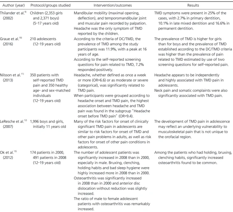
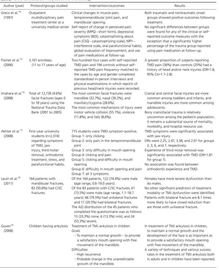
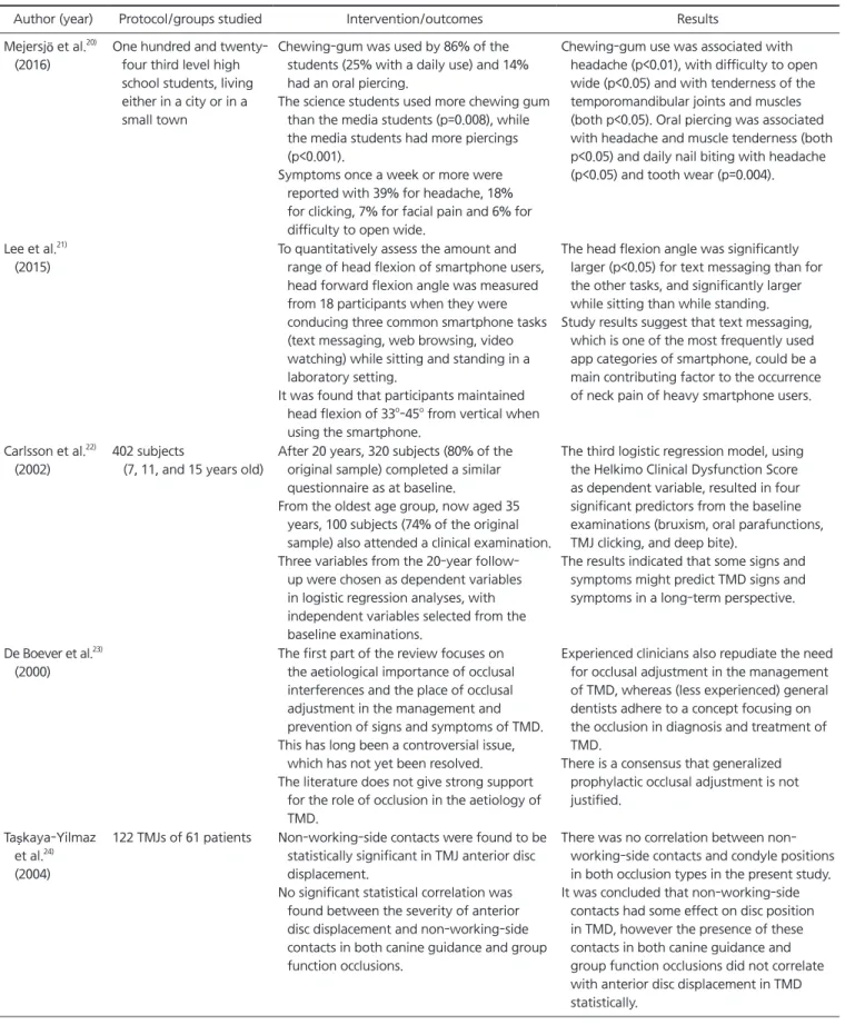
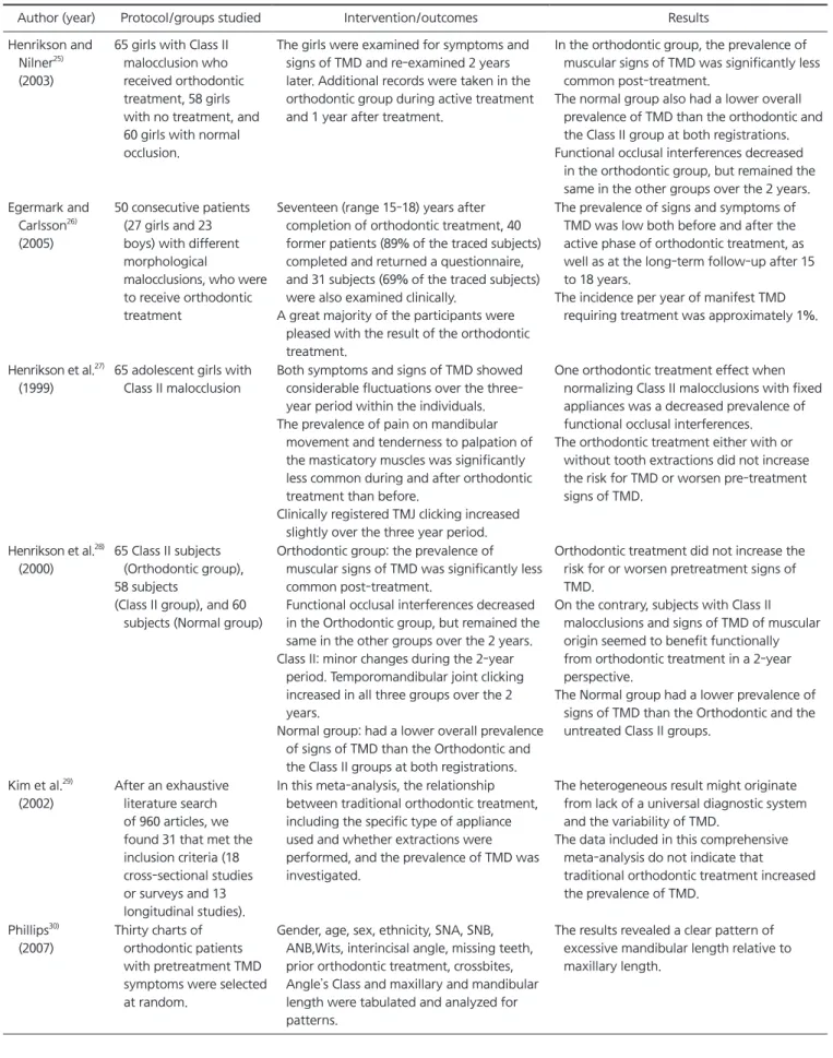
Results: According to a study conducted in Busan, the ratio of adolescents increased from 18.3% to 21% in 2008 compared to 2000, and the proportion of boys increased from 38.58% to 45.38%. One of the characteristic contributing factors for adolescents is the macrotrauma such as jaw trauma, vehicle accidents, sports, physical abuse, forceful intubation, and third molar extraction. The second is a microtrauma from parafunctional habit such as bruxism, clenching, hyperextension, wind instrument, and fingernail biting that can cause joint overload, cartilage breakdown, synovial fluid alterations, and other changes within the joint. The diagnosis of TMDs in juvenile adolescents is not significantly different from that of adults. Medical history, clinical examination and radiological examinations are required.

Conclusions: In the temporomandibular joint history and assessment, all comprehensive den- tal history examination is required, including head and neck pain, mandibular dysfunction, previous orofacial trauma, history of present illness with an account of current symptoms.

For the treatment and management of temporomandibular arthritis in juvenile adolescents, understanding the characteristics of TMDs in juvenile adolescents and thoroughly analyzing appropriate diagnosis and possible contributing factors through comprehensive history taking

& examination, conservative treatment, including fast and active cautions education, will be essential.

Key Words: Adolescent; Children; Juvenile; Temporomandibular disorder; TMD

Correspondence to:

Soo-Min Ok

Department of Oral Medicine, Pusan National University, School of Dentistry, 49 Busandaehak-ro, Mulgeum-eup, Yangsan 50612, Korea Tel: +82-55-360-5243

Fax: +82-51-510-8241 E-mail: [email protected] This study was supported by Clinical Research Grant, Pusan National University Dental Hospital (2017).

JOMP Journal of Oral Medicine and Pain

Copyright Ⓒ 2017 Korean Academy of Orofacial Pain and Oral Medicine. All rights reserved.

CC

This is an open-access article distributed under the terms of the Creative Commons Attribution Non-Commercial License (http://creativecommons.org/licenses/by-nc/4.0/),

INTRODUCTION

Temporomandibular disorder (TMD) refers to the clinical symptoms that appear in the masticatory muscles, temporo- mandibular joints (TMJs), and surrounding tissues, and is often referred to as a functional abnormality of the masti- catory system. 1) This is mainly due to stimulation beyond

the limits of the physiological adaptation ability of the mas- ticatory system. There are various differences in the contri- bution factors, physiological adaptive ability, the symptoms and treatment results are also various.

TMJs are a major contributor to the length and height

formation of mandibular growth as a result of intraosseous

ossification of the deep surface of the mandibular condyle

(2)

and cartilage. 2)

This remodeling ability is responsible for the adaptation of the condyle to a changed circumstance, and has been shown extensively in animal studies. 3,4)

In addition, adult rat studies have shown that the me- chanical strain generated by mandibular advancement can induce neovascularization and osteogenesis in mandibu- lar condyles, and induce adaptive growth of the condyle. 5,6) These results emphasize the remodeling capacities of the mandibular condyle according to the changes in the biome- chanical environment. Also, as age increases, TMJ’s ability to adapt to altered function decreases, indicating that adap- tive capacity of the mandibular condyle for mechanical stimuli can vary between adolescents and adults. 7)

Therefore, children and adolescents in growing age are different in their adaptive ability compared to adults, and respond differently to treatment. 5,7,8) The purpose of this study is to investigate the prevalence of TMDs in children and adolescents, their characteristic contributing factors, the characteristic features of symptoms and symptoms, and the response to treatment.

MATERIALS AND METHODS

We studied the researches, that were the results of the searches for words such as temporomandibular disorders, TMD, children, adolescents, and juvenile through PubMed and DBpia. We also searched and screened for relevance lit- erature published from 1975 through 2017 (Fig. 1).

RESULTS

1. Prevalence

The prevalence of TMD varies according to the varia- tion of the survey population, diagnostic criteria, diagnostic methods, and investigators. In 2012, a survey of 4,724 peo- ple aged 5 to 17 years in Sweden revealed that TMD symp- toms were present in 25% of the cases, with 2.7% in pri- mary dentition, 10.1% in late mixed dentition and 16.6% in permanent dentition an increasing pattern was observed. 9) In the general population of Saudi Arabia, the prevalence of TMD in juvenile adolescents was 27%, of which 15.6% were myofacial pain. In 2016, 167 people in 12-19 year olds in

Norway were examined by a protocol, and the total num- ber of D was 20, of which r (n=9) was the most frequent. 10) Headache may occur in adolescents independently of TMD and in close association with TMD, which occurs before the onset of TMD pain. 11) In both boys and girls, TMD pain tends to be proportional to age, and it is often found in girls to seek symptom or treatment. 12) According to a study conducted in Busan, the ratio of adolescents increased from 18.3% to 21% in 2008 compared to 2000, and the propor- tion of boys increased from 38.58% to 45.38%. 13) In many studies, the frequency of diagnosis varies, but most TMD- related diagnoses are identified (Table 1).

2. Etiology 1) Macrotrauma

One of the characteristic contributing factors for adoles- cents is the macrotrauma. This can occur most common- ly in adolescents, such as jaw trauma, vehicle accidents, sports, physical abuse, forceful intubation, and third mo- lar extraction. Long-term closed reduction during trauma may cause ankylosis and improperly treated fractures may cause facial asymmetry. 14-19) If only macrotrauma is present without any other contributing factor, treatment is effec- tive if appropriate treatment is performed at the right time.

However, if there are persistent factors mentioned below, adjusting these factors should increase the success rate.

Inclusion criteria

Searching PubMed & DBpia by key words of 1) "Temporomandibular disorder or TMD" and 2) "Children, or adolescents, or juvenile" and 3) "Etiology, or diagnosis, or treatment"

Abstract screening

Selection of 49 articles

Exclusion criteria Non-pertinent paper, opinion paper, non english language, duplicated paper

Fig. 1. A flow sheet for the article screening. TMD, temporo-

mandibular disorders.

(3)

2) Microtrauma (parafunctional habit)

The second is a microtrauma from parafunctional habit.

In addition to bruxism and clenching, other repetitive ha- bitual behaviors such as hyperextension, wind instrument, and fingernail biting can cause joint overload, cartilage breakdown, synovial fluid alterations, and other changes within the joint. 20) According to Mejersjö et al., 20) there is significant correlation between chewing gum use and head- ache, difficulty to open wide, tenderness of the TMJs &

muscles, and there is correlative conjunction between oral

piercings and headache, muscle tenderness, daily nail biting with headache. Recently, the usage of mobile phones and computers by youths, especially Korean youths, is increas- ing. 21) Analysis of various tasks and postures during smart phone use showed the most severe forward head posture in both sitting and standing positions during texting messag- ing. This can lead to the onset of TMD. The parafunction of adolescent adolescents was found to be the same in 20 years. 22) Early identification, improvement and control of habit as TMD initiation factor and TMD persistence factor

Table 1. Reference articles summary about the TMD prevalence

Author (year) Protocol/groups studied Intervention/outcomes Results

Thilander et al.

9)

(2002)

Children (2,353 girls and 2,371 boys) (5-17 years old)

Mandibular mobility (maximal opening, deflection), and temporomandibular joint and muscular pain recorded by palpation.

Headache was the only symptom of TMD reported by the children.

TMD symptoms were present in 25% of the cases, with 2.7% in primary dentition, 10.1% in late mixed dentition and 16.6% in permanent dentition.

Graue et al.

10)

(2016)

210 adolescents (12-19 years old)

According to the criteria of DC/TMD, the prevalence of TMD among the study participants was 11.9%, with a peak at 16 years of age.

According to the self-reported screening questions for pain related to TMD, 7.2%

responded positively.

The prevalence of TMD is higher for girls than for boys and the prevalence of TMD established according to the DC/TMD criteria was higher than the prevalence of pain related to TMD estimated by use of two screening questions for self-reported pain.

Nilsson et al.

11)

(2013)

350 patients with self-reported TMD pain and 350 healthy age- and sex-matched individuals

(12-19 years old)

Headache, whether defined as once a week or more (OR=6.6) or as moderate or severe (categorical), was significantly related to TMD pain.

When participants were grouped according to headache onset and TMD pain, the highest association between headache and TMD pain was found in the subgroup “Headache onset before TMD pain” (OR=9.4).

Headache appears to be independently and highly associated with TMD pain in adolescents.

Neck pain and somatic complaints were also significantly associated with TMD pain.

LeResche et al.

12)

(2007)

1,996 boys and girls, initially 11 years old

Many of the risk factors for onset of clinically significant TMD pain in adolescents are similar to risk factors for onset of TMD and other pain problems in adults, as well as risk factors for onset of other pain conditions in adolescents.

The development of TMD pain in adolescence may reflect an underlying vulnerability to musculoskeletal pain that is not unique to the orofacial region.

Ok et al.

13)

(2012)

174 patients in 2000, 491 patients in 2008 (12-19 years old)

The number of adolescent patients was significantly increased in 2008 than in 2000, especially in male. Bruxing, clenching, holding habits and bad sleep hygiene were highly increased more in 2008 than in 2000.

Osteoarthritis was significantly increased in 2008 than in 2000 and anterior disc dislocation without reduction was slightly increased.

The ratio of male to female adolescent patients with osteoarthritis was remarkably increased.

Among the patients who had holding, bruxing, clenching habits, significantly increased osteoarthritis found to be common.

TMD, temporomandibular disorders; DC, diagnostic criteria; OR, odds ratio.

(4)

are essential for lowering TMD prevalence and improving treatment effect.

3) Anatomical factors (skeletal & occlusal)

Occlusal factors and TMD occurrence are somewhat less correlated. 23,24) Neither of the claims that orthodontic treat- ment induces or improves TMD is supported by sufficient evidence. 17,25-29) But it is reasonable that some occlusal fac- tors may place greater adaptive demands on the mastica- tory system. For example, skeletal anterior open bite, steep articular eminence, overjet greater than 6-7 mm, class III malocclusion, posterior crossbite. 9,30,31)

4) Psychosocial factors

If emotional stress is severe, clenching and bruxism can cause and aggravate orofacial pain. 32) In the control and management of stress and anxiety in young adolescents, TMD symptoms and signs were significantly decreased. 32) In addition, it has been reported that depression, anxiety, post-traumatic stress disorder, psychologic distress, and sleep dysfunction can affect TMD prognosis and symptoms and signs. 33) The higher the degree of pain, the longer the difficulty of opening and the quality of life such as sleep- ing disorder decreases. 34) According to a study conducted in Busan in 2013, TMD adolescents had lower sleep quality, shorter sleeping time, shorter cyber leisure time, and lower physical activity than the control group. In the TMD group, the greater the degree of pain, the shorter the cyber leisure time. 35) In patients with TMD with pain, the patient com- plained of pain due to pain of low disability in about 80%

of patients with graded chronic pain scale. 36)

5) Systemic factors

These systemic diseases occur as a result of imbalance of pro-inflammatory cytokines which causes oxidative stress, free radical formation, and ultimately joint damage. 37)

Examples include rheumatoid arthritis, systemic lupus erythematosus, juvenile idiopathic arthritis, and psoriatic arthritis. 38) If these diseases are diagnosed, a prompt referral to the appropriate specialist and a concurrent treatment for the associated TMD symptoms are needed (Table 2). 39)

3. Diagnosing TMD

The diagnosis of TMD in juvenile adolescents is not sig- nificantly different from that of adults. Medical history, clinical examination, and radiological examinations are re- quired. In the process, trigeminal neuralgia, central nervous system lesions, odontogenic pain, sinus pain, ontologi- cal pain, developmental abnormalities, neoplasias, parotid diseases, vascular diseases, cervical muscle dysfunction, Eagle’s syndrome, otitis media, allergies, airway conges- tion and rheumatoid arthritis that look similar to TMD are excluded.

The more comprehensive examination (palpation of mas- ticatory and associated muscles and the TMJ’s, documen- tation of joint sounds, occlusal analysis, and assessment of range of mandibular movements including maximum opening, protrusion, and lateral excursions, etc.) is needed if positive history and/or signs and symptoms of TMD exists.

Additional imaging tests (panorama, lateral cephalogram, TMJ tomography, magnetic resonance imaging [both open and closed mouth to view disc position], cone-beam com- puted tomography [CBCT]) may be required for history of trauma or developing facial asymmetry, or when hard-tis- sue grinding or crepitus failed to respond to conservative TMD treatment. 39)

TMJ arthography is not recommended as a routine di- agnostic test procedure. 40,41) The easy-to-use panoramic ra- diograph is reliable for assessing the shape and angulation of the head, but it cannot assess joint space, soft tissue, or condyle movements. Panograph can evaluate bone chang- es, but negative findings cannot rule out TMJ pathology. 42) CBCT can be used to detect abnormal bone tissues and frac- tures and evaluate asymmetry, but it generates much more radiation burden than panoramic images. 40-42) Magnetic res- onance imaging provides the visualization of soft tissue, es- pecially the location and contours of TMJ discs and can be used to detect inflammation. 41,43)

Occurrence and radiographic signs of adolescent TMJ os- teoarthritis (OA) on CBCT showed OA in 40.7% of juvenile TMD patients and 30.6% of 44.6% of infantile girls, and the frequency of ill-defined cortical bone (31.7%, 65/205), small bony defect & extensive erosion (25.4%, 52/205), flattening

& shortening of the condyle (6.3%,13/205), sclerosis (14.6%,

30/205) was in that form.

(5)

Table 2. Reference articles summary about the TMD etiology

Author (year) Protocol/groups studied Intervention/outcomes Results

Greco et al.

14)

(1997)

Outpatient

multidisciplinary pain treatment center at a university medical center

Clinical changes in muscle pain, temporomandibular joint pain, and mandibular opening.

Self-report of change in perceived pain severity (MPQ—short form), depressive symptoms (BDI), catastrophizing about pain (CSQ—catastrophizing scale), MPI—

interference scale, oral parafunctional habits, global evaluation of improvement, and use of pain medications at follow-up.

Both traumatic and nontraumatic onset groups showed positive outcomes following treatment.

No significant differences between groups were found for any of the clinical or self- reported outcome measures with the exception that a significantly higher percentage of the trauma group reported using pain medication at follow-up.

Fischer et al.

15)

(2006)

3,101 enrollees (11 to 17 years of age)

Two hundred four cases with self-reported TMD pain and 194 controls without self- reported TMD pain frequency-matched to the cases by age and gender completed standardized in-person interviews and physical examinations in which reports of previous head/neck injuries were recorded.

A greater proportion of subjects reporting TMD pain (36%) than controls (25%) had a history of head and/or neck injuries (OR=1.8, 95% CIs=1.1-2.8).

Imahara et al.

16)

(2008)

Total of 12,739 (4.6%) facial fractures (ages 0 to 18 years) using the National Trauma Data Bank (2001 to 2005)

The most common facial fractures were mandible (32.7%), nasal (30.2%), and maxillary/zygoma (28.6%).

The most common mechanisms of injury were motor vehicle collision (55.1%), violence (11.8%), and falls (8.6%).

Cranial and central facial injuries are more common among toddlers and infants, and mandible injuries are more common among adolescents.

Bony craniofacial trauma is relatively uncommon among the pediatric population, it remains a substantial source of mortality, morbidity, and hospital resource use.

Akhter et al.

17)

(2008)

First-year university students (n=2,374) regarding symptoms of TMD, jaw injury, third molar removal, orthodontic treatment, stress, and parafunctional habits.

715 students were TMD symptom-positive.

Group 1: only clicking

Group 2: only pain in the temporomandibular joint

Group 3: only difficulty in mouth opening Group 4: clicking and pain

Group 5: clicking and difficulty in mouth opening

Group 6: difficulty in mouth opening and pain Group 7: all 3 symptoms

TMD symptoms were significantly associated with jaw injury.

ORs were 2.25, 2.47, 3.38, and 2.01 for groups 2, 3, 6, and 7, respectively.

Experience of third molar removal was significantly associated with TMD (OR=1.81 for group 1).

No association was found between orthodontic experience and TMD.

Leuin et al.

18)

(2011)

164 patients with mandibular fractures, 83 (50.6%) had C/SC fractures.

Of the 164 patients, 122 (74.4%) were male (age range, 0.6-19.0 years).

Of the 83 patients with C/SC fractures, 61 (73.5%) were male (age range, 1.1-18.7 years); 66 (79.5%) had unilateral fractures and 17 (20.5%) had bilateral fractures.

The A(i) distribution of the 45 patients who completed the questionnaire was as follows:

15 (33.3%) none, 6 (13.3%) mild, and 24 (53.3%) severe.

Females have more severe dysfunction than do males.

No other significant predictors of treatment modality or TMJ dysfunction were identified.

Patients with bilateral fracture are 8.1 times more likely to have closed reduction than are those with unilateral fracture.

Güven

19)

(2008)

Children having ankylosis Treatment of TMJ ankylosis in children Goals

- To maintain a normal growth - to provide a satisfactory mouth opening with free movement of the mandible.

Difficulties - High recurrence

- Probable change in the unpredictable growth of the mandible.

In treatment of TMJ ankylosis in children, to maintain a normal growth and the development of the face is as important as to provide a satisfactory mouth opening with free movement of the mandible.

A variety of techniques and various success

rates in the treatment of TMJ ankylosis both

in adults and in children have been reported.

(6)

Table 2. Continued

Author (year) Protocol/groups studied Intervention/outcomes Results

Mejersjö et al.

20)

(2016)

One hundred and twenty- four third level high school students, living either in a city or in a small town

Chewing-gum was used by 86% of the students (25% with a daily use) and 14%

had an oral piercing.

The science students used more chewing gum than the media students (p=0.008), while the media students had more piercings (p<0.001).

Symptoms once a week or more were reported with 39% for headache, 18%

for clicking, 7% for facial pain and 6% for difficulty to open wide.

Chewing-gum use was associated with headache (p<0.01), with difficulty to open wide (p<0.05) and with tenderness of the temporomandibular joints and muscles (both p<0.05). Oral piercing was associated with headache and muscle tenderness (both p<0.05) and daily nail biting with headache (p<0.05) and tooth wear (p=0.004).

Lee et al.

21)

(2015)

To quantitatively assess the amount and range of head flexion of smartphone users, head forward flexion angle was measured from 18 participants when they were conducing three common smartphone tasks (text messaging, web browsing, video watching) while sitting and standing in a laboratory setting.

It was found that participants maintained head flexion of 33

o

-45

o

from vertical when using the smartphone.

The head flexion angle was significantly larger (p<0.05) for text messaging than for the other tasks, and significantly larger while sitting than while standing.

Study results suggest that text messaging, which is one of the most frequently used app categories of smartphone, could be a main contributing factor to the occurrence of neck pain of heavy smartphone users.

Carlsson et al.

22)

(2002)

402 subjects

(7, 11, and 15 years old)

After 20 years, 320 subjects (80% of the original sample) completed a similar questionnaire as at baseline.

From the oldest age group, now aged 35 years, 100 subjects (74% of the original sample) also attended a clinical examination.

Three variables from the 20-year follow- up were chosen as dependent variables in logistic regression analyses, with independent variables selected from the baseline examinations.

The third logistic regression model, using the Helkimo Clinical Dysfunction Score as dependent variable, resulted in four significant predictors from the baseline examinations (bruxism, oral parafunctions, TMJ clicking, and deep bite).

The results indicated that some signs and symptoms might predict TMD signs and symptoms in a long-term perspective.

De Boever et al.

23)

(2000)

The first part of the review focuses on the aetiological importance of occlusal interferences and the place of occlusal adjustment in the management and prevention of signs and symptoms of TMD.

This has long been a controversial issue, which has not yet been resolved.

The literature does not give strong support for the role of occlusion in the aetiology of TMD.

Experienced clinicians also repudiate the need for occlusal adjustment in the management of TMD, whereas (less experienced) general dentists adhere to a concept focusing on the occlusion in diagnosis and treatment of TMD.

There is a consensus that generalized prophylactic occlusal adjustment is not justified.

Taşkaya-Yilmaz et al.

24)

(2004)

122 TMJs of 61 patients Non-working-side contacts were found to be statistically significant in TMJ anterior disc displacement.

No significant statistical correlation was found between the severity of anterior disc displacement and non-working-side contacts in both canine guidance and group function occlusions.

There was no correlation between non- working-side contacts and condyle positions in both occlusion types in the present study.

It was concluded that non-working-side

contacts had some effect on disc position

in TMD, however the presence of these

contacts in both canine guidance and

group function occlusions did not correlate

with anterior disc displacement in TMD

statistically.

(7)

Table 2. Continued

Author (year) Protocol/groups studied Intervention/outcomes Results

Henrikson and Nilner

25)

(2003)

65 girls with Class II malocclusion who received orthodontic treatment, 58 girls with no treatment, and 60 girls with normal occlusion.

The girls were examined for symptoms and signs of TMD and re-examined 2 years later. Additional records were taken in the orthodontic group during active treatment and 1 year after treatment.

In the orthodontic group, the prevalence of muscular signs of TMD was significantly less common post-treatment.

The normal group also had a lower overall prevalence of TMD than the orthodontic and the Class II group at both registrations.

Functional occlusal interferences decreased in the orthodontic group, but remained the same in the other groups over the 2 years.

Egermark and Carlsson

26)

(2005)

50 consecutive patients (27 girls and 23 boys) with different morphological

malocclusions, who were to receive orthodontic treatment

Seventeen (range 15-18) years after completion of orthodontic treatment, 40 former patients (89% of the traced subjects) completed and returned a questionnaire, and 31 subjects (69% of the traced subjects) were also examined clinically.

A great majority of the participants were pleased with the result of the orthodontic treatment.

The prevalence of signs and symptoms of TMD was low both before and after the active phase of orthodontic treatment, as well as at the long-term follow-up after 15 to 18 years.

The incidence per year of manifest TMD requiring treatment was approximately 1%.

Henrikson et al.

27)

(1999)

65 adolescent girls with Class II malocclusion

Both symptoms and signs of TMD showed considerable fluctuations over the three- year period within the individuals.

The prevalence of pain on mandibular movement and tenderness to palpation of the masticatory muscles was significantly less common during and after orthodontic treatment than before.

Clinically registered TMJ clicking increased slightly over the three year period.

One orthodontic treatment effect when normalizing Class II malocclusions with fixed appliances was a decreased prevalence of functional occlusal interferences.

The orthodontic treatment either with or without tooth extractions did not increase the risk for TMD or worsen pre-treatment signs of TMD.

Henrikson et al.

28)

(2000)

65 Class II subjects (Orthodontic group), 58 subjects

(Class II group), and 60 subjects (Normal group)

Orthodontic group: the prevalence of muscular signs of TMD was significantly less common post-treatment.

Functional occlusal interferences decreased in the Orthodontic group, but remained the same in the other groups over the 2 years.

Class II: minor changes during the 2-year period. Temporomandibular joint clicking increased in all three groups over the 2 years.

Normal group: had a lower overall prevalence of signs of TMD than the Orthodontic and the Class II groups at both registrations.

Orthodontic treatment did not increase the risk for or worsen pretreatment signs of TMD.

On the contrary, subjects with Class II malocclusions and signs of TMD of muscular origin seemed to benefit functionally from orthodontic treatment in a 2-year perspective.

The Normal group had a lower prevalence of signs of TMD than the Orthodontic and the untreated Class II groups.

Kim et al.

29)

(2002)

After an exhaustive literature search of 960 articles, we found 31 that met the inclusion criteria (18 cross-sectional studies or surveys and 13 longitudinal studies).

In this meta-analysis, the relationship between traditional orthodontic treatment, including the specific type of appliance used and whether extractions were performed, and the prevalence of TMD was investigated.

The heterogeneous result might originate from lack of a universal diagnostic system and the variability of TMD.

The data included in this comprehensive meta-analysis do not indicate that traditional orthodontic treatment increased the prevalence of TMD.

Phillips

30)

(2007)

Thirty charts of orthodontic patients with pretreatment TMD symptoms were selected at random.

Gender, age, sex, ethnicity, SNA, SNB, ANB,Wits, interincisal angle, missing teeth, prior orthodontic treatment, crossbites, Angle’ s Class and maxillary and mandibular length were tabulated and analyzed for patterns.

The results revealed a clear pattern of

excessive mandibular length relative to

maxillary length.

(8)

Table 2. Continued

Author (year) Protocol/groups studied Intervention/outcomes Results

Pahkala and Qvarnström

31)

(2004)

The subjects were examined at the ages of 7, 10, 15, and 19 years.

Multiple logistic regression models were applied in order to evaluate whether single signs of TMD at the age of 19 years were related to previous/present malocclusions or interferences, to misarticulations of speech, problems in oral motor skills, or other signs of TMD.

The effect of gender was also considered.

The results showed that excessive overjet was the only variable which seemed to consistently increase the risk of TMD.

In addition, girls seemed to be more prone to the development of TMD than boys.

Although, during growth, there were both local and central factors associated occasionally with TMD development, the predictive value of those variables in the estimation of the individual risk of TMD was rather small.

List et al.

32)

(2001)

63 patients (21 boys and 42 girls, 33% and 67%, respectively, with a mean age of 14.9 years; range 12 to 18 years)

64 healthy control subjects (17 boys and 47 girls, 27% and 73%, respectively, with a mean age of 14.8 years)

Subjects in the TMD group had to report pain once a week or more and to have a TMD pain diagnosis according to the Research Diagnostic Criteria for TMD.

Participants were clinically examined and filled out a questionnaire in which self-reported psychosocial functioning was assessed on standardized measures, including the YSR, somatic complaints, and stress.

No significant differences were found in dental factors among adolescents in the TMD group compared with those in the control group.

Multiple pains in the body and fatigue were significantly more common in the TMD group compared with the control group.

Adolescents with TMD also reported significantly higher levels of stress, somatic complaints, and aggressive behavior than their counterparts in the control group.

In particular, young adolescents with TMD reported high levels of psychosocial problems.

Karibe et al.

34)

(2012)

Group 1: 6-12 years (juvenile)

Group 2: 13-15 years (early adolescent) Group 3: 16-18 years

(late adolescent)

No significant gender differences were found in the symptoms among the groups, except for headache and neck pain in group 3.

Pain intensity and tightness in the jaw/face, headache, and neck pain, as well as the ADL- related difficulty in prolonged jaw opening, eating soft/hard foods, and sleeping significantly differed among the groups (p<0.01, Kruskal-Wallis test).

Late adolescent patients with TMDs have higher pain intensity in the orofacial region and greater difficulty in ADL than do early adolescent and juvenile patients with TMDs.

Kim et al.

35)

(2013)

219 adolescents patients aged 11 to 19

90 control group, personal characteristics, physical activity, cyber leisure activities, and the relationship of the TMD symptoms, and the following results were obtained.

TMD group compared to the control group, the sleep quality was lower, cyber-leisure time is longer and more frequent.

In TMD group, the shorter sleep time was, first visit NAS was higher.

The more stress was, physical activity was less.

The more TMD symptoms were severe, cyber- leisure time was shorter.

Al-Khotani et al.

36)

(2016)

456 randomly selected children and adolescents, enrolled from 10 boy’ s- and 10 girl’ s- schools in Jeddah, between 10 and 18 years of age.

On the examination day, prior to the clinical examination according to Research Diagnostic Criteria for TMD Axis I and II, the participants first answered two validated questions about TMD pain, and after that the Arabic version of the YSR scale.

According to their clinical examination and diagnosis the participants were divided into three groups; non-TMD group, TMD-pain group, and TMD-pain free group.

The TMD-pain group presents a higher frequency of the internalizing problems anxiety, depression and somatic complaints than non-TMD group.

Regarding externalizing problems the only significant association found was for aggressive behavior in the TMD-pain group.

The TMD-pain group also shows a higher frequency of social problems than the non- TMD group.

However, no such difference was found when compared to the TMD-pain free group.

There was also a significant association with a higher frequency of thought problems in the TMD-pain group (p<0.05).

TMD, temporomandibular disorders; MPQ, McGill pain questionnaire; BDI, beck depression inventory; CSQ, coping strategy questionnaire; MPI,

multidimensional pain inventory; OR, odds ratio; CI, confidence intervals; C/SC, condylar and subcondylar; TMJ, temporomandibular joint; YSR,

Youth Self-Report; ADL, activities of daily living; NAS, numerical analog scale.

(9)

According to a study by Zhao et al. 43) (2011), the pattern I was significantly higher at 20-30 years of age than that of 11-19 years of age in all female males. Pattern II was the most in the bone patterns (pattern I: erosive bone changes, obvious destruction/ill-defined cortical bone & small con- cave defect of condyle, pattern II: poliferative changes - flattening with sclerosis, osteophytes, or deformity, pattern III: bilaterally short condylar processes/c uneven subcortical sclerosis as a result of transpharyngeal radiogram. 44)

Through listening to such medical history and diagnos- tic tests, we diagnose with the TMD, such as myalgia, local myalgia, myofascial pain with spreading, myofascial pain with referral arthralgia, headache attributed to TMD, disc displacement with reduction, disc displacement with reduc- tion intermittent locking, disc displacement without reduc- tion with limited opening, disc displacement without re- duction without limited opening, degenerative joint disease (Table 3).

4. Treatment of TMD

The goal of TMD treatment is the restoral of function, de- creased pain, and return of quality of life, simple, conser- vative and reversible treatment is most effective. 45) And it is more successful to approach combined approaches than to perform one treatment alone. And it is more success- ful to approach combined approaches than to perform one treatment alone. In reversible treatment methods, patient education, physical therapy, behavioral therapy, prescrip- tion medication occlusal splints is possible. Patient educa- tion may include relaxation training, developing behavior coping strategies, modifying inadequate perceptions about TMD, patient awareness of clenching, and bruxing hab- its. Physical therapies include jaw exercises, transcutane- ous electrical nerve stimulation, ultrasound, iontophoresis, massaging, thermotherapy, and coolant therapy. Behavioral therapy can be done with avoiding excessive chewing of hard foods or gum, voluntary avoidance of stressors, habit reversal, decreasing stress, anxiety, and/or depression, ob- taining adequate, uninterrupted sleep. In the analysis of the effectiveness of the precautionary questionnaire using the repetitive self-check questionnaire conducted by Ok et al. 45) in Busan, significant improvement was observed in limita- tion of mouth opening (LOM) and maximum comfortable

opening (MCO) when the repetitive self-check questionnaire was used in the under 30’s. The improvement was sustained until 6th visit, and there was a significant negative corre- lation between total score of self-check questionnaire and pain.

The results of this study suggest that the use of self- check questionnaires in repetitive precautionary educa- tion is helpful for the behavioral therapy of adolescents in Korea, which has to treat many patients per unit time. In addition, the questionnaire that male and 30 year old ado- lescents should reduce the stress that may cause pain was not kept well in comparison with other items, using small spoon, avoiding coffee or spicy foods, It is suggested that it should be emphasized that it is relatively low to observe the cautions to fix sleeping habits and to carry out hot springs.

Next, occlusal splints therapy can be performed. The stabi- lization splints therapy covers all of the teeth on either the maxillary or mandibular arch and all teeth are in occlusion.

Musculoskeletally stable position may be induced and vari- ous device treatments such as adults may be applied de- pending on the case. However, there is a possibility that it may be necessary to make a device frequently for growth and it is difficult to apply to a tooth exchanger.

Other irreversible treatments include selective removal of teeth, occlusal therapy using full mouth restorations, orth- odontic treatment to permanently change mandibular po- sition by controlling growth using headgear or functional appliance but there is no evidence that these treatments can prevent or alleviate the TMD. 46-48) Botulinum toxin A injec- tion has recently been approved for use in the relaxation of masticatory muscles in adults, but its use in juvenile adolescents has not been approved. 49) In addition, we are studying in animal studies that simvastatin, methotrexate, and steroid injection may be helpful in arthritis of juvenile adolescents.

It is doubtful whether the signs and symptoms of juve- nile adolescents are important for the development of the disease, what diagnosis and treatment are effective, and whether these signs and symptoms warrant treatment as a predictor of TMD in adulthood. 22)

In a comparative study of the treatment effects of ado-

lescents and adults with erosion, Kim et al. 50) Reported that

the proportion of the improved group when treated with

(10)

Table 3. Reference articles summary about the diagnosing TMD

Author (year) Protocol/groups studied Intervention/outcomes Results

Brooks et al.

40)

(1997)

Various imaging techniques for the temporomandibular joint are discussed with respect to uses, strengths, and limitations.

Various imaging techniques for the

temporomandibular joint are discussed with respect to uses, strengths, and limitations.

An imaging protocol is outlined for evaluating patients with a wide variety of temporomandibular joint related signs and symptoms.

de Senna et al.

41)

(2009)

Studies published between 1976 and 2009 that appear in the Medline database

Magnetic resonance imaging remains the “gold standard” modality for TMJ analysis.

Ultrasonography, an alternative method with increasing importance in TMJ analysis, is a simple, noninvasive, and low-cost technique that allows for the visualization of the position of the disk; however, it does not detect condylar abnormalities.

Reconstructions in three-dimensions can be obtained with computed tomography, magnetic resonance imaging, and ultrasonography and can be used to obtain rapid prototyping biomodels.

Health professionals performing TMJ imaging exams should consider clinical history and findings, exam cost, radiation exposure, results of previous exams, and whether the current result will influence diagnosis and treatment planning.

Hunter and Kalathingal

42)

(2013)

The RDC/TMD recommend arthrography and MRI for disk displacement and tomography for evaluation of bony changes.

The ability to assess details in multiplanar views makes cone beam computed tomography a unique tool for accurate and precise evaluation of dento-alveolar structures.

Advanced imaging with computed tomography or MRI may be indicated for orofacial pain patients presenting with idiopathic facial pain, headaches, or trigeminal neuralgia.

Howard

38)

(2013)

A child's difficulty in verbalizing the precise location and nature of facial pain and jaw dysfunction often results in a nondefinitive history, increasing the importance of the dentist’ s awareness of the early signs and symptoms of TMD.

A focused examination of the masticatory musculature, the temporomandibular joints, and associated capsular and ligamentous structures can reveal if a patient’ s symptoms are TMD in origin.

An accurate differential diagnosis enables timely referral to appropriate health care providers and minimizes the use of diagnostic imaging.

Zhao et al.

43)

(2011)

Patients (n=4,883) with temporomandibular disorders (age, 11 to 30 years)

Seven hundred eleven patients had radiographic signs of OA.

The frequency of OA was higher in women (563/3,360, 16.8%) than in men (148/1,523, 9.7%).

Most patients (541/711, 76.1%) with signs of OA showed proliferative changes of OA.

Moreover, 56.4% of patients with TMJOA (88/156) remained stable.

These results suggest that although OA is an age-related disease, aging is not the crucial factor in the pathogenesis of OA.

Bodner and Miller

44)

(1998)

38 pediatric patients (30: TMJ dysfunction, 2: degenerative joint disease,

2: growth disturbances, 1: tumor,

3: etiology was unclear)

The treatment modalities were: non-invasive therapy in 19 (50%) patients, occlusal therapy in 10 (26%) patients and surgical treatment in nine (24%) patients.

One year later, 33 (87%) reported no symptoms, four (10%) mild symptoms and one (3%) severe symptoms.

Maximum mouth opening 1 year after treatment as compared to the initial examination increased (p<0.05) in all three treatment modalities.

Deviation of the mandible on opening, 1 year after treatment as compared to the initial examination, decreased (p<0.05) in all three treatment modalities.

RDC, research diagnostic criteria; MRI, magnetic resonance imaging; OA, osteoarthritis.

(11)

medications, behavioral therapy, physical therapy and splint therapy was higher in adolescents than in middle- Were significantly higher than patients (p=0.013).

Analysis of the distribution of degenerative changes in the mandibular condyle showed that one side (60.2%) was most affected by adolescence and bilateral change (62.3%) was the most frequent in middle age (p=0.009). Subjective clinical symptoms and relaxation period of erosion were significantly shorter in the adolescent group than in the middle-aged patients (p=0.013, p=0.038).

The conversion rate to the normal conus shape was larger in adolescents and the conversion rate to condyle shorten- ing was higher in middle age. This shows that osteoarthritis of young adolescents has better healing ability than adults.

Based on the high incidence of unilateral arthritis in adoles- cents compared to middle-aged adolescents, if unilateral ar- thritis is not managed, the possibility of bilateral migration can be carefully estimated.

DISCUSSION

TMD is multifactorial disease. In the TMJ history and as- sessment, all comprehensive dental history examination is required, including head and neck pain, mandibular dys- function, previous orofacial trauma, history of present ill- ness with an account of current symptoms.

For the treatment and management of temporomandibu- lar arthritis in juvenile adolescents, understanding the char- acteristics of characteristic TMD in juvenile adolescents and thoroughly analyzing appropriate diagnosis and possible contributing factors through comprehensive history taking

& examination, conservative treatment, including fast and active cautions education, will be essential.

Repeated precautional education using self-check ques- tionnaire could be helpful for the behavioral therapy of adolescents as a reversible treatment method according to Korean situation.

Other methods of irreversible treatment are considered to be necessary because of lack of evidence. It is doubtful whether the symptoms and signs of juvenile adolescents are important for the development of the disease, what diag- nosis and treatment are effective, and whether these signs and symptoms warrant treatment as a predictor of TMD in

adulthood. Therefore, further study is needed.

CONFLICT OF INTEREST

No potential conflict of interest relevant to this article was reported

REFERENCES

1. de Leeuw R, Klasser GD; American Academy of Orofacial Pain.

Orofacial pain: guidelines for assessment, diagnosis, and man- agement. 5th ed. Chicago, IL: Quintessence Publishing; 2013. pp.

127-186.

2. Willems NM, Langenbach GE, Everts V, Zentner A. The micro- structural and biomechanical development of the condylar bone:

a review. Eur J Orthod 2014;36:479-485.

3. Gredes T, Mack H, Spassov A, et al. Changes in condylar cartilage after anterior mandibular displacement in juvenile pigs. Arch Oral Biol 2012;57:594-598.

4. Leung FY, Rabie AB, Hägg U. Neovascularization and bone for- mation in the condyle during stepwise mandibular advancement.

Eur J Orthod 2004;26:137-141.

5. Rabie AB, Xiong H, Hägg U. Forward mandibular position- ing enhances condylar adaptation in adult rats. Eur J Orthod 2004;26:353-358.

6. Xiong H, Rabie AB, Hagg U. Neovascularization and mandibular condylar bone remodeling in adult rats under mechanical strain.

Front Biosci 2005;10:74-82.

7. Bouvier M. Effects of age on the ability of the rat temporoman- dibular joint to respond to changing functional demands. J Dent Res 1988;67:1206-1212.

8. Zeng G, Kuang B, Xun WX, Ren GT, Wei KW. Response of man- dibular condyles of juvenile and adult rats to abnormal occlusion and subsequent exemption. Arch Oral Biol 2017;80:136-143.

9. Thilander B, Rubio G, Pena L, de Mayorga C. Prevalence of temporomandibular dysfunction and its association with mal- occlusion in children and adolescents: an epidemiologic study related to specified stages of dental development. Angle Orthod 2002;72:146-154.

10. Graue AM, Jokstad A, Assmus J, Skeie MS. Prevalence among adolescents in Bergen, Western Norway, of temporomandibular disorders according to the DC/TMD criteria and examination pro- tocol. Acta Odontol Scand 2016;74:449-455.

11. Nilsson IM, List T, Drangsholt M. Headache and co-morbid pains associated with TMD pain in adolescents. J Dent Res 2013;92:802-807.

12. LeResche L, Mancl LA, Drangsholt MT, Huang G, Von Korff M.

Predictors of onset of facial pain and temporomandibular disor- ders in early adolescence. Pain 2007;129:269-278.

13. Ok SM, Kim CY, Jeong SH, Ahn YW, Ko MY. Comparative analy- sis: the patterns of temporomandibular disorder among adoles- cents. J Oral Med Pain 2012;37:47-59.

14. Greco CM, Rudy TE, Turk DC, Herlich A, Zaki HH. Traumatic

(12)

onset of temporomandibular disorders: positive effects of a standardized conservative treatment program. Clin J Pain 1997;13:337-347.

15. Fischer DJ, Mueller BA, Critchlow CW, LeResche L. The associa- tion of temporomandibular disorder pain with history of head and neck injury in adolescents. J Orofac Pain 2006;20:191-198.

16. Imahara SD, Hopper RA, Wang J, Rivara FP, Klein MB. Patterns and outcomes of pediatric facial fractures in the United States:

a survey of the National Trauma Data Bank. J Am Coll Surg 2008;207:710-716.

17. Akhter R, Hassan NM, Ohkubo R, Tsukazaki T, Aida J, Morita M.

The relationship between jaw injury, third molar removal, and orthodontic treatment and TMD symptoms in university students in Japan. J Orofac Pain 2008;22:50-56.

18. Leuin SC, Frydendall E, Gao D, Chan KH. Temporomandibular joint dysfunction after mandibular fracture in children: a 10-year review. Arch Otolaryngol Head Neck Surg 2011;137:10-14.

19. Güven O. A clinical study on temporomandibular joint ankylosis in children. J Craniofac Surg 2008;19:1263-1269.

20. Mejersjö C, Ovesson D, Mossberg B. Oral parafunctions, piercing and signs and symptoms of temporomandibular disorders in high school students. Acta Odontol Scand 2016;74:279-284.

21. Lee S, Kang H, Shin G. Head flexion angle while using a smart- phone. Ergonomics 2015;58:220-226.

22. Carlsson GE, Egermark I, Magnusson T. Predictors of signs and symptoms of temporomandibular disorders: a 20-year follow- up study from childhood to adulthood. Acta Odontol Scand 2002;60:180-185.

23. De Boever JA, Carlsson GE, Klineberg IJ. Need for occlusal thera- py and prosthodontic treatment in the management of temporo- mandibular disorders. Part I. Occlusal interferences and occlusal adjustment. J Oral Rehabil 2000;27:367-379.

24. Taşkaya-Yilmaz N, Oğütcen-Toller M, Saraç YS. Relationship between the TMJ disc and condyle position on MRI and occlusal contacts on lateral excursions in TMD patients. J Oral Rehabil 2004;31:754-758.

25. Henrikson T, Nilner M. Temporomandibular disorders, occlusion and orthodontic treatment. J Orthod 2003;30:129-137; discussion 127.

26. Egermark I, Carlsson GE, Magnusson T. A prospective long-term study of signs and symptoms of temporomandibular disorders in patients who received orthodontic treatment in childhood. Angle Orthod 2005;75:645-650.

27. Henrikson T, Nilner M, Kurol J. Symptoms and signs of temporo- mandibular disorders before, during and after orthodontic treat- ment. Swed Dent J 1999;23:193-207.

28. Henrikson T, Nilner M, Kurol J. Signs of temporomandibular dis- orders in girls receiving orthodontic treatment. A prospective and longitudinal comparison with untreated Class II malocclusions and normal occlusion subjects. Eur J Orthod 2000;22:271-281.

29. Kim MR, Graber TM, Viana MA. Orthodontics and temporoman- dibular disorder: a meta-analysis. Am J Orthod Dentofacial Or- thop 2002;121:438-446.

30. Phillips JT. What skeletal and dental characteristics do TMD pa- tients have in common? Funct Orthod 2007;24:24-26, 28, 30.

31. Pahkala R, Qvarnström M. Can temporomandibular dysfunction signs be predicted by early morphological or functional vari- ables? Eur J Orthod 2004;26:367-373.

32. List T, Wahlund K, Larsson B. Psychosocial functioning and den- tal factors in adolescents with temporomandibular disorders: a case-control study. J Orofac Pain 2001;15:218-227.

33. de Leeuw R, Klasser GD; American Academy of Orofacial Pain.

Orofacial pain: guidelines for assessment, diagnosis, and man- agement. 5th ed. Chicago, IL: Quintessence Publishing; 2013. pp.

392-426.

34. Karibe H, Goddard G, Aoyagi K, et al. Comparison of subjective symptoms of temporomandibular disorders in young patients by age and gender. Cranio 2012;30:114-1120.

35. Kim BS, He JY, Ok SM, et al. Cyber leisure activities and physical activities in adolescents with temporomandibular disorder. J Oral Med Pain 2013;38:187-201.

36. Al-Khotani A, Naimi-Akbar A, Gjelset M, et al. The associations between psychosocial aspects and TMD-pain related aspects in children and adolescents. J Headache Pain 2016;17:30.

37. Milam SB, Zardeneta G, Schmitz JP. Oxidative stress and degen- erative temporomandibular joint disease: a proposed hypothesis.

J Oral Maxillofac Surg 1998;56:214-223.

38. Howard JA. Temporomandibular joint disorders in children. Dent Clin North Am 2013;57:99-127.

39. Clinical Affairs Committee - Temporomandibular Joint Problems in Children Subcommittee, American Academy of Pediatric Den- tistry. Guideline on acquired temporomandibular disorders in infants, children, and adolescents. Pediatr Dent 2015;37:78-84.

40. Brooks SL, Brand JW, Gibbs SJ, et al. Imaging of the temporo- mandibular joint: a position paper of the American Academy of Oral and Maxillofacial Radiology. Oral Surg Oral Med Oral Pathol Oral Radiol Endod 1997;83:609-618.

41. de Senna BR, dos Santos Silva VK, França JP, Marques LS, Pereira LJ. Imaging diagnosis of the temporomandibular joint:

Critical review of indications and new perspectives. Oral Radiol 2009;25:86-98.

42. Hunter A, Kalathingal S. Diagnostic imaging for temporo- mandibular disorders and orofacial pain. Dent Clin North Am 2013;57:405-418.

43. Zhao YP, Zhang ZY, Wu YT, Zhang WL, Ma XC. Investigation of the clinical and radiographic features of osteoarthrosis of the temporomandibular joints in adolescents and young adults. Oral Surg Oral Med Oral Pathol Oral Radiol Endod 2011;111:e27-e34.

44. Bodner L, Miller VJ. Temporomandibular joint dysfunction in children: evaluation of treatment. Int J Pediatr Otorhinolaryngol 1998;44:133-137.

45. Ok SM, Kim KH, Heo JY, Ahn YW, Jeong SH. Self-checking type, repetitive, education efficacy for temporomandibular disorder pa- tients. J Oral Med Pain 2015;40:72-78.

46. Koh H, Robinson PG. Occlusal adjustment for treating and pre- venting temporomandibular joint disorders. Cochrane Database Syst Rev 2003;(1):CD003812.

47. Luther F, Layton S, McDonald F. Orthodontics for treating tem-

poromandibular joint (TMJ) disorders. Cochrane Database Syst

Rev 2010;(7):CD006541.

(13)

48. Rey D, Oberti G, Baccetti T. Evaluation of temporomandibular disorders in Class III patients treated with mandibular cervical headgear and fixed appliances. Am J Orthod Dentofacial Orthop 2008;133:379-381.

49. Dym H, Israel H. Diagnosis and treatment of temporomandibular

disorders. Dent Clin North Am 2012;56:149-161.

50. Kim JH, Ok SM, He JY, et al. The clinical and radiographic fea-

tures of patients with temporomandibular joint osteoarthritis. J

Oral Med Pain 2014;39:2-9.

수치

Fig. 1. A flow sheet for the article screening. TMD, temporo- temporo-mandibular disorders.
Table 1. Reference articles summary about the TMD prevalence
Table 2. Reference articles summary about the TMD etiology
Table 2. Continued
+4

참조

관련 문서

Micro- and nano-sized pores were formed on the surface of the alloy using PEO and anodization methods, and the pore shape change according to the Zr

avoidance: Theory, research, and treatment. Procrastination, negative self-evaluation, and stress in depression and anxiety. Johnson, &amp; McCown

Relationship between self-reported bruxism and torus mandibularis of patients with temporomandibular disorders.. Cheon Chan-Young

Behavioral symptom differences between depressed and non-depressed groups of adolescents diagnosed with CD(Conduct Disorder) and ODD(Oppositional Defiant

• Accumulated microscopic damage develops into a crack or other  macroscopic damage

An examination of the relationship between emotion dysregulation and intolerance of uncertainty in worry and generalized anxiety disorder (Doctoral

This study aims to analyse the relations between stress and eating habits of adolescents as it was assumed that adolescents need lots of nutrients and eating habits

• Theory can extent to molten polymers and concentrated solutions.. The single-molecule bead spring models.. a)