Korean J Gastroenterol Vol. 69 No. 2, 147-150 https://doi.org/10.4166/kjg.2017.69.2.147 pISSN 1598-9992 eISSN 2233-6869
CASE REPORT
Korean J Gastroenterol, Vol. 69 No. 2, February 2017 www.kjg.or.kr
Luschka 담관 손상으로 인한 담즙 누출을 내시경적 경비 담도 배액술로 치료한 1예
고순영, 이정록, 왕준호
건국대학교 의학전문대학원 내과학교실
Endoscopic Nasobiliary Drainage for Bile Leak Caused by Injury to the Ducts of Luschka
Soon Young Ko, Jeong Rok Lee and Joon Ho Wang
Department of Internal Medicine, Konkuk University School of Medicine, Chungju, Korea
A 51-year-old man underwent laparoscopic cholecystectomy for gallbladder stones. He had developed fever, chills, and abdominal pain four days after the procedure. In the drain tube, bile was persistently observed. An endoscopic retrograde cholangiopancreatog- raphy (ERCP) showed a leakage from the small duct into the right intrahepatic duct. We determined that the bile leak was caused by an injury to the ducts of Luschka. An endoscopic sphincterotomy (ES) using a 5-F nasobiliary tube (NBT) was performed, and the leak was resolved in five days. Herein, we report a bile leak caused by an injury to the ducts of Luschka after laparoscopic cholecystectomy. The leak was treated with ES using 5-F NBT, and the resolution of the leak was confirmed without repeated endoscopy. (Korean J Gastroenterol 2017;69:147-150)
Key Words: Bile ducts; Leak; ERCP
Received November 15, 2016. Revised December 24, 2016. Accepted January 15, 2017
CC This is an open access article distributed under the terms of the Creative Commons Attribution Non-Commercial License (http://creativecommons.org/licenses/
by-nc/4.0) which permits unrestricted non-commercial use, distribution, and reproduction in any medium, provided the original work is properly cited.
Copyright © 2017. Korean Society of Gastroenterology.
교신저자: 왕준호, 27478, 충주시 충원대로 268, 건국대학교 의학전문대학원 내과학교실
Correspondence to: Joon Ho Wang, Department of Internal Medicine, Konkuk University Chungju Hospital, Konkuk University School of Medicine, 82 Gugwon-daero, Chungju 27376, Korea. Tel: +82-43-840-8207, Fax: +82-43-840-8973, E-mail: wangjoonho@kku.ac.kr
Financial support: None. Conflict of interest: None.
INTRODUCTION
Bile duct injury is a significant complication of cholecys- tectomy. Cystic duct injury is the most frequent cause of post- cholecystectomy bile leakage, followed by an injury to the ducts of Luschka. These small―1-2 mm in diameter―bile ducts are found in the gallbladder bed, adjacent to the liver, which usually communicate with the right biliary tree.1 Small bile ducts were named “the ducts of Luschka,” despite the discrepancy with Luschka’s original description.2 They are difficult to identify on preoperative imaging due to their small size. The ducts of Luschka vary in size, with various duct clas- sifications, and they also have different points of origin or
drainage.2 The clinical significance of these ducts is that they often go unnoticed during cholecystectomy and may be injured.2 They exist as either a single duct communicating with the intrahepatic bile duct or as a network of ducts on the gallbladder wall.3 Aberrant ducts of Luschka are comprised of a network of small bile ducts within the connective tissue of the gallbladder wall. These aberrant ducts are diagnosed after the cholecystectomy, based on microscopic findings;
however, the subvesical (or supravesical) ducts of Luschka are diagnosed after a postoperative bile leakage via cholangiography.3 We present a patient with postoperative bile leakage from the ducts of Luschka originating from the right intrahepatic bile duct undergoing conventional endo-
148 고순영 등. Luschka 담관 손상으로 인한 담관 누출의 내시경적 경비 담도 배액술
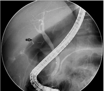
The Korean Journal of Gastroenterology Fig. 1. Cholangiogram showing a bile leak originating from a thin
duct of Luschka that drain into the right hepatic duct (the arrow shows a bile leak from the duct of Luschka).
Fig. 2. Follow-up cholangiogram showing the resolution of a bile leak after ES with NBT. ES, endoscopic sphincterotomy ; NBT, naso- biliary tube.
scopic treatment.
CASE REPORT
A 51-year-old man was admitted with severe upper ab- dominal pain. He had postprandial dyspepsia and upper ab- dominal discomfort for two months, as well as severe upper
abdominal pain for the past seven days. He was diagnosed with diabetes mellitus four years ago and has been taking a hypoglycemic agent. Physical examination revealed tender- ness in the right upper quadrant. Admission laboratory find- ings were as follows: white blood cell count of 10,000/mm3, hemoglobin level of 15.6 g/dL, platelet count of 229,000/mm3, aspartate aminotransferase (AST) level of 25IU/L, alanine ami- notransferase (ALT) level of 24IU/dL, alkaline phosphatase level of 78 IU/dL, total bilirubin of 0.6mg/dL, and albumin of 4.0 g/dL. Abdominal computed tomography revealed thick- ening of the gallbladder wall with a gallstone, consistent with calculous cholecystitis. The patient underwent laparoscopic cholecystectomy, and four days later, he developed fever, chills, and abdominal pain. Bile drainage was persistently ob- served in the drain tube (Jackson-Pratt drain), and a bile leak was suspected. He underwent endoscopic retrograde chol- angiopancreatography (ERCP). The initial cholangiography revealed bile leakage originating from an injury to the duct of Luschka that drained to the right intrahepatic duct (Fig. 1). ES using a 5-F nasobiliary tube (NBT) was performed. A bile leak- age in the drain tube was reduced from 30 mL to 7 mL by the fourth day following ERCP, and final resolution was achieved by the fifth day. A follow-up cholangiography showed a com- plete resolution of the leak (Fig. 2).
DISCUSSION
The ducts of Luschka have been described as “slender bile ducts running along the gallbladder fossa” that drain into the right biliary duct or the common duct.1,3,4 Some authors de- scribed these as aberrant small bile ducts.3 The ducts of Luschka are also known as accessory biliary ducts, vasa aberrantia, subvesicular ducts, or supravesicular ducts; and such confusion is due to the lack of definitive descriptive cri- teria in the literature.3 The ducts of Luschka are small bile ducts―1-2 mm in diameter―situated in the gallbladder bed adjacent to the liver.1,3,4 The average diameter of the bile duct is 2 mm (range 1-18 mm), and the average length is 35 mm (range 8-82 mm).2 These ducts of Luschka have various ana- tomic variations and are smaller in diameter, which makes them difficult to identify on preoperative imaging.2
The ducts of Luschka (subvesical or supravesical) are im- portant from a clinical perspective, posing a potential risk for an injury during gallbladder and hepatic operations.2 Recent
Ko SY, et al. Bile Leak Caused by Injury to the Ducts of Luschka 149
Vol. 69 No. 2, February 2017
studies suggest that clinically relevant bile leaks may cause complications in approximately 0.4-1.2% of cholecystec- tomies performed.2 The frequency of involvement of the ducts of Luschka is reportedly as high as 50%; 4.4% of all ia- trogenic bile duct injuries and 15% of type A injuries, involving a cystic duct or peripheral hepatic radicle leakage.5 A right-sided distribution is a common pattern; however, there can be variability in the points of origin or drainage.2 Variability in the anatomic location of the ducts of Luschka makes it difficult to identify a bile leak originating from duct injury during the hepato-biliary operations.2
A simple ligation is used to treat the visible leaks.5 Postoperative bile leakage is usually diagnosed because of bile tube drainage and development of fever, chills, and ab- dominal tenderness. Since any injury to the ducts of Luschka is considered as a minor leakage of the peripheral radicles, a conventional endoscopic treatment is adequate.5 An un- usual case was reported in a patient with persistent leakage and worsening symptoms despite conventional ES using 7-F double-pigtail stent that was placed in the right hepatic duct.6 The patient was treated with a fully-covered metal stent in the right hepatic duct for 6 weeks. An injury to the ducts of Lushika during laparoscopic cholecysectomy involves thin peripheral ducts around the gallbladder fossa, usually result- ing in a small leak. The aim of this treatment is to induce a low-pressure gradient of bile flow; although the papilla, which is achieved by ES alone, stent alone, or ES with a stent.5 The duration to completely resolve of bile leakages gas been re- portedly 5-12 days after endoscopic treatment.5,7 Two case of bile leaks caused by injury to the ducts of Luschka were treated using 7-F NBT and ES.5 Neuman et al. reported that the resolution of bile leakage from an injury to the ducts of Luschka was achieved by ES with insertion of a 7-F plastic stent into the common bile duct, which was removed after 6 weeks.1 Another study reported that bile leakage from an in- jury to the ducts of Luschka was successfully treated with ES and stenting in 4 cases, and small leaks were treated with on- ly a 7-F stent without ES.7 These results suggest that the type of treatment doses not influence the effectiveness in treating small leaks. Plastic stents were associated with tube ob- struction and stent migration, which required a repeat endos- copy to confirm the resolution of bile leak.5,7 An NBT has dis- advantages of discomfort and tube displacement. In this re- port, bile leakage was treated using ES with 5-F NBT on the
sixth post-operative day, and bile leakage from the drain tube was resolved by the fifth day after ERCP.
A systematic review reported that the duct of Luschka is a topographic description of a variant bile duct(s), in contact with the gallbladder fossa.2 Another variant of the duct of Luschka (aberrant type) is composed of a network of small bile ducts within the connective tissue of the gallbladder wall.
This variant was diagnosed after cholecystectomy based on microscopic findings.2,3 Microscopic examination showed that these ducts may occur as a meshwork of ductules.
Ductules are lined by a flattened-to-columnar biliary epi- thelium and are classically surrounded by a fibrous collar.3 Some ductules contain inflammatory cells with epithelial atypia.3 The findings of the ducts of Luschka (aberrant type) in the gallbladder wall suggest that the clinical significance is the differential diagnosis of invasive or metastatic carcinoma. The ducts of Luschka (aberrant type) are not known to have malignancy potential.3
In summary, conventional endoscopic treatment for post-laparoscopic bile leakage from the ducts of Luschka (subvesical or supravesical type) was effective. The use of NBT enabled successful management of bile leakage with- out the need for repeat endoscopy to verify the resolution.
ACKNOWLEDGMENTS
This paper was written as a part of the Konkuk University research support program for its faculty on sabbatical leave in 2009.
REFERENCES
1. Neumann H, Fry LC, Malfertheiner P, Mönkemüller K. Bile leak from the duct of Luschka. Z Gastroenterol 2010;48:256-257.
2. Schnelldorfer T, Sarr MG, Adams DB. What is the duct of Luschka?--A systematic review. J Gastrointest Surg 2012;16:
656-662.
3. Singhi AD, Adsay NV, Swierczynski SL, et al. Hyperplastic Luschka ducts: a mimic of adenocarcinoma in the gallbladder fossa. Am J Surg Pathol 2011;35:883-890.
4. Spanos CP, Syrakos T. Bile leaks from the duct of Luschka (subvesical duct): a review. Langenbecks Arch Surg 2006;391:
441-447.
5. Lo Nigro C, Geraci G, Sciuto A, Li Volsi F, Sciume C, Modica G. Bile leaks after videolaparoscopic cholecystectomy: duct of Luschka.
Endoscopic treatment in a single centre and brief literature re- view on current management. Ann Ital Chir 2012;83:303-312.
150 고순영 등. Luschka 담관 손상으로 인한 담관 누출의 내시경적 경비 담도 배액술
The Korean Journal of Gastroenterology 6. Hwang JC, Kim JH, Yoo BM, et al. Temporary placement of a newly
designed, fully covered, self-expandable metal stent for re- fractory bile leaks. Gut Liver 2011;5:96-99.
7. Ryan ME, Geenen JE, Lehman GA, et al. Endoscopic intervention for biliary leaks after laparoscopic cholecystectomy: a multi- center review. Gastrointest Endosc 1998;47:261-266.