大韓放射線醫學슐誌 Vo1. XJ)(, No. 3. 1983
賢周圍 B農傷의 放射線學的 所見
中央大學校 醫科大學 放射線科學敎室 金鍵相·沈炯鎭·李寬世 - Abstral't -
Radiological Diagnosis of Perinephric Abscess
Kun Sang Kim, M.D., Hyung Jin Shim, M.D. and Kwan Seh Lee, M.D.
Department of Radiology
,
Co!lege of Medicine,
Chung-Ang UniversityWith convenfional radiological approaches
,
diagnosis of perinephric abscess is difficult in many occa- slons.CT and ultrasound enable accurate detection of lesions and precise determination of their extent.
We review our cases and discuss the usefulness of these new modalities for the diagnosis and follow up of the lesions.
I.
論 絡햄周@엄8農傷은 臨 ff 的으로나 放射線學的으로나 *휴꿇한
所見을 나타내는 일이 흔치 않은 관계로 早期該斷이 비
교적 어렵고 따라서 f台續時期를 놓치는 경우가 빈번하
다1)
最近에 이르려 超音波檢좁 및 양£깎[化斷 l협겼옮影 (CT}의 활발한 臨ff 的 應用의 덕분으로 비교적 짜1뼈에 談斷이 가능하여졌을 뿐 아니라 病짧의 波及程度를 쉽게 알 수 있게 되어 治續에 큰 도움을 줄 수 있게 되었다잊 著좀 들은 自驗例를 중심으로 뽑周圍聽陽의 放射線學的 所見 을 討講하고자 한다
n.
홈例 例示:ffE例 l
V A tendemess ) 를 發見하였다~피純題部휩훌影에 서 多 量의 陽管內 '!:!.氣陰影으로 特記할 所見을 찾을 수 없었 고 經靜1l*!JJê路造影에서는 左얘~웹의 下端이 外뼈~으로 빌 려 있는 所見을 觀察할 수 있었다.
超音波짧影에서 左에 l업이 뻐大되어 있었고 잔잔한 i^1
rfg에코를 보이는 둥근 쩔間난3 有病變。l 左測뽑 下端에 {효置하여 뽑內眼傷을 확인할 수 있었으며 아울러 뽑中 心홉ß에 에코의 變形이 觀察되었다 (Fig.3a) 左測뽑 주변에는 뽑願에코의 t曾1Ju 와 함께 11: !l!~웹 下端얘없~의 뽑周圍'!:!.間에 ~llE性쨌體의 종홉積이 觀察되었다.
f훈~JII
9 歲 男兒로서 2 週H 程度에 걸친 不明熱을 主訴로
하여 來院당시 左예!I1J骨홉維角 I용痛 ( CVA tenderness) 가 있었다. 單純짧흠B振影에서 U없推썽양JilJ, 11:!l!~ /Jj씻堆 外隊 의 消失 및 까ë ßI~ 띔陰影의 t혐失이 觀察되었고 經靜lIJíU7R 路造影에서 左右에 뽑杯 및 1업굶의 早期造影이 觀쫓되었
10 歲 女兒로서 I 週日에 결친 不明熱을 主訴로 하여 으나 左ßI~의 펌杯 쩍굶의 不完숲한 充滿괴 t部 原管의 來 院당시 理學的 檢좁에서 심한 左때 曲骨홉 堆角많뼈ì
(C-딸if!所見등이 觀察되었다 갱중堆와
1i:ßlJj뽑과의 거리가 t曾
이 논운은 83 년 6 월 4 일에 채택되었음.
1Ju되어 左빼뽑이 外fJUJ 方으로 빌려있음을 일 수 있었다 (Fig. 2 a )
A .훌훌짧.,혔짧₩-쩔짧파k빼-잉&쫓γ,… ‘ B
Fig. 1. Plain 떠m of the emphysematous pyelonephritis.
a) Accumulation of abnormal gas density is demonstrated in the perirenal space and mott1ed radiolucent gas density is also noted in the renal parenchyme b) Accumulation of radiolucent rim like gas density in the right side perirenal space
is noted by gas forming infection.
B ’
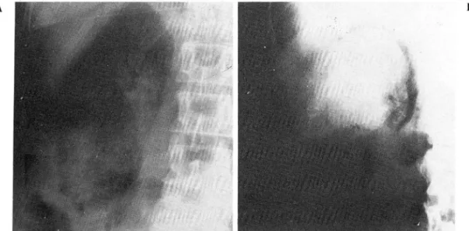
Fig. 2. Conven tional contrast studies
a) I.V.P. (supine, standing) reveal faint visualization of the left pelvis and calyces with compression of upper ureter and scoliosis with loss of left side psoas muscle shadow are also noted. Vertebra to left kidney distance is increased causing lateral displacement of left kidney.
bl) I.V.P. reveals cresentic deformity of pelvis and calyces causing large, sharply out- lined mass. (Renal ce1i carcinoma with perireilal abscess).
b2} Enema colon study of the same patient (bl) shows loss of proximal half psoas muscle shadow and lateral displacement or proximal descending colon.
超품波檢홉에 서 1iíllU 賢의 6밍大와 內部에 코가 없고 後 方陰影t홈彈의 所見을 보이는 뽑內盤場과 쩔벼心힘8에코의 뾰ill. 및 移動의 所見을 觀察할 수 있었고 뽑體에코의 增 加와 뽑周圍옐間에 충만된 쨌뽑의 i용影을 볼 수 있었다
(Fig. 3 bj
,
bz ).íÆØl
Jrn.
56 歲 女子로서 짧l 原性 좁睡로 入院하였고 觸륨ß x-線 擔影上 右..tH흉部어l 非正常的언 '!i'.氣陰影이 觀察되 었으며 댈純題部握影에서 뽑魔 및 웹周圍에 홉짧된 '!i'.氣陰影을
Fig.3.
A B B"
C
’c ’
DUltrasonography
a) Longitudinal scan reveals en1argement of left kidney and round weak echogenic area is shown in the lower pole of left kidney with distortion of central echo complex.
bl, b2) Longitudinal and transverse left kidney ultrasound reveal en1argement of kidney and echo free area with posterior enhancement in the renal parenchyme. Compression and dis- placement of central echo complex is also shown. Thickened and increased echogenicity of renal capsule with accumulation of perirenal fluid are demonstrated
cl, c2) Transverse and longitudinal scan reveal renal and perirenal gas producing reverberation artifact. Perirenal effusion is manifested as echo free area in the lower part of perirenal space
d) Supine transverse u1trasound reveals large amount of retroperitoneal fluid accumulation with ascitic fluid (Renal cell carcinoma with perirenal abscess). Renal capsule also appears thicknened.
觀察할 수 있었다 (Fig. 1 a ). CT 홉影에서 石에j 督縣및 뽑周圍空間에 多量의 空氣가 發見되 어 소위 氣睡狀뽑잦
(emphysematous nephritis) 및 뽑職周 圍잦을 確認할 수 있었다 (Fig. 4 b).
1Æ~jV .
49 歲 男子로서 血民 , H흉$lllf;澈 및 高熱을 主訴로 入 院하였다
1Æ~j IV
42 歲 男子로서 수년간 햄原病을 앓고 있었고 갑작스 런 高熱과 助骨참推角짧痛 (CVA tenderness) 를 主訴 로 X 院하였다 r힘純&훌部握影1: H없堆쩔曲파 右에띔嚴 및 뽑周圍~圖에 空氣가 觀察도l 었고(Fig.l , 6) 이들 소견 은 超흡波 (Fig. 3 cr,cz) 빚 CT檢훌 (Fig.4a) 로서 確認되었다-
單純題部 및 大|陽造 影에 서 j總外緣의 t 융이 消失된
所見괴 下行結陽 近 f立部의 外예 빼-ÜL를 볼 수 있었다 (Fig. 2 b z ).
經靜빙~*路造影에서 l엽內 陣塊의 所見을 볼 수 있었 고 (Fig. 2 b r) 超音波檢홈에서 뽑영용睡p'l에 發生한 賢
,ffij g包 의 所見과 題水 및 後題願뾰內에 옳짧된 多量의 i!U영陰影을 볼 수 있었고 뽑題이 두꺼워진 所見을 觀察 할 수 있었디 (Fig.3d). 病理組織檢훌로 뽑細8包홉이 確認되었고 뽑周@협 6農傷이.~뚱認되었다
A B
Fig.4. Computed tomography of emphysematous pyelonephritis
a) Multiple abnormal gases are demonstrated in the renal and perirenal space of right kidney.
b) Large amount of gas accumulation is noted in the right perirenal space and renal parenchyme
,
which is characteristic finding of emphysematous pyelonephritis.ill.
考 按超품波와 CT 가 臨똥的으로 利用되기 뼈에는 後題題 臨의 ~愚을 등2斷하는데 있어서 상당한 制約이 있었으나 A 體斷面相을 쉽게 얻을 수 있는 이들 談斷法들의 활발 한 應用으후 後8찢願뾰흉惠의 談斷은 울론 이 부분에 發 生한 흉뽕、의 波及程度를 評6옆하는데 큰 도움을 받을 수 있게 되었다 2)
뽑周圍8農傷은 흔치 않은 흉惠으로 흔히 臨 lf的 llE勢 냐 放射線學的 所見이 非特異的이 다 J) 이 용愚은 血 行性으로 發生하는 수도 있고 手術後遺효으로도 發~할 수 있으며 廳原病을 앓고 있거니 3) 뽑細뼈修등 기타뽑 흉愚을 앓고 있는 경우에 合혐효으로서 發生하는 일이 많은 것으로 일려져 있다 4)
單純題힘3 振影에서는 所見이 非特흥용的인 관겨l 로 진행된 llE例에서만 特徵的인 所見을 볼 수 있다. H했推의 짧曲,
體節外緣의 f법失이나 뽑驗의 輪節이 不分明해지는 所見
充浦, 뺑 f立짧ifl.등의 所見을 i을tJu로 觀察할 수 있다. 檢 좁중 呼期時 銀影과 吸期時 擬影을 하여 ’업의 üz 置를 비 교하면 웹의 移動範랩가 현저히 줍어들어 있는 所見을 觀察할 수 있다 이러한 웹固定現狀은 愚子플 몽AÜZ와 立 位에서 搬影하여 비교하여도 觀察할 수 있다.
Gallium - 67 - Citrate을 利用한 j司 üz元素走흉에서 非正常的인 同 f立元素의 集積으로 有意한 所見을 얻을 수 있으나j) 著者들은 自驗例가 숲無하여 詳細i한 記述은 省略하는 바이다.
超音波檢훌와 CT 檢훌는 이 g늦愚의 등?斷과 波及의 程 度을 얹定하는데 중요할 뿐 아니 라 該斷이 確定뭔 後f台 驚에 대한 反應、을 追顧하는데도 有用하며 穿刺등의 續 法에 길깎이로서 대 l까히 큰 역할을 한마 2,5 ,6) 이 두 檢홉에서 띔범大, 뽑의 üz 置移퍼b , 쩔I^II!홉傷의 所見 및 뽑周圍의 ~llE앤: It훗홉엽의 iit첩을 쉽게 觀察할 수 있으여 뽑體의 R巴훔등을 詳細-oj 觀察할 수 있어 wi該에 큰 도웅
을 준다. 기타 補助的인 檢좁로서는 大陽造影 및 뽑血管 등을 觀察할 수 있다. 소위 氣睡狀샘잦에 鏡發한 협周 造影퉁이 있다
圍體혔에 서 는 多量의 2E氣陰影이 觀察되 고 이 所見은 後 題睡용外陽이 나 後g힐體&효과 陽管과의 擾孔形成의 경 우를 제외하면 I업周圍 8앓傷을 確장할 수 있는 特異的인 所見이 될 수 있다. I!農없의 波及程度에 따라 同에 橫隔題의 上 昇, 뽑敵 üz 置의 移動, 또는 同예뾰뼈쨌慘出등의 所見도 觀察할 수 있다1 ,5)
經靜l!IR}}j(路造影에서는 웰純題部嚴影에서 觀察되는 所 見들을 좀 더 확실히 볼 수 있고 l협杯 및 협 j굶의 不完全
REFERENCES
l. Witten DM
,
Myeres GH,
DC Emmett’5 C/iníca/ùrography. 4th ED Vol 2:843-848
,
W.B. 5aunders,
7977.
2. Gerzof SG
,
Gale M E Computed tomography and ultrasonography for diagnosis and treatment ofrenal and retroperitoneal abscess. Urol. Clinic.
Nor. Amer. 9:785-793
,
7982.3. Sinpkins KC
,
Barradough NC Renal cortical abscess,
perinephritis and perinephric abscess in diabetes. 8j R 46:43436,
7973.4. Childs GJ, Pickelman J, Churchill RJ et al : Renal cel! carcinoma complicated by perinephric abscess
and colon perforation. Urol. clinic 4:3740
,
7982 5. WickesJ
D,
ThornburyJ
R Acute renal infections in adult. Radiol. c/inic Nor. Amer. 77:245-260,
7979.
6. Schneider M