J Korean Soc Radiol 2016;74(3):147-155 http://dx.doi.org/10.3348/jksr.2016.74.3.147
Benefits and Harms of Breast Screening: Focused on Updated Korean Guideline for Breast Cancer Screening
유방암 검진의 이득과 위해: 개정된 한국 유방암 검진 가이드라인을 중심으로
Soo-Yeon Kim, MD, Eun-Kyung Kim, MD*
Department of Radiology, Severance Hospital, Research Institute of Radiological Science, Yonsei University College of Medicine, Seoul, Korea
서론
유방암은 현재 한국 여성에서 갑상선암 다음으로 두 번째로 많이 발생하는 암으로, 1999년 국가암발생통계가 산출된 이래 매년 증가하고 있다. 1999년 유방암의 연령표준화발생률은 여 성 인구 10만 명당 24.5명이었으나, 2012년에는 44.7명으로 유 의하게 증가하였다(1). 한국 여성에서의 유방암 발생률은 서구 여성과 다른 양상을 보인다. 호발 연령이 45~49세로 서구 여성 에 비해 약 10세 정도 낮고 치밀 유방의 빈도가 더 높다(2, 3).
검진유방촬영술(screening mammography)의 유용성을 평가 한 여러 무작위 대조군 연구(randomized controlled trial)의 결 과 검진유방촬영술이 유의하게 유방암 사망률을 감소시킨다는 결과를 보고하였다(4). 그러나 최근 발표된 Canadian National Breast Screening study에서는 25년간 검진유방촬영술 대상자
를 추적 관찰한 결과 유의한 사망률 감소 효과가 없었다고 보 고하였다(5). 또한 적절한 수준의 무작위 배정이 이루어지지 않 은 몇몇 연구들이 지적되기도 하였다(4). 또한 검진유방촬영술 로 인한 과진단(overdiagnosis), 과치료(overtreatment), 위양성 (false positive) 및 위음성(false negative) 진단, 방사선 피폭 등 의 위해도 잘 알려져 있다(4). 따라서 최근에는 검진유방촬영술 의 이득과 위해를 종합적으로 고려한 판단이 필요하고 수검 대 상자에게도 균형 있는 정보를 충분히 제공하여 정보에 근거한 의사결정(informed decision)을 내릴 수 있도록 해야 한다는 주 장이 대두되고 있다(4, 6, 7). 한편, 미국에서는 유방밀도고지 법(breast density notification legislation)이 빠른 속도로 확산 되고 있고 2016년 1월 현재 50개 주 중 26개 주에서 법률을 채 택하여 시행하고 있다. 유방밀도고지법은 유방촬영술에서 높은 유방밀도(breast density)를 보일 경우 유방촬영술의 민감도 Breast cancer is the second most common malignancy among Korean women. The
incidence of breast cancer has increased since 1999, which is when the national screening program involving mammography started. Until now, the benefits of screening mammography have been emphasized, but information about its benefits and harms should be provided in a comprehensive fashion, in order to guide people toward making informed decisions. Although the main benefit of screening is reduc- tion of breast cancer mortality, harms such as overdiagnosis, overtreatment, false positive and false negative diagnoses, and radiation-induced breast cancer, can all occur as a result of screening. The 2015 Korean guideline for breast cancer screening recommends biennial screening mammography for asymptomatic women aged 40 to 69 years. This review discusses the benefits and harms of screening mammogra- phy in light of evidence-based approaches obtained from randomized trials, meta- analysis, and guidelines.
Index terms Breast Cancer Screening Mammography Ultrasonography
Received October 29, 2015 Revised December 11, 2015 Accepted January 15, 2016
*Corresponding author: Eun-Kyung Kim, MD Department of Radiology, Severance Hospital, Research Institute of Radiological Science, Yonsei University College of Medicine, 50-1 Yonsei-ro, Seodaemun-gu, Seoul 03722, Korea.
Tel. 82-2-2228-2371 Fax. 82-2-393-3035 E-mail: [email protected]
This is an Open Access article distributed under the terms of the Creative Commons Attribution Non-Commercial License (http://creativecommons.org/licenses/by-nc/3.0) which permits unrestricted non-commercial use, distri- bution, and reproduction in any medium, provided the original work is properly cited.
(sensitivity)가 감소하여 유방암을 놓칠 가능성이 있고, 높은 유 방밀도 자체가 유방암의 위험인자가 될 수 있으므로 초음파 등 의 추가적인 검진 방법을 시행할 수 있다는 내용을 수검자에게 고지해야 함을 명시하고 있다(8). 유방밀도고지법의 도입으로 미국에서는 초음파와 자기공명영상검사 등 유방촬영술 이외의 추가적인 유방암 검진법에 대한 수요가 증가하고 있다.
대한의사협회에서 2002년 발표한 유방암 검진 권고안에서 는 30세 이상 여성에서 매달 유방자가촉진(breast self-examin- ation)을 시행하고, 35~40세 여성에서는 2년마다 의사에 의한 임상유방진찰(clinical breast examination)을, 40세 이상 여성에 서는 2년마다 유방촬영술과 임상유방진찰을 시행하도록 권고 하였다. 그러나 2015년 유방암 검진 권고안이 개정 발표되었고 그 내용은 Table 1과 같다. 기존 권고안과의 차이점은 1) 검진 종 료 연령이 명시되었고, 2) 유방촬영술과 함께 일차적으로 권고 되었던 임상유방진찰이 일차적 권고 검진법에서 빠지게 되었다 는 점이다(9).
본 리뷰에서는 무작위 대조군 연구 결과와 메타 분석, 가이드 라인 등 근거 중심의 정보를 토대로 검진유방촬영술의 이득과 위해에 대해서 중점적으로 다루고자 한다. 그리고 우리나라 유 방암 검진 권고안 개정안의 자세한 내용과 배경에 대해서 살펴 보고자 한다.
검진유방촬영술의 이득
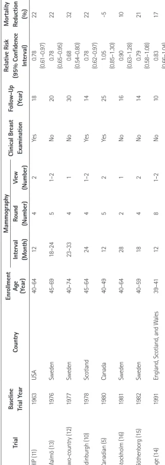
현재까지 검진유방촬영술의 효과에 대해서 8개의 무작위 대 조군 연구(randomized controlled trial)가 시행되어 결과가 발 표되었고 이 연구들의 프로토콜과 결과에 대해서 Table 2에 간 략히 정리하였다. 이 연구들은 모두 서구 여성을 대상으로 시행 되었고 한국 여성을 대상으로 한 무작위 대조군 연구는 없었다.
Canadian trial (5)을 제외한 나머지 7개의 연구에서 검진유방 촬영술은 검진을 시행하지 않은 대조군과 비교시 유방암 사망률 을 10~32%까지 감소시켰다고 보고하였다(10-16). 가장 최근
발표된 Canadian trial에서는 검진유방촬영술을 시행한 대상자 를 25년간 추적 관찰하였는데 유의한 유방암 사망률 감소 효과 가 없는 것으로 보고하였다(5). 2013년에 발표된 코크란 리뷰 에 따르면, 임상시험들 중에서 세 개의 연구는 적절하게(optimal) 무작위 배정이 되었지만(5, 13, 14), 네개의 연구는 적절한 수준 에 미치지 못했고(suboptimal)(11, 12, 15, 16), Edinburg trial (10)은 무작위 배정이 부적절하였음이(inadequate) 지적되었다 (4). 코크란 리뷰에서 Edinburg trial을 제외한 나머지 일곱 개 를 모두 포함한 메타분석 결과에서(the Canadian trial은 2002 년도에 발표된 대상자를 16년간 추적 관찰한 결과를 이용함 (17)) 유방암 사망률에 대한 상대위험도는 0.81(95% 신뢰구간:
0.74~0.87)로 유의하게 감소하였다(4). 그러나 적절하게 무 작위 배정이 되었다고 판단한 세 개의 연구만을 포함한 메타분 석 결과에서는 상대위험도가 0.90(0.79~1.02)으로 유의하게 감소하지 않았다(4). 이러한 결과를 바탕으로 코크란 리뷰에서 는 검진유방촬영술의 유방암 사망률 감소 효과가 크지 않고 몇 몇 연구들이 적절히 무작위 배정이 되지 않아서 결과를 신뢰하 기 어려운 측면이 있고, 위양성 진단 및 과진단 등 검진의 위해 가 있으므로, 검진의 득과 실을 종합적으로 고려한 판단이 필요 하다고 결론지었다(4). 2015년 대한의사협회에서 발표된 유방 암 검진 권고안 개정안에서는 무작위 배정이 부적절하였던 Ed- inburg trial을 제외한 나머지 7개의 연구를 이용하여 메타분석 을 시행하였는데, 유방암 사망률에 대한 상대위험도는 0.81 (0.73~0.91)로 검진군에서 유방암 사망률이 19% 유의하게 감소하였다(9). 이러한 결과를 바탕으로 대한의사협회에서는 40~69세 무증상 여성을 대상으로 유방촬영술을 이용한 유방 암 검진을 시행할 것을 권고하고 있다(9). 2014년 Welch와 Passow (6)가 발표한 메타분석 결과에서 10년 동안 매년 검진유 방촬영술을 시행한다고 가정하였을 때, 40세에 검진을 시작할 경우 1000명 중 0.1~1.6명이, 50세에 시작하면 0.3~3.2명이, 60세에 시작하면 0.5~4.9명이 유방암으로 인한 사망을 피할 수 있으나 유방암 사망률 감소 정도가 크지 않기 때문에 수검 Table 1. The Korean Guideline for Breast Cancer Screening
Recommendations Grade Quality of Evidence
Mammography 40–69 years Every 2 year B
≥ 70 years Recommend against routine screening.
Individual decision and patient context
C
Breast ultrasound Alone or combination with mammography Insufficient evidence I
Clinical Breast Examination Alone or combination with mammography Insufficient evidence I
Grade B: Recommend routine screening mammography based on the moderate evidence for mortality reduction of screening mammography. Grade C:
Recommend against routine screening mammography based on the low evidence for mortality reduction of screening mammography, but selectively rec- ommend according to the individual decision and patient context. Grade I: No recommendation nor recommend against, based on the insufficient evi- dence for benefits and harm.
대상자에게 이득과 위해에 대한 정보를 충분히 제공하여 정보 에 근거한 의사결정을 내릴 수 있도록 한다고 주장하였다. 그러 나 이러한 메타분석은 30여 년 전에 시행되었던 임상시험 연구 들의 결과를 토대로 한 것이므로 현재의 상황에 그대로 적용하 고 이해하는 것은 적절하지 않을 수 있다는 주장도 있다(18).
과거와 비교하였을 때 유방암 치료 방법이 매우 발전하였고 유 방암의 발생에 영향을 미치는 인자(폐경 후 호르몬 치료와 비 만 등)가 시대의 흐름에 따라 변화하였고, 유방촬영술의 방법 및 기술도 발전되었기 때문이다(18).
검진유방촬영술의 위해
과진단과 과치료
유방촬영술을 이용한 검진에서 과진단과 과치료가 발생할 수 있다. 과진단(overdiagnosis)이란 검진에서 발견된 암은 증 상이 있어 발견된 암에 비해서 크기가 작고 천천히 자라는 암일 가능성이 높아서 이번 검진에서 발견되지 않았다고 하더라도 추 후 증상이 있어 발현되거나 그 암으로 인해 사망할 가능성이 낮 다는 것을 의미한다(4). 검진으로 발견된 모든 유방암 중에서 54%는 과진단되었던 암으로 추정된다는 보고가 있다(19).
Welch와 Passow (6)에 따르면, 10년간 매년 검진유방촬영술을 시행 받는다고 가정하면, 40세에 검진을 시작할 경우 1000명당 11명까지, 50세에 시작하면 1000명당 3~14명이, 60세에 시작 하면 1000명당 6~20명이 과진단될 수 있다. 과치료(overtreat- ment)란 과진단으로 발견된 암을 불필요하게 치료하는 과정에 서 오히려 치료로 인한 여러 단기적, 장기적인 합병증이 발생할 가능성이 있다는 것을 의미한다(4). 수술로 인한 신체 변형과 흉터, 방사선 치료로 인한 심혈관 질환과 폐암, 화학요법치료로 인한 심독성 등의 부작용이 발생할 수 있다(4). 과치료에서 발 생한 부작용으로 유방암 이외의 다른 원인으로 인한 전 원인 사 망률(all-cause mortality)이 증가할 수 있다(4). 유방촬영술에 서 발견되는 암은 증상이 있어 발견되는 암에 비해서 크기가 작 고 덜 진행된 암이어서 부분 절제술(lumpectomy)로 충분하고 보조 치료 요법(adjuvant therapy)도 덜 필요할 것으로 기대되 었다(4). 그러나 검진 시행 후 오히려 유방 전절제술(mastecto- my)의 빈도가 20%가량 증가하였다(4). 미국에서 유방 검진이 시행되기 전에 관상피내암(ductal carcinoma in situ)은 전체 유방암의 5%에 불과했으나 검진 시행 후에는 새롭게 발견되 는 유방암 중 20%를 차지하고, 검진에서 발견되는 유방암의 20~40%는 관상피내암이다(20). 모든 관상피내암이 침윤성 유방암(invasive breast cancer)으로 진행되는 것은 아니기 때 문에 유방 검진으로 발견되는 관상피내암을 침윤성 유방암과
Table 2. Randomized Controlled Trials of Mammography Screening: Protocols and Results Trial Baseline Trial YearCountryEnrollment Age (Year)
Mammography Clinical Breast ExaminationFollow-Up (Year)
Relative Risk (95% Confidence Interval)
Mortality Reduction (%)Interval (Month)Round (Number)View (Number) HIP (11) 1963USA40–641242Yes180.78 (0.61–0.97)22 Malmö (13)1976Sweden45–6918–2451–2No200.78 (0.65–0.95)22 Two-country (12)1977Sweden40–7423–3341No300.68 (0.54–0.80)32 Edinburgh (10)1978Scotland45–642441–2Yes140.78 (0.62–0.97)22 Canadian (5)1980Canada40–491252Yes251.05 (0.85–1.30)-5 Stockholm (16)1981Sweden40–642821No160.90 (0.63–1.28)10 Göthenborg (15)1982Sweden40–591842No140.79 (0.58–1.08)21 Age (14)1991England, Scotland, and Wales39–411281–2No100.83 (0.66–1.04)17
동일한 방법으로 치료하는 것은 과치료일 수 있다는 주장이 있 다(21). 그러나 관상피내암의 자연사 및 침윤성 유방암으로 진 행될 가능성이 높은 관상피내암을 구별해 낼 수 있는 방법이 현 재까지 밝혀지지 않았고, 검진에서 발견된 관상피내암의 60%
가 향후 5년 이내 침윤암으로 진행될 가능성이 높은 고등급 (high grade) 암이라는 보고 등에 기반하여, 아직까지는 검진에 서 발견되는 모든 암은 치료해야 한다는 의견이 우세하다(21).
위양성 진단
모든 진단 방법이 그렇듯이, 유방촬영술의 민감도와 특이도 (specificity)는 완벽하지 않기 때문에 검진유방촬영술로 위양성 진단이 발생할 수 있다(4, 7). 이는 불필요한 추가 검사, 조직 검 사 및 추적 검사를 발생시키고, 이로 인한 의료 비용 증가와 더 불어 환자에게 상당 기간 지속되는 심리적인 고통을 야기할 수 있다(4, 7). Welch와 Passow (6)의 보고에 따르면, 10년간 매 년 검진유방촬영술을 시행 받는다고 가정하였을 때, 약 절반가 량의 환자는 한 번 이상의 위양성 진단을 받는다(40세부터 시작 하면 510~690/1000명, 50세부터 시작하면 490~670/1000명, 60세부터 시작하면 390~540/1000명). 이러한 위양성 진단은 상당하게 지속되는 심리적인 고통을 야기할 수 있는데 위양성 진단을 받았던 여성은 암이 없다고 최종 판정이 내려진 후에도 최소한 3년 이상 지속되는 심리적인 고통을 경험한다는 보고가 있었다(22, 23).
위음성 진단
침윤성 유방암을 가진 여성의 6~46%는 유방촬영술에서 유 방암이 보이지 않아서 위음성 진단을 받게 될 수 있다(24, 25).
위음성 진단은 젊은 여성, 치밀 유방, 유방암 아형이 점액암이나 소엽암인 경우, 매우 빨리 자라는 유방암인 경우에 호발하는 경 향이 있다(26). 검진유방촬영술에서 놓친 유방암은 다음 번 검 진 전에 증상이 있는 간격암(interval cancer)으로 발견될 수 있 으며, 간격암은 빨리 자라고 진단 시에 이미 진행된 단계를 보이 는 경우가 많다(26, 27). 이는 유방촬영술의 안정성에 대한 불 안감을 증가시키고 미국에서 유방밀도고지법률이 시행되게 된 배경이며 유방촬영술 이외의 추가적인 검진 방법의 필요성에 대한 문제를 대두시킨다(8).
방사선 유발 유방암
검진유방촬영술을 이른 연령에서 시행할수록, 검진 간격이 짧 을수록, 생애 누적 피폭선량이 증가할수록 방사선에 의해 발생 한 유방암(radiation-induced breast cancer)의 사망자 수와 사 망에 대한 이익 대비 위험이 증가한다(28, 29). 검진에 이용되는
유방촬영은 내외사위(mediolateral view)와 상하위(cranio- caudal view)가 포함된 두 가지 촬영을 시행하며, 이러한 유방 촬영으로 인한 방사선량은 약 2~4 mSv에 불과하다(30). 40 세 이상의 연령에서는 방사선 피폭의 위해보다 검진의 이득이 더 큰 것으로 추정되지만, 모세관 확장실조(ataxia-telangiectasia) 등 특정군에서는 방사선에 대한 위해가 더 높을 수 있으므로 주의를 요한다(28-31).
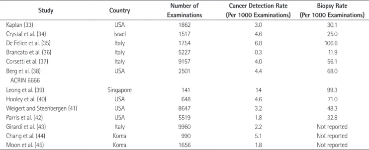
유방촬영술과 병행된 검진유방초음파
유방촬영술의 민감도는 약 85%이지만 치밀 유방에서는 47.8~64.4%까지 감소하여 침윤성 유방암을 놓칠 수 있고 치밀 유방 자체가 유방암의 위험도를 4~6배까지 높인다고 보고되 어 있다(3, 32). 유방촬영술에서 음성(negative)소견이지만 치 밀 유방을 가지는 여성에서 병행된 유방초음파의 암 발견율을 조사한 연구 문헌들에 따르면(Table 3), 유방초음파는 1000명 당 0.3~6.8명의 유방암을 추가로 발견하였다(33-45). Berg 등 (38)에 의한 American College of Radiology Imaging Net- work 6666 trial에서 유방암에 대한 높은 위험도를 가진 무증 상 여성에서 검진유방초음파를 병행함으로써 1000 검사당 4.2 개의 추가암을 발견하였고, 유방촬영술 단독과 비교시 진단능 이 유의하게 상승하였다(area under the receiver operating ch- aracteristic curve: 0.78 유방촬영술 단독, 0.80 유방초음파 단 독, 0.91 유방촬영술과 유방초음파 병행). 그러나 유방촬영술에 비해서 유방초음파 단독 혹은 유방촬영술과 유방초음파를 병 행 시에 높은 위양성률(4.4% 유방촬영술 단독, 8.1% 유방초음 파 단독, 10.4% 유방촬영술과 유방초음파 병행)과 낮은 양성 예측도[positive predictive value (이하 PPV) 1: 7.6% 유방촬영 술 단독, 6.5% 유방초음파 단독, 7.3% 유방촬영술과 유방초 음파 병행, PPV2: 22.6% 유방촬영술 단독, 8.9% 유방초음 파 단독, 11.2% 유방촬영술과 유방초음파 병행. PPV1은 추가 적인 검사 또는 조직 검사가 권유된 사람들 중에서 암이 발견된 사람의 숫자로 정의되었고 PPV2는 조직 검사가 권유된 사람들 중에서 암이 발견된 사람의 숫자로 정의되었다]에 대한 문제가 지적되었다(38). 최근에 발표된 40~49세의 무증상 여성을 대 상으로 한 일본의 무작위 대조군 연구 결과에서 유방촬영술 단 독에 비해서 유방초음파 병행 시 민감도와 조기암의 발견율이 유의하게 상승하였지만, 특이도는 유의하게 감소하였다(46).
유방초음파에서 추가적으로 발견된 암은 1 cm 미만의 림프절 전이가 없는 조기의 침윤성 유방암인 경우가 대부분이어서 부 분 절제술로 완치가 가능한 경우가 많고 이로써 생존율의 증가 및 삶의 질 향상이 예상되었다(47). 그러나 유방초음파를 시행
함으로써 추가되는 조직검사율이 1000명당 11.9~106.6개로 (Table 2) 상당한 수의 위양성 조직 검사가 발생하고 이는 의료 비의 손실과 함께 육체적, 정신적 스트레스를 발생시킨다. 검진 유방초음파에서 breast imaging reporting and data system (BI-RADS) 범주 3으로 매겨지는 양성 추정 병변(probable benign finding) 진단이 약 20%를 차지하는 것으로 보고되는 데, 이는 6개월 후 단기 추적 검사를 요하므로 검진유방초음파 의 의학적 검사에서 양성(positive) 범주로 분류된다(48, 49).
그러나 이들 중에서 추적 초음파를 시행하여 결국 암으로 밝혀 지는 것은 1% 미만으로 매우 드물기 때문에 위양성 초음파 검 사의 비율이 매우 높고, 이는 역시 의료비의 손실 및 정신적 스 트레스 등의 문제를 야기시킨다(48, 49). 이 외에도 초음파 검 사는 검사자 의존도가 높고 시간, 노동 집약적이라는 제한점을 가지고 있다.
현재까지 유방초음파 단독 또는 유방촬영술과 병행한 유방 초음파 검사가 유방암 사망률을 감소시키는지에 대한 무작위 대조군 연구나 코호트 연구는 없었기 때문에 유방초음파 검사 의 유방암 사망률 감소 효과는 평가할 수 없다(9). 유방암 검진 권고안 개정안에서는 초음파 검사를 유방암 검진으로 시행하 는 것은 근거가 불충분하다고 판단하여 무증상 여성에서 유방 초음파 검사 단독 또는 유방촬영술과 병행하여 유방초음파 검 사를 유방암 검진으로 시행하는 것을 권고하거나 반대하지는 않는다(9). 다만 고위험군의 여성(고위험군에 대한 구체적 정 의는 언급되지 않았다)에서는 임상의의 판단에 따라서 임상유 방진찰, 유방초음파 검사 등의 추가적인 조치를 시행할 수 있다 (9). 미국의 여러 유방암 검진 권고안들에서도 마찬가지로 유 방초음파 검사 단독 또는 유방촬영술과 병행한 유방초음파 검
사를 시행하는 것을 권고하거나 반대하지 않는다(47).
유방촬영술을 이용한 유방암 검진의 시작, 종결 연령과 주기
검진유방촬영술이 유방암 사망률을 낮춘다는 데에는 합의가 있지만 적절한 검진의 시작과 종결 연령 및 검진 주기에 대해서 는 아직까지 논란이 있다. Table 4에서 보여지듯이 여러 나라의 가이드 라인마다 검진의 시작과 종결 연령, 검진 주기에 약간의 차이가 있으며, 그 이유는 무작위 대조군 연구에서 보여지는 이 용 가능한 근거들의 질 및 중요도 등급을 어떻게 해석하고 판단 하였는지에 따른 결과로 생각된다. 유방암 검진 권고 개정안에 서는 캐나다 권고안(50)과 코크란 리뷰(4)의 메타분석 결과와 한국의 국가유방암 검진 대상자 자료를 이용한 코호트 내 환자 대조군 연구 결과(51)를 기반으로 하여 40세(시작 연령)~69세 (종결 연령)의 여성을 대상으로 유방촬영술을 이용한 유방 검 진을 2년마다(주기) 시행하는 것이 유방암 사망률을 유의하게 감소시키므로 이 연령군에서 2년마다 유방촬영술을 권고한다 (9). Table 5에서 연령별에 따른 유방암 사망률 감소에 대하여 캐나다 권고안과 코크란 리뷰의 메타분석 결과가 간단히 제시 되었다(4, 50). 30대 여성에서 검진유방촬영술이 유방암 사망 률을 낮추는지에 대한 연구는 없었다. 40~49세와 50~69세 여 성에서 검진 유방촬영술을 시행함으로써 유방암 사망률이 유의 하게 감소하였다(Table 5). 캐나다 권고안에서 70세 이상 여성 에 대한 유방암 사망률의 상대위험도는 0.68로 통계적으로 유 의하지 않았고, 메타분석에 포함된 2개의 연구 모두 무작위 배 정과 비정밀도에 문제점이 있어 근거 수준이 낮았다(Table 5).
Table 3. Performances of Hand-Held Screening Ultrasound in Women with Negative but Dense Breasts on Mammography
Study Country Number of
Examinations
Cancer Detection Rate (Per 1000 Examinations)
Biopsy Rate (Per 1000 Examinations)
Kaplan (33) USA 1862 3.0 30.1
Crystal et al. (34) Israel 1517 4.6 25.0
De Felice et al. (35) Italy 1754 6.8 106.6
Brancato et al. (36) Italy 5227 0.3 11.9
Corsetti et al. (37) Italy 9157 4.0 56.1
Berg et al. (38) ACRIN 6666
USA 2501 4.4 68.0
Leong et al. (39) Singapore 141 14 99.3
Hooley et al. (40) USA 648 4.6 71.0
Weigert and Steenbergen (41) USA 8647 3.2 48.3
Parris et al. (42) USA 5519 1.8 32.8
Girardi et al. (43) Italy 9960 2.2 Not reported
Chang et al. (44) Korea 990 5.1 Not reported
Moon et al. (45) Korea 1656 1.8 Not reported
검진의 주기에 따른 유방암 사망률 감소에 대한 메타분석은 캐나다 권고안에서만 시행되었고 코크란 리뷰에서는 시행되지 않았다. 캐나다 권고안의 메타분석 결과에서 전체 연령에서 검 진유방촬영술의 유방암 사망률에 대한 상대위험도는 주기가 24개월 미만일 때는 0.83(0.76~0.92)으로 통계적으로 유의하 였으나, 24개월 이상일 때는 0.77(0.57~1.03)로 유의한 효과 가 없었다(50). 한국의 국가 유방암 검진 대상자 자료를 이용한 코호트 내 환자 대조군 연구 결과에서도 40~69세의 연령에서 2년 주기의 검진유방촬영술은 유방암 사망률을 통계적으로 유 의하게 감소하게 하였으나, 70세 이상 연령에서는 어떤 주기에 서도 사망률 감소 효과가 없었다(51). 이에 근거하여 유방암 검 진 권고 개정안에서는 70세 이상에서 유방촬영술을 이용한 유 방암 검진은 개인별 위험도에 대한 임상적 판단과 수검자의 선 호도를 고려하여 선택적으로 시행할 것을 권고한다(9).
임상유방진찰과 유방자가촉진
2002년에 발표된 기존 권고안에서는 30세 이상에서 매월 유 방자가촉진, 35~40세에서 2년마다 의사에 의한 임상유방진
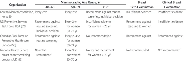
찰, 40세 이상에서 2년마다 유방촬영술과 임상유방진찰을 시 행하도록 권고하였다. 2015년에 발표된 개정 권고안에서는 임 상유방진찰과 유방자가촉진을 단독으로 또는 유방촬영술과 병 행하여 시행하는 것은 유방암 사망률 감소 효과에 대한 근거가 불충분한 것으로 판단하여 이를 권고하거나 반대하지 않는다 (9). Table 4에서 보여지듯이 여러 다른 나라의 가이드 라인에 서도 임상유방진찰과 유방자가촉진을 권고하지 않거나(rec- ommend against), 근거가 불충분하므로 권고하거나 반대하지 않는 입장(insufficient evidence)을 취하고 있다(50, 52, 53).
결론
현재까지는 검진유방촬영술의 이득에 대해서만 강조되어온 측면이 있었으나, 유방촬영술의 득과 실에 대해서 균형 있는 정 보가 대중에게 제공되어야 할 필요가 있다. 검진유방촬영술은 유방암 사망률을 감소시키는 효과가 있지만 그 효과가 크지는 않고 최근에 시행된 무작위 대조군 연구 결과가 부족하다. 한 편 검진으로 인하여 과진단과 과치료, 위양성과 위음성 진단, 방사선 피폭으로 인한 유방암 등의 위해가 발생할 수 있다. 현 Table 4. Comparison of Recommendations for Breast Cancer Screening
Organization Mammography, Age Range, Yr Breast
Self-Examination
Clinical Breast Examination
40–49 50–69 ≥ 70
Korean Medical Association, Korea (9)
Every 2 yr Every 2 yr Recommend against routine screening. Individual decision
Insufficient evidence Insufficient evidence
US Preventive Services Task Force, USA (52)
Recommend against routine screening.
Individual decision
Every 2 yr for women 50–74 yr
Insufficient evidence for women > 75 yr
Recommend against teaching to women
Insufficient evidence
Canadian Task Force on Preventive Health care, Canada (50)
Recommend against routine screening
Every 2–3 yr for women 50–74 yr
No recommendation Recommend against Recommend against
National Health Service breast cancer screening program, UK (53)
No active recruitment*
Every 3 yr for women 50–70 yr
No routine recruitment for women > 70 yr*
Not recommended Not recommended
*Program is expanding to extend screening mammography every 3 years to women aged 47–73 yr.
Table 5. Meta-Analysis Results of the Benefits in Reduction of Breast Cancer Mortality According to the Age
Relative Risk (95% Confidence Interval) Grade Quality of Evidence Breast cancer mortality for ages 40–49 yr
The Canadian Task Force Review (50) 0.85 (0.75–0.96) Moderate
Cochrane review (4)* 0.84 (0.73–0.96) Moderate
Breast cancer mortality for ages 50–69 yr
The Canadian Task Force Review (50) 0.79 (0.68–0.90) Moderate
Cochrane review (4)* 0.77 (0.69–0.86) Moderate
Breast cancer mortality for ages at least 70 yr
The Canadian Task Force Review (50) 0.68 (0.45–1.01) Low
*In Cochrane review (4), meta-analysis was not performed for women aged at least 70 yr.
재 유방암 검진 권고안에서는 40~69세의 여성에서 2년마다 검 진유방촬영술을 시행하는 것은 유방암 사망률 감소효과가 있 고, 위해보다 이득이 크다고 판단되므로 40~69세의 무증상 여 성에서 검진유방촬영술을 2년 주기로 시행하는 것을 권고한다.
한국에서 시행된 무작위 대조군 연구 결과는 없고, 한국 여성에 서 유방암 발생률의 시기 및 치밀 유방의 비율 등은 서구와는 다 른 양상을 보인다. 따라서 국가유방암검진사업을 효과적으로 시행하기 위해서는 한국 여성을 대상으로 한 유방암 검진의 효 과와 검진의 질 향상을 위한 연구가 필요하겠다.
REFERENCES
1. Jung KW, Won YJ, Kong HJ, Oh CM, Cho H, Lee DH, et al.
Cancer statistics in Korea: incidence, mortality, survival, and prevalence in 2012. Cancer Res Treat 2015;47:127-141 2. Kim SH, Kim MH, Oh KK. Analysis and comparison of breast
density according to age on mammogram between Korean and Western women. J Korean Radiol Soc 2000;42:1009- 1014
3. Shin HJ, Ko ES, Yi A. Breast cancer screening in Korean woman with dense breast tissue. J Korean Soc Radiol 2015;
73:279-286
4. Gøtzsche PC, Jørgensen KJ. Screening for breast cancer with mammography. Cochrane Database Syst Rev 2013;6:
CD001877
5. Miller AB, Wall C, Baines CJ, Sun P, To T, Narod SA. Twenty five year follow-up for breast cancer incidence and mor- tality of the Canadian National Breast Screening Study:
randomised screening trial. BMJ 2014;348:g366
6. Welch HG, Passow HJ. Quantifying the benefits and harms of screening mammography. JAMA Intern Med 2014;174:
448-454
7. Løberg M, Lousdal ML, Bretthauer M, Kalager M. Benefits and harms of mammography screening. Breast Cancer Res 2015;17:63
8. Freer PE, Slanetz PJ, Haas JS, Tung NM, Hughes KS, Arm- strong K, et al. Breast cancer screening in the era of den- sity notification legislation: summary of 2014 Massachu- setts experience and suggestion of an evidence-based management algorithm by multi-disciplinary expert panel.
Breast Cancer Res Treat 2015;153:455-464
9. Lee EH, Park B, Kim NS, Seo HJ, Ko KL, Min JW, et al. The
Korean guideline for breast cancer screening. J Korean Med
Assoc 2015;58:408-419
10. Alexander FE, Anderson TJ, Brown HK, Forrest AP, Hepburn W, Kirkpatrick AE, et al. 14 years of follow-up from the Ed- inburgh randomised trial of breast-cancer screening. Lancet 1999;353:1903-1908
11. Habbema JD, van Oortmarssen GJ, van Putten DJ, Lubbe JT, van der Maas PJ. Age-specific reduction in breast cancer mortality by screening: an analysis of the results of the Health Insurance Plan of Greater New York study. J Natl
Cancer Inst 1986;77:317-320
12. Tabár L, Vitak B, Chen TH, Yen AM, Cohen A, Tot T, et al.
Swedish two-county trial: impact of mammographic screen- ing on breast cancer mortality during 3 decades. Radiology 2011;260:658-663
13. Andersson I, Aspegren K, Janzon L, Landberg T, Lindholm K, Linell F, et al. Mammographic screening and mortality from breast cancer: the Malmö mammographic screening trial.
BMJ 1988;297:943-948
14. Moss SM, Cuckle H, Evans A, Johns L, Waller M, Bobrow L;
Trial Management Group. Effect of mammographic screen- ing from age 40 years on breast cancer mortality at 10 years’ follow-up: a randomised controlled trial. Lancet 2006;
368:2053-2060
15. Bjurstam N, Björneld L, Warwick J, Sala E, Duffy SW, Nys- tröm L, et al. The Gothenburg Breast Screening Trial. Cancer 2003;97:2387-2396
16. Frisell J, Lidbrink E, Hellström L, Rutqvist LE. Followup after 11 years--update of mortality results in the Stockholm mam- mographic screening trial. Breast Cancer Res Treat 1997;
45:263-270
17. Miller AB, To T, Baines CJ, Wall C. The Canadian National Breast Screening Study-1: breast cancer mortality after 11 to 16 years of follow-up. A randomized screening trial of mammography in women age 40 to 49 years. Ann Intern
Med 2002;137(5 Part 1):305-312
18. Elmore JG, Harris RP. The harms and benefits of modern screening mammography. BMJ 2014;348:g3824
19. Jørgensen KJ, Gøtzsche PC. Overdiagnosis in publicly or- ganised mammography screening programmes: systematic review of incidence trends. BMJ 2009;339:b2587
20. Feig SA. Ductal carcinoma in situ. Implications for screening
mammography. Radiol Clin North Am 2000;38:653-668, vii 21. Feig SA. Screening mammography benefit controversies:
sorting the evidence. Radiol Clin North Am 2014;52:455- 480
22. Salz T, Richman AR, Brewer NT. Meta-analyses of the ef- fect of false-positive mammograms on generic and specific psychosocial outcomes. Psychooncology 2010;19:1026- 1034
23. Brodersen J, Siersma VD. Long-term psychosocial conse- quences of false-positive screening mammography. Ann
Fam Med 2013;11:106-115
24. Rosenberg RD, Hunt WC, Williamson MR, Gilliland FD, Wiest PW, Kelsey CA, et al. Effects of age, breast density, ethnicity, and estrogen replacement therapy on screening mammo- graphic sensitivity and cancer stage at diagnosis: review of 183,134 screening mammograms in Albuquerque, New Mexico. Radiology 1998;209:511-518
25. Kerlikowske K, Grady D, Barclay J, Sickles EA, Ernster V.
Likelihood ratios for modern screening mammography. Risk of breast cancer based on age and mammographic inter- pretation. JAMA 1996;276:39-43
26. Porter PL, El-Bastawissi AY, Mandelson MT, Lin MG, Khalid N, Watney EA, et al. Breast tumor characteristics as pre- dictors of mammographic detection: comparison of inter- val- and screen-detected cancers. J Natl Cancer Inst 1999;
91:2020-2028
27. Hakama M, Holli K, Isola J, Kallioniemi OP, Kärkkäinen A, Visakorpi T, et al. Aggressiveness of screen-detected breast cancers. Lancet 1995;345:221-224
28. Beemsterboer PM, Warmerdam PG, Boer R, de Koning HJ.
Radiation risk of mammography related to benefit in screen- ing programmes: a favourable balance? J Med Screen 1998;
5:81-87
29. Bijwaard H, Brenner A, Dekkers F, van Dillen T, Land CE, Boice JD Jr. Breast cancer risk from different mammography screening practices. Radiat Res 2010;174:367-376
30. Kopans D. Mammography and radiation risk. In Janower ML, Linton OW. Radiation risk: a primer. Reston, VA: Ameri- can College of Radiology, 1996:21-22
31. Swift M, Morrell D, Massey RB, Chase CL. Incidence of can- cer in 161 families affected by ataxia-telangiectasia. N Engl
J Med 1991;325:1831-1836
32. Brem RF, Lenihan MJ, Lieberman J, Torrente J. Screening breast ultrasound: past, present, and future. AJR Am J Roent-
genol 2015;204:234-240
33. Kaplan SS. Clinical utility of bilateral whole-breast US in the evaluation of women with dense breast tissue. Radiolo-
gy 2001;221:641-649
34. Crystal P, Strano SD, Shcharynski S, Koretz MJ. Using so- nography to screen women with mammographically dense breasts. AJR Am J Roentgenol 2003;181:177-182
35. De Felice C, Savelli S, Angeletti M, Ballesio L, Manganaro L, Meggiorini ML, et al. Diagnostic utility of combined ultra- sonography and mammography in the evaluation of wom- en with mammographically dense breasts. J Ultrasound 2007;10:143-151
36. Brancato B, Bonardi R, Catarzi S, Iacconi C, Risso G, Taschini R, et al. Negligible advantages and excess costs of routine addition of breast ultrasonography to mammography in dense breasts. Tumori 2007;93:562-566
37. Corsetti V, Houssami N, Ferrari A, Ghirardi M, Bellarosa S, Angelini O, et al. Breast screening with ultrasound in wom- en with mammography-negative dense breasts: evidence on incremental cancer detection and false positives, and associated cost. Eur J Cancer 2008;44:539-544
38. Berg WA, Blume JD, Cormack JB, Mendelson EB, Lehrer D, Böhm-Vélez M, et al. Combined screening with ultrasound and mammography vs mammography alone in women at elevated risk of breast cancer. JAMA 2008;299:2151-2163 39. Leong LC, Gogna A, Pant R, Ng FC, Sim LS. Supplementary
breast ultrasound screening in Asian women with negative but dense mammograms-a pilot study. Ann Acad Med Sin-
gapore 2012;41:432-439
40. Hooley RJ, Greenberg KL, Stackhouse RM, Geisel JL, Butler RS, Philpotts LE. Screening US in patients with mammo- graphically dense breasts: initial experience with Connect- icut Public Act 09-41. Radiology 2012;265:59-69
41. Weigert J, Steenbergen S. The connecticut experiment: the role of ultrasound in the screening of women with dense breasts. Breast J 2012;18:517-522
42. Parris T, Wakefield D, Frimmer H. Real world performance of screening breast ultrasound following enactment of Con- necticut Bill 458. Breast J 2013;19:64-70
43. Girardi V, Tonegutti M, Ciatto S, Bonetti F. Breast ultrasound
in 22,131 asymptomatic women with negative mammogra- phy. Breast 2013;22:806-809
44. Chang JM, Koo HR, Moon WK. Radiologist-performed hand-held ultrasound screening at average risk of breast cancer: results from a single health screening center. Acta
Radiol 2015;56:652-658
45. Moon HJ, Jung I, Park SJ, Kim MJ, Youk JH, Kim EK. Com- parison of cancer yields and diagnostic performance of screening mammography vs. supplemental screening ul- trasound in 4394 women with average risk for breast can- cer. Ultraschall Med 2015;36:255-263
46. Ohuchi N, Suzuki A, Sobue T, Kawai M, Yamamoto S, Zheng YF, et al. Sensitivity and specificity of mammography and adjunctive ultrasonography to screen for breast cancer in the Japan Strategic Anti-cancer Randomized Trial (J-START):
a randomised controlled trial. Lancet 2015 Nov 4 [Epub].
http://dx.doi.org/10.1016/S0140-6736(15)00774-6 47. Scheel JR, Lee JM, Sprague BL, Lee CI, Lehman CD. Screen-
ing ultrasound as an adjunct to mammography in women with mammographically dense breasts. Am J Obstet Gyne-
col 2015;212:9-17
48. Berg WA, Zhang Z, Lehrer D, Jong RA, Pisano ED, Barr RG,
et al. Detection of breast cancer with addition of annual screening ultrasound or a single screening MRI to mam- mography in women with elevated breast cancer risk. JAMA 2012;307:1394-1404
49. Barr RG, Zhang Z, Cormack JB, Mendelson EB, Berg WA.
Probably benign lesions at screening breast US in a popula- tion with elevated risk: prevalence and rate of malignancy in the ACRIN 6666 trial. Radiology 2013;269:701-712 50. Canadian Task Force on Preventive Health Care, Tonelli M,
Connor Gorber S, Joffres M, Dickinson J, Singh H, et al. Rec- ommendations on screening for breast cancer in average- risk women aged 40-74 years. CMAJ 2011;183:1991-2001 51. Cho B. Evaluation of the validity of current national health
screening program and plan to improve the system. Cheon-
gju: Korea Centers for Disease Control and Prevention, 2013 52. US Preventive Services Task Force. Screening for breastcancer: U.S. Preventive Services Task Force recommendation statement. Ann Intern Med 2009;151:716-726, W-236 53. GOV.UK. National Health Service. Available from: http://
www.cancerscreening.nhs.uk/breastscreen/. Acccessed 2015 October 15
유방암 검진의 이득과 위해: 개정된 한국 유방암 검진 가이드라인을 중심으로
김수연 · 김은경*
유방암은 한국 여성에서 두 번째로 많은 암으로, 1999년부터 유방촬영술을 이용한 유방암 검진이 시행된 이래 매년 발생 률이 증가하였다. 현재까지는 검진유방촬영술의 이득에 대해서만 강조되어온 측면이 있었으나, 유방촬영술의 이득과 위해 에 대해서 균형 있는 정보를 제공하여 정보에 근거한 의사결정을 내릴 수 있도록 해야 한다. 검진유방촬영술은 유방암 사 망률을 감소시키는 효과가 있지만 과진단과 과치료, 위양성과 위음성 진단, 방사선 피폭으로 인한 유방암 등의 위해가 발 생할 수 있다. 대한의사협회에서 2015년에 발표한 유방암 검진 권고안 개정안에서는 40~69세의 무증상 여성에서 검진유 방촬영술을 2년 주기로 시행하는 것을 권고한다. 이 리뷰에서는 무작위 대조군 연구 결과와 메타분석, 가이드 라인 등 근 거 중심의 정보를 토대로 검진유방촬영술의 이득과 위해에 대해서 중점적으로 다루고자 한다.
연세대학교 의과대학 세브란스병원 영상의학교실, 방사선의과학연구소