Histomorphometric study of rabbit’s maxillary sinus augmentation with various graft materials
전체 글
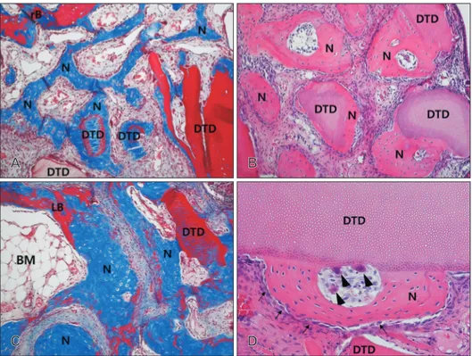
수치

관련 문서
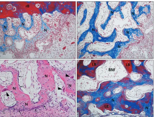
Histopathologic findings of control group at 4 weeks show little bone- implant contact (BIC) around the implant (asterisks) and new-bone formation in the defect
From the results of this study, we concluded that two different sized graft materials have positive effects on new bone formation.. Additionally, smaller
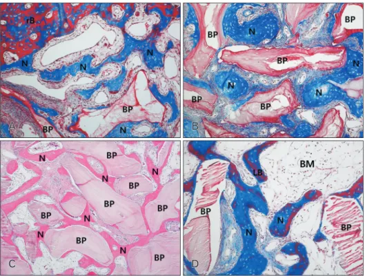
The grafted particulated tooth driven allografts were partially surrounded with new bone matrix and fairly supported graft associated new bone formation with some
Alveolar ridge augmentation with titanium mesh and a combination of autogenous bone and anorganic bovine bone: a 2-year prospective study.. Corinaldesi G, Pieri F, Sapigni
The aim of this study was to compare the effect on bone regeneration relative to maintenance period of PTFE membrane in rabbit calcarial defects.. Eight adult
The OSFE (osteotome sinus floor elevation) technique has been used for maxillary sinus augmentation.. The implants were clinically and radiographically followed
success rates of dental implants placed at the time of or after alveolar ridge augmentation with an autogenous mandibular bone graft and titanium mesh: a 3-to
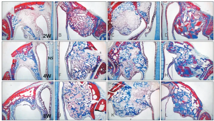
In 4-week group, the group filled with bone graft with decortication revealed larger new bone formation area than shown in the group that had a defect area
