JAK2 V617F, MPL, and CALR Mutations in Korean Patients with Essential Thrombocythemia and Primary Myelofibrosis
Mutations in the calreticulin gene, CALR, have recently been discovered in subsets of patients with essential thrombocythemia (ET) or primary myelofibrosis (PMF). We investigated Korean patients with ET and PMF to determine the prevalence, and clinical and laboratory correlations of CALR/JAK2/MPL mutations. Among 84 ET patients, CALR mutations were detected in 23 (27.4%) and were associated with higher platelet counts (P = 0.006) and lower leukocyte counts (P = 0.035) than the JAK2 V617F mutation.
Among 50 PMF patients, CALR mutations were detected in 11 (22.0%) and were also associated with higher platelet counts (P = 0.035) and trended to a lower rate of cytogenetic abnormalities (P = 0.059) than the JAK2 V617F mutation. By multivariate analysis, triple-negative status was associated with shorter overall survival (HR, 7.0; 95%
CI, 1.6-31.1, P = 0.01) and leukemia-free survival (HR, 6.3; 95% CI, 1.8-22.0, P = 0.004) in patients with PMF. The type 1 mutation was the most common (61.1%) type among all patients with CALR mutations, and tended toward statistical predominance in PMF patients. All 3 mutations were mutually exclusive and were never detected in patients with other myeloid neoplasms showing thrombocytosis. CALR mutations characterize a distinct group of Korean ET and PMF patients. Triple-negative PMF patients in particular have an unfavorable prognosis, which supports the idea that triple-negative PMF is a molecularly high-risk disease.
Keywords: CALR; JAK2 V617F; MPL; Thrombocythemia, Essential; Primary Myelofibrosis Bo Hyun Kim,1 Young-Uk Cho,1
Mi-Hyun Bae,1 Seongsoo Jang,1 Eul-Ju Seo,1 Hyun-Sook Chi,1 Yunsuk Choi,2 Dae-Young Kim,2 Jung-Hee Lee,2 Je-Hwan Lee,2 Kyoo-Hyung Lee,2 Young-Mi Park,3 Jong-Keuk Lee,3 and Chan-Jeoung Park1
1Department of Laboratory Medicine and
2Department of Internal Medicine, University of Ulsan, College of Medicine and Asan Medical Center, Seoul; 3Asan Institute for Life Sciences, University of Ulsan College of Medicine, Seoul, Korea
Received: 13 December 2014 Accepted: 19 March 2015 Address for Correspondence:
Young-Uk Cho, MD
Department of Laboratory Medicine, University of Ulsan, College of Medicine and Asan Medical Center, 88 Olympic-ro 43-gil, Songpa-gu, Seoul 138-736, Korea Tel: +82.2-3010-4501, Fax: +82.2-478-0884 E-mail: yucho@amc.seoul.kr
http://dx.doi.org/10.3346/jkms.2015.30.7.882 • J Korean Med Sci 2015; 30: 882-888
INTRODUCTION
Although the JAK2 V617F gain-of-function mutation has a piv- otal role in the diagnosis of Philadelphia chromosome-negative myeloproliferative neoplasms (Ph-MPN), it only occurs in 50%- 60% in patients with essential thrombocythemia (ET) and pri- mary myelofibrosis (PMF). Gain-of-function mutation in MPL, which is another molecular marker for ET and PMF, has been reported in 5%-10% of patients with these diseases with nonmu- tated JAK2 (1). Therefore, the diagnosis of other patients with findings suggestive of ET or PMF but negative for JAK2 and MPL mutations, must be based on the clinical exclusion of reactive thrombocytosis and histopathological examinations that focus on morphologic changes in megakaryocytes or bone marrow fibrosis. Histopathological evaluation is inherently susceptible to interobserver variation, requires an experienced hematopa- thologist, and lacks standardization. Therefore, it is very inter- esting that 60% to 88% of ET and PMF patients negative for JAK2 V617F and MPL mutations have been found to harbor novel mu- tations of the calreticulin gene, CALR (1-8).
Calreticulin is a functionally complex Ca2+-binding protein that is localized primarily to the endoplasmic reticulum (9-11).
All CALR mutations reported so far are located in exon 9 and
are somatic insertions or deletions. There are two major vari- ants: type 1 (L367fs*46), resulting from a 52-bp deletion, and type 2 (K385fs*47), from a 5-bp (TTGTC) insertion (1-8). In par- ticular, the clinical course of PMF patients with CALR altera- tions has been found to be more indolent than the courses of patients with the JAK2 V617F mutation and patients who are triple-negative (5, 6).
The aim of this study was to determine the prevalence, bio- logical characteristics, and clinical correlations of these novel CALR mutations, in addition to the well-established JAK2 V617F and MPL mutations, in a single-center cohort of patients with ET and PMF.
MATERIALS AND METHODS Patients
The study included 150 patients seen at the Asan Medical Cen- ter, Seoul, Korea from January 2003 to April 2013. There were 84 patients with ET, 50 patients with PMF, 7 patients with post-ET or post-polycythemia vera (PV) myelofibrosis, and 9 patients with other myeloid neoplasms with thrombocytosis, as follows:
4 refractory anemia with ring sideroblasts associated with mark- ed thrombocytois, 1 refractory anemia with multilineage dys-
plasia, 1 myelodysplastic syndrome with isolated del(5q), 1 chro- nic myeloid leukemia, 1 myelodysplastic/myeloproliferative neo plasm, unclassifiable, and 1 acute myeloid leukemia. It should be stressed that the study patients, who were selected, were pri- marily those with available archived samples. Furthermore, pa- tients with available data for JAK2 and MPL mutations were pre- ferentially analyzed.
The median age of study patients was 60 yr (range, 19-90 yr);
and 74 (49.3%) patients were male. ET, PMF, and other myeloid malignances were diagnosed based on 2008 World Health Or- ganization (WHO) criteria. Post-ET or -PV myelofibrosis and leukemic transformation were also diagnosed according to 2008 WHO criteria. Major thromboembolic events at presentation or in the 2 preceding years or anytime during follow-up were re- corded if definitely documented. Bone marrow aspirations and biopsies were independently reviewed by 2 hematopathologists who were blinded to the patient’s mutation profile. Discrepant cases were resolved by consensus between the 2 hematopathol- ogists. The clinical and laboratory parameters and cytogenetic findings at the time of initial presentation were reviewed. Karyo- types were designated as unfavorable based on the Dynamic International Prognostic Scoring System (DIPSS) risk categori- zation, as described previously; and included a complex karyo- type (the presence of 3 or more distinct numeric or structural cytogenetic abnormalities) or 1 or 2 abnormalities that includ- ed +8, -7/7q-, i(17q), inv(3), -5/5q-, 12p- or 11q23 rearrange- ment (12).
Mutation and cytogenetic analysis
Study samples were either stored DNA extracted from periph- eral blood or bone marrow of patients at initial presentation, or genomic DNA extracted from archived bone marrow smears according to a standard protocol and our laboratory’s internal guidelines. The JAK2 V617F mutation was assessed using a poly- merase chain reaction (PCR)-based amplification refractory mutation system, as previously described (13). The MPL W515L/
K mutations were assessed by real-time PCR (Real-Q MPL W515L/
K Screening Kit, BioSewoom Inc., Seoul, Korea) according to the manufacturer’s instructions. All positive samples by real-time PCR were subsequently analyzed by Sanger sequencing. Exon 10 of MPL was amplified using the following primers: F, 5´-TTC TGTACATGAGCATTTCATCA-3´and R, 5´-GACAGGCTGTGT- GTGTGTACCTCT-3´. The CALR mutations were assessed by PCR followed by Sanger sequencing. Exon 9 of CALR was am- plified using the following primers: F, 5´-GAGGAGTTTGGCAA CGAGAC-3´and R, 5´-AACCAAAATCCACCCCAAAT-3´. The PCR conditions consisted of initial denaturation step at 94°C for 5 min; followed by 35 cycles of 94°C for 45 sec, 57°C for 30 sec, and 72°C for 1 min; and a final extension step at 72°C for 10 min.
Purified PCR fragments were sequenced. Cytogenetic analysis was performed using conventional G-banding techniques.
Statistical analysis
Differences in the distributions of continuous variables between categories were analyzed using either the Wilcoxon rank-sum test or the Kruskal-Wallis test. Patient groups with nominal val- ues were compared using the chi-square test or Fisher exact test.
Overall survival (OS) was calculated from the date of diagnosis of ET or PMF to date of death (uncensored) or last contact (cen- sored). Date of leukemic transformation replaced date of death (uncensored) to evaluate leukemia-free survival (LFS). OS and LFS were plotted using Kaplan-Meier curves and compared by a log-rank test. The Cox proportional hazard regression model was used for multivariate analysis. P values < 0.05 were consid- ered to indicate statistically significant differences. Statistical analyses were performed using MedCalc program version 12.4.0.0 (MedCalc Software, Acacialaan, Belgium).
Ethics statement
This study was approved by the institutional review board of the Asan Medical Center (2014-0974). The board waived informed consent from participants. The study was conducted in accor- dance with the Declaration of Helsinki.
RESULTS
Frequencies and distribution of mutations
CALR mutations were detected in 36 patients (25.5%) of pati- ents with ET or PMF or post-ET or PV myelofibrosis. Among these patients, type 1 (L367fs*46), type 2 (K385fs*47) and other types of mutations were detected in 22 (61.1%), 10 (27.8%), and 4 (11.1%) patients, respectively. The type 1 mutation was found more frequently in patients with PMF than the type 2 mutation, with marginal statistical significance (P = 0.076, Table 1).
Of the 84 patients with ET, 43 (51.2%) harbored the JAK2 V617F mutation, 23 (27.4%) had CALR mutations, and 1 (1.2%) had MPL W515 mutation. Seventeen (20.2%) patients were negative for all 3 mutations (Fig. 1A). ET patients with CALR mutations accounted for 57.5% of patients that had nonmutated JAK2 and MPL. Of the ET patients with CALR mutations, 11 (47.8%) had type 1 mutations, 9 (39.1%) had type 2 mutations, and 3 (13.0%) had other types of mutations (Table 1).
Of the 50 patients with PMF, 27 (54.0%) harbored the JAK2 Table 1. Frequency of the CALR mutation subtypes detected in this study of Korean patients with essential thrombocythemia and primary myelofibrosis
Type ET (n = 23) PMF (n = 11) Post-ET or PV MF (n = 2)
Type 1 11 (47.8%) 9 (81.8%) 2 (100.0%)*
Type 2 9 (39.1%) 1 (9.1%) 0
Others† 3 (13.0%) 1 (9.1%) 0
*These patients had post-ET myelofibrosis, not post-PV myelofibrosis; †Other types included c.1091_1124del (p.E364fs*55), c.1101_1105delAGG (p.K368del), c.1113_
1143del (p.E372fs*48), and c.1115_1145del (p.D373fs*47). ET, essential thrombo- cythemia; PMF, primary myelofibrosis; PV, polycythemia vera; MF, myelofibrosis.
V617F mutation, 11 (22.0%) had CALR mutations, and 2 (4.0%) harbored the MPL W515 mutations. Ten (20.0%) patients were negative for all 3 mutations (Fig. 1B). PMF patients with CALR mutations accounted for 52.4% of the patients with nonmutated JAK2 and MPL. Of the patients with CALR mutations, 9 (81.8%) patients had type 1 mutations, 1 (9.1%) patient had a type 2 mu- tation, and 1 (9.1%) patient had another type of mutation (Table 1).
Of the 7 patients with post-ET or PV myelofibrosis, 5 (71.4%) harbored the JAK2 V617F mutation and the remaining 2 (28.6%) had CALR mutations. The 2 CALR mutations were only found
in patients with post-ET myelofibrosis (Table 1), whereas the 2 patients with post-PV myelofibrosis who were included in this study had the JAK2 V617F mutation. These 3 mutations (CALR/
JAK2/MPL) were mutually exclusive and were never detected in patients with other myeloid neoplasms with thrombocytosis.
Clinical, hematologic and molecular correlates
Tables 2 and 3 summarize the presenting clinical characteristics of study patients stratified by mutational status as follows: mu- tated CALR, JAK2, MPL and triple-negative. Univariate analysis Table 2. Clinical and laboratory features of Korean patients with essential thrombocythemia, stratified by the presence or absence of JAK2 V617F, MPL, and CALR mutations*
Variables CALR mutations
(n = 23) (A) JAK2 V617F mutation
(n = 43) (B) Triple-negative (n = 17) (C)
P value
(A) vs. (B) (A) vs. (C) (B) vs. (C)
Males (%) 11 (47.8) 20 (46.5) 5 (29.4) 0.562 0.332 0.260
Age in years, median (range) 55 (19-84) 59 (26-90) 46 (22-73) 0.531 0.428 0.135
Leukocyte, × 109/L, median (range) 8.4 (5.3-17.8) 9.5 (3.0-40.9) 9.9 (4.3-29.4) 0.035 0.039 0.611
Hemoglobin, g/dL, median (range) 13.4 (7.0-16.3) 14.5 (7.5-18.4) 12.4 (8.1-16.4) 0.105 0.404 0.021
Platelet, × 109/L, median (range) 931 (546-1,516) 651 (453-2,435) 761 (485-2,271) 0.006 0.299 0.536
LD, IU/L, median (ragne) 281 (189-441) 240 (98-590) 196.5 (103-762) 0.072 0.004 0.025
Cytogenetic abnormality (%) 4/14 (28.6) 1/37 (2.7) 3/13 (23.1) 0.017 1.000 0.049
Thromboembolic events (%) 1 (4.3) 7 (16.3) 1 (5.9) 0.154 1.000 0.420
P values with statistically significance are shown in bold type. *A single patient harboring MPL W515 mutation was excluded from the analysis. LD, lactate dehydrogenase.
Table 3. Clinical and laboratory features of Korean patients with primary myelofibrosis, stratified by the presence or absence of JAK2 V617F, MPL, and CALR mutations*
Variables CALR mutations
(n = 11) (A) JAK2 V617F mutation
(n = 27) (B) Triple-negative (n = 10) (C)
P value
(A) vs. (B) (A) vs. (C) (B) vs. (C)
Males (%) 5 (45.4) 16 (59.3) 8 (80.0) 0.491 0.183 0.439
Age in years, median (range) 65 (49-77) 64 (43-78) 57 (32-80) 0.936 0.181 0.137
Leukocyte, × 109/L, median (range) 7.2 (2.3-81.6) 12.4 (1.1-100.2) 3.4 (0.7-559.7) 0.394 0.218 0.101
Hemoglobin, g/dL, median (range) 9.5 (6.4-13.8) 11.2 (7.8-15.3) 8.1 (4.7-10.9) 0.171 0.038 0.0001
Platelet, × 109/L, median (range) 410 (47-1351) 216 (7-963) 90 (22-618) 0.035 0.020 0.383
Circulating blast, %, median (range) 0 (0-8) 1 (0-8) 0 (0-6) 0.463 0.876 0.639
LD, IU/L, median (range) 563 (256-916) 523 (231-2,000) 313 (191-1,976) 0.666 0.057 0.037
Cytogenetic abnormality (%) 2/9 (22.2) 16/26 (61.5) 6/9 (66.7) 0.059 0.153 1.000
Unfavorable karyotype (%) 2/9 (22.2) 7/26 (26.9) 4/9 (44.4) 1.000 0.620 0.416
DIPSS score, Int-2 or High (%) 5 (45.5) 8 (29.6) 6 (60.0) 0.351 0.670 0.132
Leukemic transformation (%) 1 (9.1) 3 (11.1) 3 (30.0) 0.667 0.311 0.313
P values with statistically significance are shown in bold type. *Two patients harboring MPL W515 mutation was excluded from the analysis. LD, lactate dehydrogenase; DIPSS, Dynamic International Prognostic Scoring System; Int, intermediate.
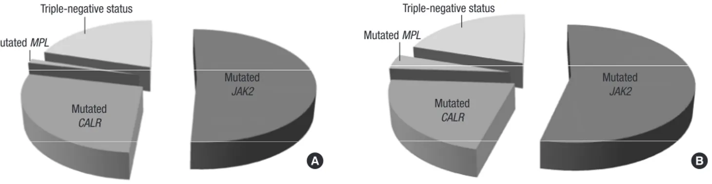
Fig. 1. Distribution of JAK2 V617F, MPL, and CALR mutations in Korean patients with essential thrombocythemia (ET) and primary myelofibrosis (PMF). (A) In patients with ET, 51.2% harbored JAK2 V617F and 27.4% had mutations in CALR, and 1.2% had mutations in MPL, and 20.2% were negative for all 3 mutations (triple-negative). (B) In patients with PMF, 54.0% harbored JAK2 V617F, 22.0% had CALR mutations, 4.0% had a mutation in MPL, and 20.0% were triple-negative.
Triple-negative status Triple-negative status
Mutated CALR
Mutated CALR Mutated
JAK2
Mutated JAK2
Mutated MPL Mutated MPL
A B
of patients with ET found that CALR mutations were associated with a higher platelet count (P = 0.006), lower leukocyte count (P = 0.035) and higher frequency of cytogenetic abnormalities (P = 0.017), compared to the JAK2 V617F mutation. CALR mu- tations were associated with a higher lactate dehydrogenase (LD) level (P = 0.004) and lower leukocyte count (P = 0.039) com- pared with triple-negative status. A triple-negative status was associated with a lower hemoglobin level (P = 0.021), lower LD level (P = 0.025) and higher frequency of cytogenetic abnormal- ities (P = 0.049), compared with the JAK2 mutation (Table 2).
Thromboembolic events seemed to be more frequent in pati- ents with the JAK2 V617F mutation compared to patients with CALR mutation or triple-negative status, although statistical sig- nificance was not achieved. Neither post-ET myelofibrosis or nor leukemic transformation was documented during follow-up.
Of patients with PMF, CALR mutations were associated with a higher platelet count (P = 0.035), and there was a trend toward lower frequency of cytogenetic abnormalities (P = 0.053), com- pared to the JAK2 V617F mutation. CALR mutations were also associated with a higher platelet count (P = 0.020) compared with triple-negative status. A triple-negative status was associ- ated with a lower hemoglobin level (P = 0.001) and lower LD level (P = 0.037), compared to the JAK2 V617F mutation. CALR mutations had lower frequency of leukemic transformation com- pared to the JAK2 V617F mutation or triple-negative status, al- though statistical significance was not achieved (Table 3).
Prognostic impacts of mutations
The median follow-ups of the study population were 29.8 months (range, 0.2-120.2 months) for ET patients and 13.7 months (range, 0.2-81.2 months) for PMF patients. Four (4.8%) ET patients died, and 9 (18.0%) PMF patients died. The mortality rates of PMF patients with the JAK2 V617F mutation, CALR mutations, and triple-negative status were 7.4% (2/27), 18.2% (2/11), and 50.0%
(5/10), respectively.
OS and LFS times were analyzed according to the various para- meters, including mutational status. Mutational status, includ- ing CALR and JAK2 V617F mutations, had no significant prog- nostic impact on patients with ET (P = 0.124). However, among PMF patients, the median OS time of those with CALR muta- tions, the JAK2 V617F mutation, and triple-negative status was 15.7, 14.3, and 13.0 months, respectively. Univariate analysis found significant survival differences between patients with JAK2 and CALR mutations and triple-negative patients (P = 0.011 for OS and P = 0.026 for LFS). Triple-negative status was associated with shorter OS than JAK2 V617F mutations (hazards ratio [HR], 7.7; 95% confidence interval [CI], 1.4-42.9) and CALR mutations (HR, 3.6; 95% CI, 0.8-16.0; Fig. 2A). Triple-negative status was also associated with shorter LFS than JAK2 V617F mutations (HR, 4.3; 95% CI, 1.0-17.7) and CALR mutations (HR, 2.9; 95%
CI, 0.8-10.8; Fig. 2B). Among the other clinical parameters, uni- variate analysis found that an intermediate-2 or high DIPSS score was associated with significantly shorter OS (HR, 10.8; 95% CI, 2.4-49.5; P=0.0001) and LFS (HR, 6.5; 95% CI, 1.8-23.1; P=0.0001) than an intermediate-1 or low score. An unfavorable karyotype was associated with shorter LFS (HR, 4.0; 95% CI, 1.1-14.7; P = 0.005), but not shorter OS.
By multivariate analysis, triple-negative status (HR, 7.0; 95%
CI, 1.6-31.1, P = 0.01) and intermediate-2 or high DIPSS score (HR, 22.4; 95% CI, 2.6-196.0, P = 0.005) were associated with shor- ter OS times. Triple-negative status (HR, 6.3; 95% CI, 1.8-22.0, P = 0.004), intermediate-2 or high DIPSS score (HR, 9.7; 95% CI, 2.5-37.3, P = 0.001), and unfavorable karyotypes (HR, 4.5; 95%
CI, 1.4-14.6, P = 0.013) were associated with shorter LFS.
DISCUSSION
The most consistent finding of this study was that among ET and PMF patients, those with CALR mutations had significantly higher platelet counts than those with the JAK2 V617F mutation.
Fig. 2. Kaplan-Meier estimate of overall survival (A) and leukemia-free survival (B) of Korean patients with primary myelofibrosis according to mutational status. Two patients harboring MPL W515 mutations were excluded from the analysis.
P = 0.011
JAK2 V617F CALR
Triple-negative
Survival probability (%)
Time (months)
0 20 40 60 80 100 100
90 80 70 60 50 40 30
P = 0.011
JAK2 V617F CALR
Triple-negative
A
P = 0.026
JAK2 V617F CALR
Triple-negative
Survival probability (%)
Time (months)
0 20 40 60 80 100 100
90 80 70 60 50 40 30 20
P = 0.026
JAK2 V617F CALR
Triple-negative
B
Previous studies revealed that CALR mutations were not found in patients with PV (1, 2), and that CALR mutations were found in few, if any, patients with myeloid neoplasm with thombocy- tosis (1). Both these observations and our results are consistent with a recent study that used measurements of the levels of cal- reticulin RNA and immunostaining by an antibody specific for mutated CALR. It found that calreticulin expression is restricted to megakaryocytes (14). In the endoplasmic reticulum (ER), cal- reticulin has important functions in directing the proper con- formation of proteins and glycoproteins, as well as in homeo- static control of cytosolic and ER calcium levels (9-11). Because transient ER stress activation is thought to trigger the apoptotic- like phase of the thrombopoiesis process (11), the effects of cal- reticulin as a calcium-buffering chaperone of the ER and ulti- mately as a contributor to platelet production might vary, de- pending on the presence or absence of a CALR mutation. The key region during the molecular pathogenesis of Ph-MPN is the acidic C-domain that terminates in a KDEL sequence, and which is involved in the homeostasis of cellular calcium and is lost in mutated CALR. The absence of this sequence in mutated CALR results in altered intracellular localization and impairment of the calcium-binding function (1, 2, 15).
Overall, the other findings of our study also confirm the re- sults of previous studies. Our study found that the frequency of CALR mutations was 27.4% in patients with ET and 22.0% in pa- tients with PMF. The reported frequencies of CALR mutations range from 15.5% to 33% in ET patients and from 22.7% to 25%
in PMF patients (3-8). The lower leukocyte counts in the CALR- mutated ET patients in this study, compared to the JAK2-mu- tated ET patients, has also been reported in previous studies ( 3, 4, 7, 8). Among patients with PMF, the triple-negative patients in our study had lower hemoglobin levels and trended toward higher prevalence of cytogenetic abnormalities and acute leu- kemic transformation than the JAK2- and CALR-mutated PMF patients. Ultimately, the triple-negative PMF patients had the shortest OS and LFS. These findings for triple-negative patients have also been reported by previous studies, and corroborate the adverse effects that triple-negative mutations had on the sur- vival times of patients with PMF in our study (5, 6, 8). It is not surprising, therefore, that Tefferi et al. have emphasized that tri- ple-negative PMF should be thought of a molecularly high-risk disease (5).
Molecular variations resulting from genetic alterations may be highly diverse between different ethnicities. Since the stud- ies referred to in our discussion were principally performed in Western countries, a review of studies on Asian populations is needed. We found 4 very recent reports in the PubMed database on CALR mutations in studies of Chinese populations (16-19).
On the whole, the results of these Chinese studies are in line with the results of the Western studies, regarding the frequencies of mutations and associated clinical characteristics and prognos-
tic impacts. Of note, the results of a single study on Chinese PMF patients reported by Li et al. indicate that CALR mutations may have different effects in different populations (19). First, their study found that 43.8% of patients with no detectable mutations in JAK2 or MPL had CALR mutations. This frequency is substan- tially lower than the frequencies for PMF patients of predomi- nantly European descent, which range from 69.4% to 74.2% (5, 6, 8), but is similar to the frequency in our study (52.4% of JAK2- or MPL-unmutated PMF patients). The reasons accounting for the discrepant findings include the different characteristics of the study patients, different sensitivities of the methods used, and ethnicity-based differences in genetic profiles. The last rea- son may be a reasonable explanation, because of the slightly lower frequencies of JAK2 V617F and MPL mutations in Asian patients compared to Western patients. The frequencies of the JAK2 V617F mutation in a Chinese study (19) and our study were 50% and 54%, respectively, versus 58% and 64.7% in Western studies (5, 6). The frequency of MPL mutations in a Chinese study (19) and our study was 3% and 4%, respectively, versus 8.3% and 7% in Western studies (5, 8). Second, the ratio between type 1 and type 2 CALR mutations was reversed in the Chinese PMF patients (32% were type 1 and 64% were type 2 mutations) com- pared with PMF patients of predominantly European descent (19). A large study of Italian PMF patients found frequencies of type 1 and type 2 mutations of 72% and 16%, respectively (6).
Our results also found a predominance of type 1 mutation in PMF patients compared with ET patients. Therefore, the pre- dominance of the CALR type 2 mutation in Chinese PMF pa- tients should be confirmed in a larger Asian cohort.
The identification of disease-specific mutations justifies, in certain cases, the use of mutation analysis for determining the diagnosis, prognosis and assessment of response to treatment.
CALR mutations are frequent in JAK2/MPL-unmutated ET/PMF and therefore are important clonal markers for such cases. There- fore, Tefferi et al. have urged that these mutations should be in- cluded as clonal markers, along with JAK2 and MPL mutations, in the list of major diagnostic criteria for ET and PMF in the up- coming WHO classification system (20).
The major limitations of this study include the relatively low numbers of patients in our cohort and relatively short follow- up period. This might be major causes for the lack of data dem- onstrating the clinical relevance of CALR mutations, such as longer thromboembolic event-free survival and lower rate of thrombotic events for ET patients with CALR mutations than for patients with JAK2 V617F mutations (3, 4, 8), or better OS for PMF patients with CALR mutations compared to PMF patients with JAK2 V617F mutations (5, 6, 8). In addition, type 1 and type 2 CALR mutations have manifested as distinctly different clini- cal phenotypes. The type 1 mutation was found to cause in vitro cytokine-independent growth as a result of its activation of the signal transducer and activator of transcription 5, while the type
2 mutation was found to be associated with increased platelet counts and probably has an important role in the maturation of megakaryocytes (1, 7). Furthermore, in the previously mention- ed study on Chinese PMF patients, the unfavorable prognostic impact of CALR mutations was limited to patients with type 2 mutations, and type 2 mutations or no detectable mutation was an independent unfavorable prognostic factor by multivariate analysis (19). In our study, we could not detect differences be- tween the 2 types of CALR mutations, mainly because of the relatively small number of study patients.
Although a larger study cohort is definitely needed to validate the results of this study, our results should not be underestimat- ed because, to the best of our knowledge, this is the first study that has investigated the frequency of CALR/JAK2/MPL muta- tions and related biologic and clinical features in Korean Ph-MPN patients. Recently, two Korean studies on CALR mutations were reported. However, the clinical usefulness of these studies seems to be limited, either by lack of prognostic data (21) or exclusion of MPL mutation and small size of each subgroup (22). More- over, CALR mutations will probably be included in the major criteria for ET and PMF of the new WHO classification system (20). We have confirmed that CALR mutations occur in Korean patients with Ph-MPN that lack JAK2 V617F or MPL mutations.
Therefore, diagnosis of ET or PMF can now be facilitated by an- alyzing CALR mutations in Korean patients. In addition to the diagnostic value of mutation profiling, it is also useful for detect- ing high-risk PMF patients who are triple-negative for mutations.
In conclusion, we demonstrated that CALR mutations charac- terize a subset of Korean Ph-MPN patients who have distinct phe- notypes similar to those reported for patients in Western coun- tries. Because molecular markers are still unknown for approxi- mately 50% of JAK2- and MPL-unmutated cases, morphological examination of bone marrow remains the cornerstone for accu- rate diagnosis for a substantial number of Korean patients sus- pected of having Ph-MPN. CALR mutations provide additional information on clinical outcomes, especially for patients with PMF.
DISCLOSURE
The authors have no conflicts of interest to disclose.
AUTHOR CONTRIBUTION
Conceived and designed the study: Cho YU. Prepared the data:
Kim BH, Bae MH. Analyzed the data: Kim BH, Bae MH, Cho YU. Wrote the first draft of the manuscript: Kim BH. Wrote the paper: Kim BH, Cho YU. Performed molecular analysis: Park YM, Lee JK, Cho YU. Contributed to the interpretation of the data and critically reviewed the draft: Jang S, Park CJ, Seo EJ, Chi HS, Choi Y, Kim DY, Lee JH, Lee JH, Lee KH. Manuscript appro- val: all authors.
ORCID
Bo Hyun Kim http://orcid.org/0000-0003-4456-5612 Young-Uk Cho http://orcid.org/0000-0002-4403-8989 Mi-Hyun Bae http://orcid.org/0000-0003-2818-342X Seongsoo Jang http://orcid.org/0000-0002-0045-1747 Eul-Ju Seo http://orcid.org/0000-0002-8247-3746 Hyun-Sook Chi http://orcid.org/0000-0001-9596-3296 Yunsuk Choi http://orcid.org/0000-0002-7983-8089 Dae-Young Kim http://orcid.org/0000-0002-2894-0462 Jung-Hee Lee http://orcid.org/0000-0002-3127-0068 Je-Hwan Lee http://orcid.org/0000-0002-7060-1675 Kyoo-Hyung Lee http://orcid.org/0000-0002-2841-0779 Young-Mi Park http://orcid.org/0000-0003-2843-3505 Jong-Keuk Lee http://orcid.org/0000-0003-1125-4017 Chan-Jeoung Park http://orcid.org/0000-0003-4396-8348 REFERENCES
1. Klampfl T, Gisslinger H, Harutyunyan AS, Nivarthi H, Rumi E, Milose- vic JD, Them NC, Berg T, Gisslinger B, Pietra D, et al. Somatic mutations of calreticulin in myeloproliferative neoplasms. N Engl J Med 2013; 369:
2379-90.
2. Nangalia J, Massie CE, Baxter EJ, Nice FL, Gundem G, Wedge DC, Avezov E, Li J, Kollmann K, Kent DG, et al. Somatic CALR mutations in myelo- proliferative neoplasms with nonmutated JAK2. N Engl J Med 2013; 369:
2391-405.
3. Rumi E, Pietra D, Ferretti V, Klampfl T, Harutyunyan AS, Milosevic JD, Them NC, Berg T, Elena C, Casetti IC, et al. JAK2 or CALR mutation sta- tus defines subtypes of essential thrombocythemia with substantially dif- ferent clinical course and outcomes. Blood 2014; 123: 1544-51.
4. Rotunno G, Mannarelli C, Guglielmelli P, Pacilli A, Pancrazzi A, Pieri L, Fanelli T, Bosi A, Vannucchi AM; Associazione Italiana per la Ricerca sul Cancro Gruppo Italiano Malattie Mieloproliferative Investigators.
Impact of calreticulin mutations on clinical and hematological pheno- type and outcome in essential thrombocythemia. Blood 2014; 123: 1552-5.
5. Tefferi A, Lasho TL, Finke CM, Knudson RA, Ketterling R, Hanson CH, Maffioli M, Caramazza D, Passamonti F, Pardanani A. CALR vs JAK2 vs MPL-mutated or triple-negative myelofibrosis: clinical, cytogenetic and molecular comparisons. Leukemia 2014; 28: 1472-7.
6. Rumi E, Pietra D, Pascutto C, Guglielmelli P, Martínez-Trillos A, Casetti I, Colomer D, Pieri L, Pratcorona M, Rotunno G, et al.; Associazione Itali- ana per la Ricerca sul Cancro Gruppo Italiano Malattie Mieloprolifera- tive Investigators. Clinical effect of driver mutations of JAK2, CALR, or MPL in primary myelofibrosis. Blood 2014; 124: 1062-9.
7. Tefferi A, Wassie EA, Guglielmelli P, Gangat N, Belachew AA, Lasho TL, Finke C, Ketterling RP, Hanson CA, Pardanani A, et al. Type 1 versus Type 2 calreticulin mutations in essential thrombocythemia: a collabor- ative study of 1027 patients. Am J Hematol 2014; 89: E121-4.
8. Andrikovics H, Krahling T, Balassa K, Halm G, Bors A, Koszarska M, Ba- tai A, Dolgos J, Csomor J, Egyed M, et al. Distinct clinical characteristics of myeloproliferative neoplasms with calreticulin mutations. Haemato- logica 2014; 99: 1184-90.
9. Gold LI, Eggleton P, Sweetwyne MT, Van Duyn LB, Greives MR, Naylor SM, Michalak M, Murphy-Ullrich JE. Calreticulin: non-endoplasmic re- ticulum functions in physiology and disease. FASEB J 2010; 24: 665-83.
10. Michalak M, Groenendyk J, Szabo E, Gold LI, Opas M. Calreticulin, a multi-process calcium-buffering chaperone of the endoplasmic reticu- lum. Biochem J 2009; 417: 651-66.
11. Lopez JJ, Palazzo A, Chaabane C, Albarran L, Polidano E, Lebozec K, Dally S, Nurden P, Enouf J, Debili N, et al. Crucial role for endoplasmic reticulum stress during megakaryocyte maturation. Arterioscler Thromb Vasc Biol 2013; 33: 2750-8.
12. Tefferi A. Primary myelofibrosis: 2014 update on diagnosis, risk-stratifi- cation, and management. Am J Hematol 2014; 89: 915-25.
13. Chen Q, Lu P, Jones AV, Cross NC, Silver RT, Wang YL. Amplification re- fractory mutation system, a highly sensitive and simple polymerase chain reaction assay, for the detection of JAK2 V617F mutation in chronic my- eloproliferative disorders. J Mol Diagn 2007; 9: 272-6.
14. Vannucchi AM, Rotunno G, Bartalucci N, Raugei G, Carrai V, Balliu M, Mannarelli C, Pacilli A, Calabresi L, Fjerza R, et al. Calreticulin muta- tion-specific immunostaining in myeloproliferative neoplasms: patho- genetic insight and diagnostic value. Leukemia 2014; 28: 1811-8.
15. Guglielmelli P, Nangalia J, Green AR, Vannucchi AM. CALR mutations in myeloproliferative neoplasms: hidden behind the reticulum. Am J He- matol 2014; 89: 453-6.
16. Wu Z, Zhang X, Xu X, Chen Y, Hu T, Kang Z, Li S, Wang H, Liu W, Ma X,
et al. The mutation profile of JAK2 and CALR in Chinese Han patients with Philadelphia chromosome-negative myeloproliferative neoplasms.
J Hematol Oncol 2014; 7: 48.
17. Chen CC, Gau JP, Chou HJ, You JY, Huang CE, Chen YY, Lung J, Chou YS, Leu YW, Lu CH, et al. Frequencies, clinical characteristics, and out- come of somatic CALR mutations in JAK2-unmutated essential throm- bocythemia. Ann Hematol 2014; 93: 2029-36.
18. Shen H, Chao H, Ding Z, Feng Y, Cen J, Pan J, He J, Zhou M, Chen Z, Chen S. CALR and ASXL1 mutation analysis in 190 patients with essential thrombocythemia. Leuk Lymphoma 2015; 56: 820-2.
19. Li B, Xu J, Wang J, Gale RP, Xu Z, Cui Y, Yang L, Xing R, Ai X, Qin T, et al.
Calreticulin mutations in Chinese with primary myelofibrosis. Haema- tologica 2014; 99: 1697-700.
20. Tefferi A, Thiele J, Vannucchi AM, Barbui T. An overview on CALR and CSF3R mutations and a proposal for revision of WHO diagnostic crite- ria for myeloproliferative neoplasms. Leukemia 2014; 28: 1407-13.
21. Ha JS, Kim YK. Calreticulin exon 9 mutations in myeloproliferative neo- plasms. Ann Lab Med 2015; 35: 22-7.
22. Park SH, Kim SY, Lee SM, Yi J, Kim IS, Kim HH, Chang CL, Lee EY, Song MK, Shin HJ, et al. Incidence, clinical features, and prognostic impact of CALR exon 9 mutations in essential thrombocythemia and primary my- elofibrosis: an experience of a single tertiary hospital in Korea. Ann Lab Med 2015; 35: 233-7.