www.krspine.org
Delayed-Onset Leg Weakness Caused by Posterior Migration of a Herniated Disc to the Thecal Sac Mimicking Epidural Hematoma in a Flexion-Distraction
Injury of the Lumbar Spine - A Case Report -
Chang-Hoon Jeon, M.D., Nam-Su Chung, M.D., Han-Dong Lee, M.D., Hee-Woong Chung, M.D.
J Korean Soc Spine Surg 2019 Sep;26(3):105-110.
Originally published online September 30, 2019;
https://doi.org/10.4184/jkss.2019.26.3.105
Korean Society of Spine Surgery
SMG-SNU Boramae Medical Center, 20, Boramae-ro 5-gil, Dongjak-gu, Seoul 07061, Korea Tel: +82-2-831-3413 Fax: +82-2-831-3414
©Copyright 2017 Korean Society of Spine Surgery pISSN 2093-4378 eISSN 2093-4386
The online version of this article, along with updated information and services, is located on the World Wide Web at:
http://www.krspine.org/DOIx.php?id=10.4184/jkss.2019.26.3.105
This is an Open Access article distributed under the terms of the Creative Commons Attribution Non-Commercial License (http://
creativecommons.org/licenses/by-nc/4.0) which permits unrestricted non-commercial use, distribution, and reproduction in any medium, provided the original work is properly cited.
Journal of Korean Society of
Spine Surgery
Delayed-Onset Leg Weakness Caused by Posterior Migration of a Herniated Disc to the Thecal Sac Mimicking Epidural Hematoma in a Flexion-
Distraction Injury of the Lumbar Spine - A Case Report -
Chang-Hoon Jeon, M.D., Nam-Su Chung, M.D., Han-Dong Lee, M.D., Hee-Woong Chung, M.D.
Department of Orthopaedic Surgery, Ajou University School of Medicine, Suwon, Republic of Korea Study Design: Case report.
Objectives: To document the first known case of posterior migration of a herniated disc in a lumbar flexion-distraction injury.
Summary of Literature Review: Lumbar disc herniation is sometimes confused with epidural hematoma, especially when the disc migrates posterior to the thecal sac. There has been no report of posterior migration of a herniated disc after a lumbar flexion-distraction injury.
Materials and Methods: A 47-year-old woman with no pertinent medical history was diagnosed with a flexion-distraction injury of the L2–L3 vertebrae after a motor vehicle accident. The patient had no neurological deficit initially. Magnetic resonance imaging (MRI) showed a space-occupying lesion with T2 hyperintensity and T1 isointensity on the dorsal side of the thecal sac at L2–L3. After posterior lumbar fixation and fusion, progressive leg weakness occurred 1 week postoperatively.
Results: A second operation revealed no evidence of epidural hematoma, but a sequestrated disc. Decompression and sequestrectomy were performed, and the patient’s neurological status had recovered fully at 4 months postoperatively.
Conclusions: This case highlights the potential for posterior migration of a herniated disc with flexion-distraction injuries of the thoracolumbar spine. Discontinuity of the posterior annulus fibrosus on MRI may aid the distinction of posterior migration of a herniated disc from epidural hematoma. Because posterior migration of a herniated disc is associated with progressive neurological deficits, surgeons must consider decompression surgery when such herniation is suspected, even in the absence of neurological symptoms.
Key Words: Posterior migration, Herniated nucleus pulposus, Flexion-distraction injury, Motor weakness, Epidural hematoma
Received: June 26, 2019 Revised: July 29, 2019 Accepted: August 27, 2019
Published Online: September 30, 2019 Corresponding author: Han-Dong Lee, M.D.
ORCID ID: Han-Dong Lee: https://orcid.org/0000-0001-6604-7715 Chang-Hoon Jeon: https://orcid.org/0000-0003-0092-5022 Nam-Su Chung: https://orcid.org/0000-0003-2790-765X Hee-Woong Chung: https://orcid.org/0000-0001-9865-0623 Department of Orthopaedic Surgery, Ajou University School of Medicine 164, World Cup rd, Yeongtong-gu, Suwon, 16499, Republic of Korea
TEL: +82-31-219-5220, FAX: +82-31-219-5229 E-mail: handonglee@gmail.com
Disc herniation in the lumbar spine is rarely seen in acute trauma cases.1) Biomechanically, the vertebral endplate is weaker than the intervertebral disc under compressive forces.2) Failure of the intervertebral disc occurs only when a huge force is applied to the vertebral column, such as anterior flexion accompanied by a posterior distraction force. Although rare in the lumbar spine, disc herniation does occur with flexion-distraction injuries of the cervical spine, where the intervertebral discs are smaller. Because disc herniation is rare in acute trauma cases, it is sometimes confused with epidural hematoma.1, 3) Although the magnetic resonance imaging (MRI) findings associated with disc herniation and epidural
hematoma are well described in the literature, distinguishing
Chang-Hoon Jeon et al Volume 26 • Number 3 • September 30 2019
www.krspine.org 106
between these conditions can be difficult due to the similarity of their radiological features.1, 3, 4)
In the absence of neurological deterioration, disc herniation and epidural hematoma are usually benign and responsive to conservative treatment.5, 6) However, the migration of disc herniation posterior to the thecal sac is frequently associated with neurological deficit,7, 8) and surgical treatment is known to yield favorable outcomes in such cases. Thus, differential diagnosis of a space-occupying lesion (SOL) posterior to thecal sac as disc herniation or epidural hematoma is critical to determine the correct treatment and to predict prognosis.
To our knowledge, no case report has described posteriorly migrated disc herniation mimicking epidural hematoma in a flexion-distraction injury of the lumbar spine. This case report also describes the occurrence of cauda equina syndrome 1 week after posterior instrumentation and fusion.
Case Report
A 47-year-old female patient presented to the emergency department with severe abdominal and back pain. The patient had no pertinent medical history. She had been in a motor vehicle accident 1 hour before admission to the hospital. On primary trauma survey, the patient was found to be hypotensive (96/54 mmHg) and to have a decreased hemoglobin level (8.5 mg/dl). Abdominal computed tomography showed intra-abdominal air, and the patient underwent emergent
abdominal exploration surgery. Injuries to the small bowel and mesenteric vessel were found, and the patient was diagnosed with panperitonitis. After treatment, the patient’s condition stabilized and further work-up was performed. Her back pain persisted after abdominal surgery. On a lumbar radiograph, a compression fracture of L3 and increased distance between the L2 and L3 spinous processes were found (Fig. 1). Lumbar MRI showed a signal change in the L3 vertebral body and rupture of the posterior ligamentous complex (PLC) of L2–L3, leading to classification as an L2–L3 flexion-distraction injury (AO type B2). The patient had no neurological deficit, such as radiating pain, numbness, or weakness, but the SOL was located on the dorsal side of the thecal sac in continuity with the ruptured PLC. The SOL showed T2 hyperintensity and T1 isointensity, which differed slightly different from an intervertebral disc signal (Fig. 2). No change was observed in the intervertebral disc signal or height. The patient had any neither prior bleeding disorder nor any anticoagulant treatment. Prothrombin time (12.9 sec), partial thromboplastin time (34 sec), international
Fig. 1. Anteroposterior (A) and lateral (B) radiographs of the lumbar spine at the initial assessment show a compression fracture of L3 (arrow) and increased distance between the L2 and L3 spinous processes (bidi- rectional arrow).
A B
Fig. 2. T2-weighted sagittal (A) and axial (B) magnetic resonance images of the lumbar spine at the initial assessment show a hyperintense lesion (white arrow). T1-weighted sagittal (C) and axial (D) images show an isointense lesion (gray arrow).
A
C
B
D
normalized ratio (1.03), and platelet count (397,000/µL) were normal. In accordance with previous literature, which defines the radiological characteristics of epidural hematoma in terms of location, shape, and signal intensity, a provisional diagnosis of a spinal epidural hematoma was made. After explaining the comparative advantages and disadvantages of long-level and short-level instrumentation, the possibility of delayed-onset neurological symptoms, and the risks of decompressive surgery to the patient and her caregivers, we performed single-level posterior fusion with instrumentation of the L2–L3 flexion- distraction injury without decompression surgery (Fig. 3).
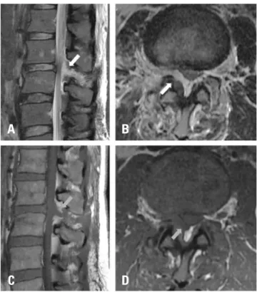
One week postoperatively, the patient complained of bilateral weakness with ankle dorsiflexion. Physical examination showed motor grade 4 for bilateral knee extension and ankle dorsiflexion and plantarflexion. One day later, the patient complained of pain radiating down the right leg. Physical examination showed motor grade 0 for right hip flexion, knee extension, and ankle dorsiflexion and plantar flexion. Left-side hip flexion, knee extension, and ankle dorsiflexion and plantarflexion were grades 1, 4, 0, and 0 respectively. The patient’s deep tendon reflexes were decreased. Sensation in both extremities and anal tone were intact. Repeat MRI showed compression of the spinal canal by an epidural hematoma, and emergency surgery was planned for decompression and bleeding control (Fig. 4). After clearing the graft material, total laminectomy was performed. Under the epidural fat, no evidence of a hematoma was found;
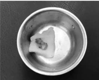
instead, a large disc extrusion was removed, which was later confirmed by histological examination (Fig. 5). The annular
defect was palpated with a Penfield probe. Posterior fusion using an H-shaped autologous cancellous iliac bone graft was performed. On postoperative day 2, the patient’s neurological status began to improve. At 3 months postoperatively, she could walk unaided; at 4 months postoperatively, her neurological status was fully recovered.
This study was approved by our institutional review board.
(MED-EXP-19-247)
Discussion
Intervertebral discs are very resistant to compression, distraction, flexion, and extension, but are vulnerable to rotational and horizontal forces. As compression forces increase, the annulus fibrosus bulges slightly, with no alteration of the shape of the nucleus pulposus; the majority of the
A B
Fig. 3. Anteroposterior (A) and lateral (B) radiographs of the lumbar spine after the first operation show single-level fusion with instrumenta- tion and the correction of spinal alignment.
Fig. 4. T2-weighted sagittal (A) and axial (B) magnetic resonance images of the lumbar spine after the first operation show increased compression of the cauda equina (white arrow).
A B
Fig. 5. Sequestrated disc fragment.
Chang-Hoon Jeon et al Volume 26 • Number 3 • September 30 2019
www.krspine.org 108
distortion occurs at the vertebral endplate. If the compression continues to increase, the endplate bulges more and finally cracks, leaving the compression forces to be absorbed by the vertebral body.2) Nucleus pulposus herniation (NPH) associated with spinal trauma is rare, with a reported incidence of 0.4%, and it is usually combined with fracture-dislocation injury caused by rotational and horizontal forces. Flexion- distraction forces can cause NPH in the cervical vertebrae, which have small surface areas, whereas this condition is very rare in the thoracolumbar spine. Only three case reports have described NPH caused by flexion-distraction injuries in the thoracolumbar spine.1, 9)
In the acute phase of trauma, NPH can be confused with epidural hematoma. The reported incidence of epidural hematoma after trauma is 0.5–11.4%.6) Generally, hematomas are located dorsal to the thecal sac and appear isointense (in acute cases) to hyperintense (in chronic cases) relative to the spinal cord on T1-weighted images. On T2-weighted images, hematomas are “heterogeneously hyperintense,” or have mixed high and low signals.3) Traumatic NPH has a low signal on T1-weighted images and a hyperintense signal on T2-weighted images.1) Due to the similarity of the MRI features of the two diseases, Dorsay et al.4) established the following radiological criteria for the identification of epidural hematoma: (1) retro- somatic epidural location, (2) teardrop or egg-like shape in the sagittal plane, (3) plasticity (close conformation with the contours of bone), (4) size greater than half the height of the
vertebral body in the craniocaudal dimension, (5) high signal on T1-weighted images (6) signal distinct from that of the disc, and (6) little or no disk space narrowing. The current case met most of these criteria; discontinuity of the posterior margin of the annulus was the only sign possibly indicating NPH (Fig. 6).
In the absence of coagulopathy or neurological deficit, NPH and epidural hematoma are known to resolve and heal spontaneously. Therefore, differentiation between the two diseases may not be necessary. However, posteriorly migrated NPH is frequently combined with neurological deficit and requires emergent surgical decompression.7, 8) Cauda equina syndrome is rare, with a reported incidence of 2–6% in typical NPH cases and about 50% in cases of posteriorly- migrated NPH.8) Symptoms usually include intense low back pain; we found no previously reported case involving delayed- onset neurological deficit resulting from posterior migration of NPH.7) In the current case, the patient showed no neurological deficit initially or immediately postoperatively. However, progressive leg weakness occurred, which may be a unique feature of posteriorly migrated NPH. The surgical treatment of this condition has favorable outcomes, with full neurological recovery. In the current case, the patient’s neurological status began to improve immediately after decompression; she could walk at 3 months postoperatively and her neurological deficit had disappeared completely at 4 months postoperatively.
We performed single-level fixation and fusion without decompression for a flexion-distraction injury at L2–
L3. Finkelstein et al.10) performed single-level fixation of 22 flexion-distraction injuries, with good outcomes.10) We decided to perform single-level surgery to minimize comorbidity after considering several factors: the patient was a young woman, she had no neurological symptom, and she had just survived life-threatening panperitonitis. The delayed-onset leg weakness in this patient may have been an indicator of posteriorly migrated NPH or a result of the single-level fixation, which may not provide sufficient stability to prevent further disc herniation. In addition, decompression may be required when correction of a kyphotic deformity is predicted to result in spinal canal compression, even in the absence of neurological pathology.
Here, we have presented a very rare case of posteriorly migrated NPH mimicking epidural hematoma accompanying a flexion-distraction injury of the lumbar spine.
Fig. 6. T2-weighted sagittal magnetic resonance image shows disconti- nuity of the posterior annulus (arrow).
Posteriorly migrated NPH and epidural hematoma may be indistinguishable in some cases; discontinuity of the posterior annulus may aid their differentiation. Posteriorly migrated NPH is frequently combined with neurological deficit, the onset of which may be delayed. Physicians must warn patients and caregivers about the risk of delayed-onset neurological deficit, even when patients have no initial symptoms. If posteriorly migrated NPH is suspected, early decompression surgery must be considered, even in the absence of neurological symptoms.
The outcomes of early surgical treatment are usually good, with full neurological recovery.
REFERENCES
1. Kim JH, Kim SH, Lee SK, et al. Traumatic lumbar disc herniation mimicking epidural hematoma: A case report and literature review. Medicine. 2019 May;98(18):E15438.
DOI: 10.1097/MD.0000000000015438.
2. Roaf R. A study of the mechanics of spinal injuries. The Journal of Bone and Joint Surgery. British volume. 1960 Nov;42:810-23. DOI: 10.1302/0301-620X.42B4.810.
3. Jain N, Crouser N, Yu E. Lumbar intervertebral disc her- niation masquerading as an epidural hematoma: A case re- port and review of the literature. JBJS case connector. 2018 Jul-Sep;8(3):E59. DOI: 10.2106/JBJS.CC.17.00300.
4. Dorsay T, Helms C. Mr imaging of epidural hematoma in the lumbar spine. Skeletal radiology. 2002 Dec;31(12):677- 85. DOI: 10.1007/s00256-002-0584-y.
5. Weinstein J, Lurie J, Tosteson T, et al. Surgical vs nonop- erative treatment for lumbar disk herniation: The spine patient outcomes research trial (sport) observational co- hort. Jama. 2006 Nov;296(20):2451-9. DOI: 10.1001/
jama.296.20.2451k.
6. Tamburrelli F, Meluzio M, Masci G, et al. Etiopathogenesis of traumatic spinal epidural hematoma. Neurospine. 2018 Mar;15(1):101-7. DOI: 10.14245/ns.1834938.469.pp 7. Akhaddar A, El-Asri A, Boucetta M. Posterior epidural mi-
gration of a lumbar disc fragment: A series of 6 cases: A re- view. Journal of Neurosurgery: Spine. 2011 Jul;15(1):117- 28. DOI: 10.3171/2011.3.SPINE10832.
8. Elsharkawy A, Hagemann A, Klassen P. Posterior epidural migration of herniated lumbar disc fragment: A litera- ture review. Neurosurgical review. 2019 Jan:1-13. DOI:
10.1007/s10143-018-01065-1.
9. Lewis S, Amritanand R. Traumatic disc herniation follow- ing flexion-distraction injury of the thoracolumbar spine:
A rare presentation. Journal of Trauma & Treatment. 2016 Jan;5:293. DOI: 10.4172/2167-1222.1000293.
10. Finkelstein J, Wai E, Jackson S, et al. Single-level fixation of flexion distraction injuries. Clinical Spine Surgery. 2003 Jun;16(3):236-42.
110
J Korean Soc Spine Surg. 2019 Sep;26(3):105-110. https://doi.org/10.4184/jkss.2019.26.3.110
Case Report
© Copyright 2019 Korean Society of Spine Surgery
Journal of Korean Society of Spine Surgery. www.krspine.org. pISSN 2093-4378 eISSN 2093-4386
This is an Open Access article distributed under the terms of the Creative Commons Attribution Non-Commercial License (http://creativecommons.org/licenses/by-nc/4.0/) which permits unrestricted non-commercial use, distribution, and reproduction in any medium, provided the original work is properly cited.
요추부의 굴곡 신연 손상에서 동반된 경막외 혈종과 유사한 경막낭의 후방으로 전위된 추간판 탈출증과 지 연성 하지 근력 약화에 대한 - 증례 보고 -
전창훈 • 정남수 • 이한동 • 정희웅 아주대학교 의과대학 정형외과학교실
연구 계획: 증례 보고
목적: 요추부 굴곡 신연 손상에서 동반된 후방 전위된 추간판 탈출증에 대해서 처음으로 보고하고자 한다.
선행 연구문헌의 요약: 요추부 추간판 탈출증은 경막외 혈종과 혼동되는 경우가 있으며, 특히 경막 후방으로 전위되는 경우에 감별하기 어렵다. 요추부 굴곡 신연 손상에서 동반된 경막 후방으로 전위된 추간판 탈출증에 대해서는 보고된 바가 없다. .
대상 및 방법: 47세 여환은 특이 병력 없으며, 교통사고 이후 발생한 요통으로 내원하였으며, 제 2-3요추간 굴곡 신연 손상으로 진단되었다. 신경학적 이 상은 관찰되지 않았다. 요추부 자기공명영상에서 T2 고신호 및 T1 등신호 병변이 제 2-3요추간 경막 후방에서 관찰되었다. 후방 기기 고정 및 유합술 시 행 1주 후 양측 하지 근력 약화가 발생되었다.
결과: 이차 수술에서 감압을 시행하였고, 경막외 혈종 소견은 없었으며 탈출된 추간판이 격리되어 경막 후방에서 관찰되었다. 충분한 감압 및 디스크 제 거술 후 환자는 약 수술 4개월째 신경학적으로 완전히 회복되었다.
결론: 본 증례보고는 흉요추부 굴곡 신연손상에서 후방으로 전위되는 추간판 탈출증이 동반될 수도 있음을 보여주었다. 자기 공명 영상에서 관찰되는 외 측 섬유륜의 후방의 단절은 후방 전위된 추간판 탈출을 경막외 혈종과 감별하는데 도움이 될 수 있다. 후방 전위된 추간판 탈출증은 신경학적 이상이 자 주 동반되기 때문에, 후방 전위된 추간판 탈출증이 흉요추부 신연 굴곡 손상에서 동반되었다면, 신경학적 이상이 없더라도 적극적인 감압을 조기에 시행 하는 것이 좋을 것으로 생각된다.
색인 단어: 후방 전위, 추간판 탈출증, 굴곡 신연 손상, 근력 약화, 경막외 혈종 약칭 제목: 외상성 후방 전위 추간판 탈출증
접수일: 2019년 6월 26일 수정일: 2019년 7월 29일 게재확정일: 2019년 8월 27일 교신저자: 이한동
경기도 수원시 영통구 월드컵로 164 아주대학교병원 정형외과학교실
TEL: 031-219-5220 FAX: 031-219-5229 E-mail: handonglee@gmail.com