Printed in the Republic of Korea DOI 10.5012/jkcs.2011.55.3.418
N
1,N
2-bis(3-((3-hydroxynaphthalen-2-yl)methylene-amino)propyl)phthalamide 의 크롬(III), 망간(II), 철(III), 코발트(II), 니켈(II), 구리(II), 루테늄(III) 및 산화 지르
코늄(II) 착물에 대한 합성과 분광학 및 생물학적 연구
Ahmed N. Al-Hakimi†, Mohamad M. E. Shakdofa‡, Ahemd M. A. El-Seidy‡, and Abdou S. El-Tabl*,§
†Department of chemistry, Faculty of science, Ibb University, Ibb, Yemen
‡Inorganic Chemistry Department, National Research Center, P.O. 12622 Dokki, Cairo, Egypt
§Department of chemistry, Faculty of science, El-Menoufia University, Shebin El-Kom, Egypt.
(접수 2010. 8. 20; 수정 2010. 10. 8; 게재확정 2010. 12. 31)
Synthesis, Spectroscopic, and Biological Studies of Chromium(III), Manganese(II), Iron(III), Cobalt(II), Nickel(II), Copper(II), Ruthenium(III), and Zirconyl(II)
Complexes of N
1,N
2-Bis(3-((3-hydroxynaphthalen-2-yl)methylene-amino)pro- pyl)phthalamide
Ahmed N. Al-Hakimi†, Mohamad M. E. Shakdofa‡, Ahemd M. A. El-Seidy‡, and Abdou S. El-Tabl*,§
†Department of chemistry, Faculty of science, Ibb University, Ibb, Yemen
‡Inorganic Chemistry Department, National Research Center, P.O. 12622 Dokki, Cairo, Egypt
§Department of chemistry, Faculty of science, El-Menoufia University, Shebin El-Kom, Egypt.
*E-mail: [email protected]
(Received August 20, 2010; Revised October 8, 2010; Accepted December 31, 2010)
요 약. N1,N2-bis(3-((3-hydroxynaphthalen-2-yl)methylene-amino)propyl)phthalamide (H4L, 1) 의 새로운 크롬(III), 망간(II), 철(III), 코발트(II), 니켈(II), 구리(II), 루테늄(III) 및 산화 지르코늄(II) 착물을 합성하여 원소분석, 물리적 성질 및 분광학적 으로 특성을 규명하였다. 분광학적 결과를 통해 이 리간드는 [H4LMX2(H2O)]·nH2O (M = Cu(II), Ni(II), Co(II), X = Cl 또 는 NO3)의 일반식을 갖는 착물2-5에서는 중성의 삼배위 리간드로 행동한다. 또는 [H4L(ZrO)2Cl2]·8H2O 의 일반식을 갖는 착물 6-9 에서는 이염기성 육배위 리간드로 행동한다. DMF 용액에서의 몰전기전도도 실험결과 이들 착물은 비이온성을 나타낸다. 고체 구리착물 2, 5 및 6 의 ESR 스펙트럼에서 g||>g>ge 을 보이는데, 이는 일그러진 팔면체구조와 큰 공유결합 성을 갖는 dx2
-y2오비탈에 비공유 전자쌍이 존재함을 의미한다. 이합체 구리(II) 착물 [H2LCu2Cl2(H2O)4]·3H2O (6)에 대해 두 구리원자 사이의 거리를 ESR 스펙트럼으로부터 추정한 parallel component 의 field zero splitting 파라메타를 이용하여 계 산하였다. 이들 화합물의 항박테리아 및 항균 활성도를 측정한 결과, 몇가지 금속 착물의 경우 표준시약인 tetracycline (박 테리아) 및 Amphotricene B (균류)보다 더 큰 저해효과를 보였다.
주제어: 착물, 합성, Schiff 염기, 자성, 생물학적 연구
ABSTRACT. Novel chromium(III), manganese(II), iron(III), cobalt(II), nickel(II), copper(II), ruthenium(III), and zirconyl(II) complexes of N1,N2-bis(3-((3-hydroxynaphthalen-2-yl)methylene-amino)propyl)phthalamide (H4L, 1) have been synthesized and characterized by elemental, physical, and spectral analyses. The spectral data showed that the ligand behaves as either neutral tridentate ligand as in complexes 2-5 with the general formula [H4LMX2(H2O)]·nH2O (M=Cu(II), Ni(II), Co(II), X
= Cl or NO3), neutral hexadentate ligand as in complexes 10-12 with the general formula [H4LM2Cl6]·nH2O (M=Fe(III), Cr(III) or Ru(III)), or dibasic hexadentate ligand as in complexes 6-9 with the general formula [H2LM2Cl2(H2O)4]·nH2O (M
= Cu(II), Ni(II), Co(II) or Mn(II), and 13 with general formula [H4L(ZrO)2Cl2]·8H2O. Molar conductance in DMF solution indicated the non-ionic nature of the complexes. The ESR spectra of solid copper(II) complexes 2, 5, and 6 showed g||>g>ge, indicating distorted octahedral structure and the presence of the unpaired electron in the dx2-y2 orbital with significant covalent bond character. For the dimeric copper(II) complex [H2LCu2Cl2(H2O)4]·3H2O (6), the distance between the two copper centers was calculated using field zero splitting parameter for the parallel component that was estimated from the ESR spectrum. The antibacterial and antifungal activities of the compounds showed that, some of metal complexes exhibited a greater inhibitory effect than standard drug as tetracycline (bacteria) and Amphotricene B (fungi).
Keywords: Complexes, Synthesis, Schiff base, Magnetism, Biological studies
INTRODUCTION
Schiff base transition metal complexes have been of great interest for many years since they are becoming increasingly important as biochemical, analytical and antimicrobial reagents.1 Many of metal complexes were showed anticancer and antimicrobial activities.2-4 It was reported that, some drugs have greater activity when administered as metal complexes than that as free organic compounds.5 So, Schiff base complexes might an untapped reservoir for drugs. Synthetic model studies involving magnetically coupled binuclear transition metal systems had attracted much interest because these studies pro- vided deeper insights into complex biological processes.
Also, homo binuclear lanthanide(III) complexes with isonicotinoyl hydrazone ligand had been prepared and characterized.6 Polyamino carboxylate groups had been used for the design of polydentate ligands such as ethyl- enediamine tetraacetic acid and diethylenetriamine penta acetic acid, generally showed high affinity for metal cat- ions.7 These ligands were widely used as chelating agents in fundamental research8 or as diagnostic tools in the phar- maceutical industry9 and they are particularly suitable for magnetic resonance imaging (MRI).10,11 A majority of Schiff base complexes incorporating five- or six-mem- bered metallocycles are represented by N. O- ligand envi- ronment have been published.12 The aim of this work is the preparation, characterization and antimicrobial activity of chromium(III), manganese(II), iron(III), cobalt (II), nickel(II), copper(II), ruthenium(III), and zirconyl (II) complexes of N1,N2-bis(3-((3-hydroxynaphthalen-2- yl)methylene amino)propylphthalamide (H4L).
EXPERIMENTAL
All chemicals and solvents were reagent grade com- mercial material and used as received. Elemental analy- ses for (C, H, N, Cl) were determined at the Analytical Unit of Cairo University, Egypt. Standard analytical meth- ods were used to determine the metal ion content.13 IR spectra of the ligand and its metal complexes were mea- sured using KBr discs with a Jasco FT/IR 300E Fourier transform infrared spectrophotometer covering the range 400-4000 cm-1 and in the 500-100 cm-1 region using poly- ethylene-sandwiched Nujol mulls on a Perkin Elmer FT- IR 1650 spectrophotometer. Mass spectrum of the ligand recorded using a JEOL JM-S-AX-500 massspectrometer.
Electronic absorption spectra in the 200-900 nm regions were recorded on a Perkin-Elmer 550 spectrophotometer.
Magnetic susceptibilities were measured at 25oC by the Gouy method using mercuric tetrathiocyanato-cobal- tate(II) as the magnetic susceptibility standard. The mag- netic moments were calculated from the equation: µeff.=
2.84 . Molar conductance was measured on a Tacussel type CD6NG conductivity bridge using 10-3 M DMF solutions. Thermal analyses (DTA and TG) were carried out in air using a Shimadzu DT-30 thermal ana- lyzer. The ESR spectra of the solid complexes at room temperature and at 77 K were recorded using a Varian E- 109 spectrophotometer (Leicester University, Engeland).
DPPH was used as the marker. TLC was used to confirm the purity of the prepared compounds.
Preparation of the ligand
Ethylphathalate was prepared using a published proce- dure.14
Preparation of N1,N2-bis(3-aminopropyl)phthalamide:
The N1,N2-bis(3-aminopropyl)phthalamide was prepared (Fig. 1) by the dropwise addition of ethanol solution (50 cm3) of ethyl phthalate (5.0 g, 0.22 mol) to the ethanol- solution (25 cm3) of 1,3-diaminopropane (3.33 g, 0.044 mol) with gentle warming and stirring for one hour. The white precipitate formed was filtered off, washed with
χMcorr⋅T
Fig. 1.
Fig. 2. Structure representation of the ligand.
ethanol, and dried under vacuum over anhydrous CaCl2. Preparation of the ligand (H4L, 1): The ligand (H4L, 1) (Fig. 2) was prepared by adding hot ethanol solution (50 cm3) of N1,N2-bis(3-aminopropyl)phthalamide (5.0 g, 0.018 mol) to a hot ethanol solution (50 cm3) of naph- thaldehyde (6.19 g, 0.036 mol). The mixture was refluxed for two hours and left to cool at room temperature. The yellow product was filtered off, washed with ethanol and dried under vacuum over anhydrous CaCl2.
Preparation of metal complexes
Complexes 2-5 were prepared by the dropwise addition of a hot (60oC) methanol solution of CuCl2·2H2O, NiCl2· 6H2O, CoCl2·6H2O or Cu(NO3)2·2.5H2O to a hot (70oC) ethanol solution of the ligand in molar ratio 1:1M/L (Metal/Ligand). The mixture was refluxed for four hours.
The precipitates formed after coaling were filtered, washed with ethanol, then with diethyl ether and dried under vac- uum over anhydrous CaCl2.
Complexes 6-13 were prepared by mixing a hot (60 oC) methanol solution of CuCl2·2H2O, NiCl2·6H2O, CoCl2· 6H2O, Mn(Cl)2·4H2O, FeCl3.6H2O, RuCl3.3H2O, CrCl3. 6H2O or ZrOCl2.8H2O with a hot (70 oC) ethanol solution of the ligand in molar ratio 2:1 M/L (Metal/Ligand). The mixture was refluxed for four hours. The precipitates formed after coaling were filtered, washed with ethanol, then with diethyl ether and dried under vacuum over anhydrous CaCl2.
In-vitro antibacterial and antifungal activities The investigation of the biological activities of the newly synthesized Schiff base ligand, its metal com- plexes and their corresponding metal salts were carried out in the Botany Department Lab. of microbiology, Fac- ulty of Science, El-Menoufia University. The antibacte- rial and antifungal activities were investigated by disc diffusion method.15,16 The antibacterial activities were done using Escherichia coli and Aspergillusnigerat 2000 ppm concentrations in DMSO. DMSO poured disc was used as negative control. The bacteria were subcultured in nutrient agar medium which was prepared using (g·L-1 dis- tilled water) NaCl (5 g), peptone (5 g), beef extract (3 g), agar (20 g). The fungus was subcultured in Dox’s medium which was prepared using (g·L-1 distilled water) yeast extract (1g), sucrose (30 g), NaNO3, agar (20 g), KCl (0.5 g), KH2PO4 (1 g), MgSO4·7H2O (0.5 g) and trace of FeCl3· 6H2O. These mediums were then sterilized by autoclav- ing at 120oC for 15 min. After cooling to 45oC the medium was poured into 90 mm diameter Petri dishes and incu-
bated at 37oC or 28oC, respectively. After few hours, Petri dishes were stored at 4oC. Microorganisms were spread over each dish by using sterile bent loop rod. The test is carried out by placing filter paper disks with a known concentration of the compounds on the surface of agar plates inoculated with a test organism. Standard anti- bacterial drug (tetracycline), antifungal drug (Amphot- ricene B) and solution of metal salts were also screened under similar conditions for comparison. The Petri dishes were incubated for 48 h at 37 or 28oC, respectively. The zone of inhibition was measured in millimeters carefully.
All determinations were made in duplicated manner for each of the compounds. An average of the two indepen- dent readings for each compound was record.
RESULT AND DISCUSSION
All the prepared compounds are colored, non-hygro- scopic, crystalline solid and stable at room temperature.
The complexes are insoluble in non-polar solvents but sol- uble in polar coordinating solvents such as DMSO and DMF. Elemental analyses, physical data, Table 1, and spectral data, Tables 2 and 3 are compatible with the pro- posed structures shown in Fig. 4. To date, no diffractable crystals have been grown.
N1, N2-bis(3-((3-hydroxynaphthalen-2-yl)methylene- amino)propyl)phthalamide (H4L) can exist in the keto form (I), keto-enol form (II), enol form (III), Fig. 3.
1H- and 13C-NMR spectra of 1
The 1H-NMR spectrum of the ligand (H4L, 1) in d6- DMSO shows signals consistent with the proposed struc- ture (Fig. 2) and indicated that, the ligand is present in its ketonic form.17 This conclusion was supported by the presence of peaks due to NH groups appearing at 8.62 ppm (s, 2H)18,19 and azomethine group HC=N appearing at 8.12 ppm (s, 2H).20 The peak appeared at 10.1 ppm, may be due to the proton of hydroxyl groups (s, 2H).21 The peaks appearing in the 7.04-7.85 ppm range may corre- spond to protons of the aromatic hydrogen (16H). The sig- nals at 3.96, 3.51 and 2.1 ppm may be assigned to terminal -CH2-N=C, terminal -CH2-NH and middle -CH2- groups.
The 13C-NMR spectrum showed different peaks appear- ing at 182.1 and 160.23 ppm. These peaks can be due to -CH=N- and -C-OH groups respectively,21 however the peaks at 127.63-140.2 ppm range assigned to the carbon of aromatic ring.21 The peaks appearing at 60.22 and 31.52 ppm may be due to terminal -CH2-N- and middle -CH2- group carbons.22
Mass spectra
The mass spectrum of the ligand reveals the molecular ion peak (m/z) 587 a.m.u consistent with the molecular
weight of the ligand. The most fragments appear at (m/z) 77, 144, 163, 170, 248 and 417 a.m.u corresponding to [C6H5], [C10H8O], [C10H13NO], [C11H8NO], [C14H20N2O2], and [C25H27N3O3], respectively (Fig. 5). However cop- per(II) complexes 2 and 6 show molecular ion peaks (m/z) at 739 and 908a.m.u., consisted with the molecular weights of 739.15 and 908.79 respectively.
Conductivity measurements
The molar conductance values of the complexes in DMF (10-3 M), lie in the 12-23.2 Ω-1 mol-1cm2 range, Table 1, indicating that, all the complexes are not electrolytes. These confirmed that the anion is coordinated to metal ion.23,24
IR spectra
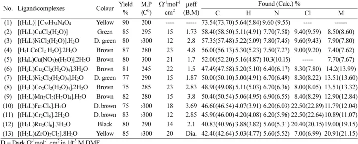
The IR spectral data for the ligand (H4L, 1) and its metal Table 1. Elemental analyses and physical properties of the ligand (H4L) and its metal complexes
No. Ligand\complexes Colour Yield
% M.P (C0)
Ω-1mol-1 cm2
µeff (B.M)
Found (Calc.) %
C H N Cl M
(1) [(H4L)] [C36H34N4O4 Yellow 90 200 ---- --- 73.54(73.70) 5.64(5.84) 9.60 (9.55) ---- --- (2) [(H4L)CuCl2(H2O)] Green 85 295 15 1.73 58.40(58.50) 5.11(4.91) 7.70(7.58) 9.40(9.59) 8.50(8.60) (3) [(H4L)NiCl2(H2O)].H2O D. green 80 ›300 12 2.8 57.35(57.48) 5.22(5.09) 7.80(7.45) 9.60(9.43) 7.90(7.80) (4) [H4LCoCl2 H2O].2H2O Brown 87 280 23 4.8 56.00(56.13) 5.30(5.23) 7.50(7.27) 9.00(9.20) 7.40(7.62) (5) [(H4L)Cu(NO3)2(H2O)].2H2O Brown 80 300 21 1.7 52.00(52.20) 5.16(4.87) 10.3(10.15) --- 7.70(7.67) (6) [(H2L)Cu2Cl2(H2O)4].3H2O Brown 81 245 22 1.5 47.49(47.58) 5.20(5.10) 6.40(6.17) 8.30(7.80) 14.2(13.99) (7) [(H2L)Ni2Cl2(H2O)4].H2O D. green 77 290 15 1.87 50.00(50.10) 5.00(4.91) 6.70(6.49) 8.30(8.22) 13.51(13.60) (8) [(H2L)Co2Cl2(H2O)4].2H2O Brown 75 285 23 2.83 48.90(49.08) 5.11(5.03) 6.70(6.36) 8.00(8.05) 13.51(13.32) (9) [(H2L)Mn2Cl2(H2O)4].H2O Brown 82 280 15 3.8 50.40(50.54) 5.06(4.95) 6.90(6.55) 8.40(8.29) 12.90(12.84) (10) [(H4L)Fe2Cl6].H2O D. brown 75 ›300 18 3.69 46.60(46.54) 4.07(3.91) 6.20(6.03) 22.50(22.89) 11.79(12.04) (11) [(H4L)Cr2Cl6].2H2O D. brown 83 ›300 12 2.85 45.90(46.00) 4.20(4.08) 6.20(5.96) 22.50(22.64) 10.89(11.07) (12) [(H4L)Ru2Cl6].3H2O Black 80 290 14 2.1 40.83(40.96) 3.88(3.82) 5.60(5.31) 20.40(20.15)19.00(19.15) (13) [(H2L)(ZrO)2Cl2].8H2O Yellow 85 ›300 20 Dia. 42.40(42.64) 5.03(4.77) 5.60(5.52) 7.00(6.99) 20.91(21.15) D = Dark Ω-1mol-1 cm2 in 10-3 M DMF
Table 2. IR spectra (assignments) of the ligand (1) and its metal complexes
NO. H2Ohydr./coord. υ(OH) υ(NH) υ(C=O) υ(C=N) υ(C-O) υ(M-O) υ(M-N) υ(M-Cl)
(1) - 3360s 3252s 1656v.s 1620s 1315m --- --- ---
(2) 3452br 3361s, 3341m 3254s, 3239m 1655v.s 1621s, 1609m 1317m, 1325m 680m 570m 402w (3) 3580-3445br 3362s, 3338m 3250s, 3237m 1654v.s 1623s, 611m 1316m, 1325m 656m 565 382w (4) 3620-3450br 3358s, 340m 3249s, 3241m 1655v.s 1621s, 607m 1314m, 1321m 690m 565m 398w (5) 3610-3440br 3359s, 3346m 3250s, 3235m 1653v.s 1624s,1606m 1315m, 1328m 660m 560m ---
(6) 3625-3454br - 3239m 1655v.s 1605m 1325m 670m 550m 385m
(7) 3605-3449br - 3235m 1657v.s 1608m 1330m 690m 590m 410m
(8) 3600-3455br - 3225m 1656v.s 1611m 1338m 675m 565m 380w
(9) 3612-3450br - 3231m 1655v.s 1604m 1335m 680m 565m 380w
(10) 3580br 3349m 3236m 1657v.s 1606m 1330m 670m 605m 375m
(11) 3590br 3344m 3239m 1656v.s 1605m 1325m 650m 570m 365m
(12) 3585br 3347m 3239m 1658v.s 1610m 1329m 670m 595m 405m
(13) 3605br - 3235m 1655v.s 1612m 1339m 650m 570m 413m
Fig. 3.
complexes are presented in Table 2. The IR spectrum of the ligand showed bands at 3360 and 3252 cm-1may be due to u(OH) and u(NH) group.19 However, broad, medium bands were observed in the 3450-3200 and 3180- 2600 cm-1 ranges, attributed to intra-and intermolecular hydrogen bonding between -OH and -C=N, -NH and -C=O groups respectively19,25 thus, the higher frequency band is associated with a weaker hydrogen bond and the lower frequency band with a stronger hydrogen bond. Also, the spectrum shows bands at 1656 and 1620 and 1586 cm-1 which were assigned to υ(C=O), υ(C=N) and (C=C)Ar
respectively.26,27 The spectra of solid complexes are com-
pared with those of the ligand in order to know the mode of bonding. The spectra showed that the ligand behaved either as:
Neutral tridentate ligand, coordinating through OH, C=N, and NH of one arm of the ligand as in case of com- plexes 2-5, the mode of coordination was suggested by the following evidence: i) the bands due toone OH, C=N, and NH were shifted to lower wave number with decreasing their intensities, while the other ones found almost at their original place, indicating that, only one of each pair were involved in the coordination,19,25,28-30 ii) one band of the two C-O bands was shifted to a higher wave number while the other is found almost at its original place, indicating that, only one phenolic oxygen was involved in the coor- dination,28 iii) the band of both carbonyl groups found as one band almost at its original place in the ligand indi- cating that they are not involved in the coordination,18,26,27 iv) the simultaneous appearance of new bands in the 656- 690 and 560-570 cm-1 regions are due to the υ(M-N) and υ(M-O) vibrations,31,32 respectively.
Bibasichexadentate ligand, coordinating through all O-, C=N and NH groups as in case of complexes 6-9 and 13, the mode of coordination was suggested by the following evidence: i) the disappearance of the band of the two OH groups,28 ii) the bands of C=N and NH groups were shifted to lower wave number with decreasing their intensities, indi- cating that, all C=N and NH groups were involved in the coordination,19,25,29,30 iii) the band of two C-O groups were shifted to higher wave number, indicating that, both phenolic oxygen atoms were involved in the coordination28 iv) the bands of both carbonyl groups were observed as single band at its original place in the ligand indicating that, they are not involved in the coordination,18,26,27 iv) the simultaneous appearance of new bands in the 650-690 and 550-590 cm-1 regions are due to the υ(M-N) and υ(M- O) vibrations, respectively.31,32
Neutral hexadentate ligand, coordinating through all OH, C=N and NH groups as in case of complexes 10-12, the mode of coordination was suggested by the following evi- dence: i) the bands of C=N, OH and NH groups were shifted to lower wave number with decreasing their inten- sities, indicating that, all C=N, OH and NH groups were involved in the coordination,19,25,28-30 iii) the band of both C-O groups were shifted to higher wave number, indicating that, all hydroxyl groups were involved in the coordination28 iv) the bands of both carbonyl groups were observed as single band at its original place in the ligand indicating that, they are not involved in coordination,18,26,27 iv) the simultaneous appearance of new bands in the 650-670 and Fig. 4. Structure representation of metal complexes.
570-605 cm-1 regions are due to the υ(M-N) and υ(M-O) vibrations, respectively.31,32 All complexes except com- plex 5, show band in the 375-413 region, assignable to υ(M-Cl).19,28 The broad bands in the 3600-3400 cm-1 region are due to coordinated water or water of crystal- lization. Complexes 2-9 showed a band in the 400-600 cm-1 region indicating the presence of coordinated water, but the absence of these bands in the spectra of complexes 10-13 indicate the presence of hydrated water rather than coordinated ones. The presence of water molecules within the coordination sphere in the hydrated complexes 2-9 is
further supported by the presence of bands in the 3480- 3494, 1605-1610, 940-950 and 613-630 cm-1 regions due to OH stretching, HOH deformation, H2O rocking and H2O wagging, respectively.33,34 The spectrum of the com- plex 5 showed bands at 1465 cm-1 (υ1), 1050 cm-1 (υ2), 1377 cm-1 (υ4) and 710 cm-1 (υ5) with υ1-υ4 separation of 88 cm-1, characteristic of monodentate nitrato group.35 Zirconyl(II) complex 13 shows band at 825 cm-1 assigned to Zr=O.19
Electronic spectra
The electronic spectral data of the ligand (H4L, 1) and Fig. 5. The fragmentation pattern of the ligand.
its metal complexes in DMF solution are summarized in Table 3. The ligand (H4L, 1) showed two bands at 370 and 320 nm which may be assigned to n→π* and π→π* tran- sitions respectively.32 In the metal complexes, the spectra showed bands in the 345-290 nm range, due to intraligand transitions. Copper(II) complexes 2, 5, and 6 in DMF solution showed bands in the 430-460, 580-590 and 625- 640 nm range, which were assigned to ligandcopper(II) change transfer, 2B1→2E and 2B1→2B2 transitions indi- cating a distorted octahedral structure.36,37 Nickel(II) com- plexes 3 and 7 showed bands in 490-510, 600-610 and 865-870 nm ranges respectively, which are attributable to
3A2g(F)→3T1g(P) (ν3), 3A2g(F)→3T1g(F) (ν2) and 3A2g(F)→
3T2g(F) (ν1) transitions indicating octahedral nickel(II) complexes.38,39 The ν2/ν1 ratio for these complexes were 1.41 and 1.45 respectively, which is less than the usual range of 1.5-1.75, indicating distorted octahedral nickel(II) complexes.37 The cobalt(II) complexes 4 and 8 showed bands at 455-475, 590-610 and 650-665 nm ranges, which were assigned to 4T1g(F)→4T1g(P), 4T1g(F)→4A2g and
4T1g(F)→4T2g(F) transitions respectively, corresponding to high spin cobalt(II) octahedral complexes.19,40 Manga- nese(II) complex 9 showed bands at 430, 480 and 590 nm, these bands were corresponding to 6A1g→4Eg, 6A1g→4T2g
and 6A1g→4T1g transitions which are compatible to an octahedral structure for manganese(II) complexes.41,42 Iron(III) complex 10 gave bands at 400 and 600 nm are due to charge transfer and 6A1→4T1 transitions, suggest- ing octahedral structure.19,36,42 Chromium(III) complex 11 showed bands at 460 and 540 nm which are attributed to charge transfer and 4A2→4T1 transitions of six coordinate chromium(III) complex.43,44 Ruthenium(III) complex 12 showed bands at 460 and 665 nm, are due to LMCT tran-
sition and the other one is assigned to 2T2g→2A2g transition.
These band were similar to those observed for octahedral ruthenium(III) complexes.19,45,46 Zirconyl(II) complex 13 shows bands (Table 3) were due to intraligand transitions.
Magnetic moments
The room temperature magnetic moments of complexes 2-14 are present in Table 1. Copper(II) complexes 2, 5 and 6 show values 1.73, 1.7 and 1.5 B.M.. These values are correspond to one unpaired electron in an octahedral structure.19 Nickel(II) complexes 3 and 7 show values 2.8 and 1.87 B.M., indicating octahedral nickel(II) com- plexes.19,47,48 Cobalt(II) complexes 4 and 8 show values 4.8 and 2.83 B.M., indicating high spin octahedral cobalt (II) complexes.19,38 Manganese(II) complex 9 shows 3.8 B.M., suggesting octahedral geometry around the man- ganese(II) ion.19,38 Iron(III) complex shows 3.69 B.M., indicating high spin iron(III) octahedral geometry.19,50 Chromium(III) complex 11 shows 2.85 B.M., which is lower than the spin-only value, implying an operation of spin-exchange interactions take place between chromium (III) ions.44 The ruthenium(III) complex 12 shows a mag- netic value of 1.67 B.M., indicating an octahedral ruthe- nium(III) structure.19,51 Zirconyl(II) complex 13 shows diamagnetic property.19 The complexes 6-11 showed low magnetic moment values indicating spin exchange inter- actions take place between the two ion centers.19,47,49
ESR spectra
The ESR spectra of solid copper(II) complexes 2, 5 and 6 at room and at liquid nitrogen temperatures were char- acteristic of a d9 system and having an axial symmetry type of a dx2-y2 ground state.52 On lowering the tempera- Table 3. UV-Vis. spectra of the ligand, (H4L) and its metal complexes
No. Compounds λmax, nm (ε mol-1 cm-1)
(1) [(H4L)] [C36H34N4O4 320(3684), 370(2787)
(2) [(H4L)CuCl2(H2O)] 315(2320), 345(2966), 450(791), 590(232), 640(58)
(3) [(H4L)NiCl2(H2O)].H2O 305(1699), 345(2295), 490(476), 580(188), 610(151), 865(189) (4) [H4LCoCl2 H2O].2H2O 315(2360), 455(3654), 610(370), 665(190)
(5) [(H4L)Cu(NO3)2(H2O)].2H2O 315(2464), 345(1947), 430(992), 585(66)
(6) [(H2L)Cu2Cl2(H2O)4].3H2O 305(3497), 345(2057), 460(764), 580(257), 625(170) (7) [(H2L)Ni2Cl2(H2O)4].H2O 305(3236), 345(2330), 510(497), 600(388), 870(104) (8) [(H2L)Co2Cl2(H2O)4].2H2O 300(2457), 475(1654), 590(296), 650(135)
(9) [(H2L)Mn2Cl2(H2O)4].H2O 325(3714), 430(2844), 480(636), 590(85) (10) [(H4L)Fe2Cl6].H2O 290(2204), 310(5176), 400(1390), 600(92) (11) [(H4L)Cr2Cl6].2H2O 315(2153), 460(3700), 500(429), 540(38) (12) [(H4L)Ru2Cl6].3H2O 305(4097), 430(3465), 460(969), 665(25) (13) [(H2L)(ZrO)2Cl2].8H2O 320(2885), 370 (4464), 430(594)
ture to liquid nitrogen, the spectra were not changed, sug- gesting that, the geometry of the complexes is not changed on cooling. The g-values suggest octahedral geometry for complexes 2, 5 and 6. The complexes show g||>g⊥>ge, indicating a dx2-y2 ground state, and spectral features were characteristic of axial symmetry.53 The ESR parameters for 2 is g|| = 2.27, g⊥ = 2.05, giso = 2.13, G = 5.4, K⊥2 = 0.64, K||2 = 0.68, K2 = 0.65 and K = 0.81, for 5 is g|| = 2.24, g⊥ = 2.06, giso = 2.12, G = 4.0, K⊥2 = 0.81, K|2 = 0.61, K2 = 0.74 and K = 0.86, for 6 is g|| = 2.26, g⊥ = 2.08, giso = 2.14, G = 3.25, K⊥2 = 1.02, K||2 = 0.67, K2 = 0.9 and K = 0.95. Kiv- elson and Neiman54 show that, the g||-value in the cop- per(II) complexes can be used as a measure of covalent character of the metal-ligand bond. If this value is greater than 2.3, the environment is essentially ionic and the value less than this limit indicate a covalent environment. All complexes showed covalent bond character.19,54,55 The g- values were related by the expression,56 G = (g||-2)/(g⊥-2), if G > 4.0 then local tetragonal axes were aligned parallel or only a slightly misaligned, if G < 4.0, significant exchange coupling is present. Complexes 2 and 5 show value ≥ 4.0, however complex 6 shows G = 3.25, indi- cating spin-exchange interactions take place between the copper(II) ions, which is furthered confirmed from the magnetic moment value (Table 1). Also, the g-values of copper(II) complexes with a 2B1g ground state (g||>g⊥) may be expressed.35,57
K⊥2= (g⊥− 2.002)∆Exz/2λo (1) (2) K2= (K112
+ 2K⊥2)/3 (3)
Where K|| and K⊥ were the parallel and perpendicular components respectively of the orbital reduction factor (K), λo is the spin-orbit coupling constant for the free cop- per, ∆Exy and ∆Exz were the electron transition energies.
From the above relations, the orbital reduction of covalency35,57] can be calculated for an ionic environ- mental, K=1 and for a covalent environment K<1, the lower the value of K, the greater is the covalent character.
The K-values of the complexes 2, 5, and 6 were lower than 1.0, confirming covalent bond character.19,35,58 The ESR spectra of 4, 9, and 10 showed isotropic type with giso= 2.21, 2.012 and 2.0035, indicating octahedral geometry around Co(II), Mn(II), and Fe(III) ions respectively.54,57
The zero field splitting parameter (D) for the parallel components of the dimmer complex 6 was estimated from the spectrum, and is equal to 414. The distance between two copper centers was calculated using the following
equation.59
D = (3µB / 2R3)*(3 cos2θ - 1) (4) Where mB is the magnetic moment of the electron and R is the distance (Å) between two electrons. For parallel com- ponent (D), θ=0, by substitution in equation 4, D is equal to 3µB/2R3. For a diradical system in the triplet-state, it is found that, D for the parallel components is equal to 402 G and the distance between the two radicals is equal to 5.2 Å. From these data, the distance between the two cop- per(II) centers was calculated and is equal to 5.3 Å. This value is close to that for a dimeric copper(II) compound in the triplet state.60
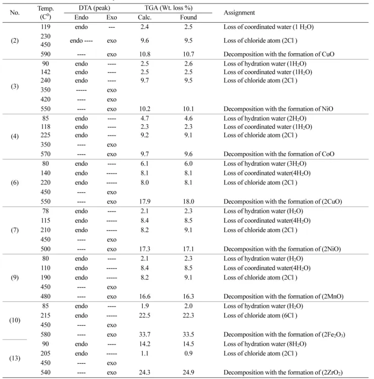
Thermal analyses (DTA and TG)
The results of TG and DTA analyses of complexes were shown in Table 4. The results showed good agreement with theoretical formula as suggested from the analytical data (Table 1). All complexes except complex 2, lost hydration water molecules in the temperature 78-90oC range and were accompanied with an endothermic peak.
The coordinated water molecules were eliminated from these complexes at relatively higher temperature; 110- 142oC, than those of the hydrate water molecules (Table 4).
The removal of HCl molecules was observed for allcom- plexes in the temperature 190-240oC range, which was accompanied by an endothermic peak. The complexes decompose through degradation of the Schiff base ligand at a temperature over than 400oC leaving metal oxides (480-590) range.
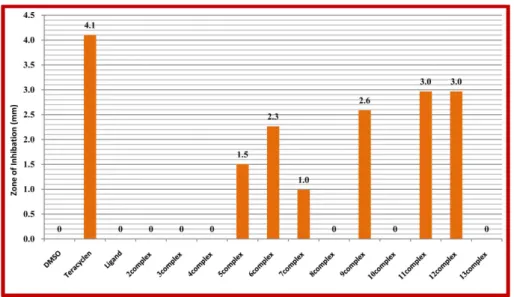
Biological activity
The antibacterial and antifungal activities of the ligand and its metal complexes were screened on bacterial and fungal strains using the disk diffusion method. It is impor- tant to note that the ligand and most of its metal com- plexes exhibit more antifungal inhibitory effects than the Standard antifungal drug (amphotricene B) and most of the metal complexes more active than parent ligands and the solution of metal ions. It is also clear that the ligand and its metal complexes have more antifungal activity than antibacterial activity. The inhibition zone diameter of the compounds is shown in Figs. 6 and 7. The order of antifungal activity of the compounds is 9, 10 > 6, 11 > 12 >
5 > Ligand > Amphotricene B > 7, however, the order of antibacterial activity of the compounds is Tetracycline >
11, 12 > 9 > 6 > 5 > 7. The increased activity of the metal complexes can be explained on the basis of chelation the- ory.61 It is known that the chelation tends to make the K112 =(g11–2002)∆Exy/8λo
ligand act as more powerful and potent fungicidal and bactericidal agents, thus killing more fungi and bacteria than the ligand. It is known that, in a complex, the positive charge of the metal is partially shared with the donor atoms present in the ligands, and there may be π-electron delocalization over the whole chelating system,62 this increases the lipophilic character of the metal chelate and favors its permeation through the lipoid layer of the mem- branes. There are other factors which also increase the
activity, which are solubility, conductivity, coordination mode and bond length between the metal and the ligand.
The variation in the effectiveness of different compound against different organisms also depends either on the impermeability of the cell of the microbes or differences in ribosomes of microbial cells.63,64 The variation of bio- logical activity of the complexes may be due to change in electronic configuration of the metal and also, the envi- ronment around the metal ion.
Table 4. Thermal data for the metal complexes
No. Temp.
(C0)
DTA (peak) TGA (Wt. loss %)
Assignment
Endo Exo Calc. Found
(2)
119 endo --- 2.4 2.5 Loss of coordinated water (1 H2O)
230
450 endo ---- exo 9.6 9.5 Loss of chloride atom (2Cl )
590 ---- exo 10.8 10.7 Decomposition with the formation of CuO
(3)
90 142 240
endo endo endo
---- ---- ----
2.5 2.5 9.7
2.6 2.5 9.5
Loss of hydration water (1H2O) Loss of coordinated water (1H2O) Loss of chloride atom (2Cl )
350 --- exo
420 ---- exo
550 ---- exo 10.2 10.1 Decomposition with the formation of NiO
(4)
85 118 225
endo endo endo
---- ---- ----
4.7 2.3 9.2
4.6 2.3 9.1
Loss of hydration water (2H2O) Loss of coordinated water (1H2O) Loss of chloride atom (2Cl )
350 ---- exo
570 ---- exo 9.7 9.6 Decomposition with the formation of CoO
(6)
80 endo ---- 6.1 6.0 Loss of hydration water (3H2O)
140 endo --- 8.1 8.1 Loss of coordinated water(4H2O)
220 endo --- 8.0 8.1 Loss of chloride atom (2Cl )
450 ---- exo
550 ---- exo 17.9 18.0 Decomposition with the formation of (2CuO)
(7)
78 endo ---- 2.1 2.3 Loss of hydration water (H2O)
115 endo --- 8.4 8.5 Loss of coordinated water(4H2O)
210 endo --- 8.2 9.1 Loss of chloride atom (2Cl )
450 ---- exo
500 ---- exo 17.3 17.1 Decomposition with the formation of (2NiO)
(9)
80 endo ---- 2.1 2.3 Loss of hydration water (H2O)
110 endo --- 8.4 8.5 Loss of coordinated water(4H2O)
190 endo --- 8.2 9.1 Loss of chloride atom (2Cl )
450 ---- exo
480 ---- exo 16.6 16.3 Decomposition with the formation of (2MnO)
(10)
85 endo ---- 1.9 2.0 Loss of hydration water (H2O)
215 endo --- 22.5 22.3 Loss of chloride atom (6Cl )
450 ---- exo
580 ---- exo 33.7 33.5 Decomposition with the formation of (2Fe2O3)
(13)
90 endo ---- 14.2 14.5 Loss of hydration water (8H2O)
205 endo --- 1.1 0.9 Loss of chloride atom (2Cl )
450 ---- exo
540 ---- exo 24.3 24.9 Decomposition with the formation of (2ZrO2)
SUMMARY
The chromium(III), manganese(II), iron(III), cobalt(II), nickel(II), copper(II), ruthenium(III) and zirconyl(II) com- plexes of N1,N2-bis(3-((3-hydroxynaphthalen-2-yl)meth- yleneamino)propyl) phthalamide has been synthesized and characterized by elemental and thermal analyses as well as spectroscopic techniques. The analyses data showed that, the ligand behaves as a neutral tridentate, neutral hexadentate, dibasic tridentate or dibasic hexadentate ligand bonded to the metal ion/ions through azomethine nitrogen atoms, protonated or deprotonated hydroxyl groups
and protonated or deprotonated amine groups. The metal complexes have a distorted octahedral, square planer or octahedral geometry. The ESR spectra of solid copper(II) complexes 2, 5, and 6 show g||>g⊥>ge(2.0023), indicating octahedral structure with significant covalent bond char- acter. The biological studies showed that the ligand bio- logically in active against Gram negative bacterium (Escherichia coli), and its metal complexes have mild activity in comparing with Standard antibacterial drug (Tetracycline) but has strongly biological activity against Fungus (Aspergillusniger) in comparing with Standard antifungal drug (Amphotricene B).
Fig. 6. Antibacterial activity of the ligand and its metal complexes against gram-negative bacterium (E. coli).
Fig. 7. Antifungal activity of the ligand and its metal complexes against Fungus (Aspergillusniger).
REFERENCES
1. You, Z.-L.; Shi, D.-H.; Xu, C.; Zhang, Q.; Zhu, H.-L.
European Journal of Medicinal Chemistry 2008, 43, 862.
2. Bekhit, A. A.; El-Sayed, O. A.; Al-Allaf, T. A. K.; Aboul- Enein, H. Y.; Kunhi, M.; Pulicat, S. M.; Al-Hussain, K.;
Al-Khodairy, F. Arif, J. Eur. J. Med. Chem. 2004, 39, 499.
3. Golcu, A.; Tumer, M.; Demirelli, H.; Wheatley, R. A.
Inorg. Chim. Acta. 2005, 35, 8.
4. Singh, K.; Barwa, M. S.; Tyagi, P. Eur. J. Med. Chem.
2006, 41, 147.
5. Chakraborty, J.; Patel, R. N. J. Indian Chem. Soc. 1996, 73, 191.
6. Bu, X. H.; Du, M.; Zhang, L.; Song, X. B.; Zhang, R. H.
Inorg. Chim. Acta 2000, 308, 143.
7. Carraquilleo, J. A.; White, J. D.; Paik, C. H.; Raubitschek, A. N.; Rotman, M.; Brechbiel, M.; Gansow, C. A.; Top, L. E.; Peretesis, P.; Reynolds, J. C.; Nelson, D. L.; Wald- mann, T. A. J. Nucl. Med. 1999, 40, 268.
8. Guo, Z.; Sadler, P. J. Angew. Chem. Int. Ed. 1999, 38, 1512.
9. Mortellaro, M. A.; Nocera, D. G. J. Am. Chem. Soc. 1996, 118, 7414.
10. Zhang, S.; Wu, K.; Sherry, A. D. Angew. Chem. Int. Ed., 1999, 38, 3192.
11. Caravan, P.; Ellison, J. J.; Mcmurry, T. J.; Lauffer, R. B.
Chem. Rev. 1999, 99, 2293.
12. Garnovskii, A. D.; Vasilchenko, I. S.; Garnovskii, D. A.;
Kharisov, B. I. J. Coord. Chem. 2009, 62, 151.
13. G. Svehla; Vogel’s textbook of macro and semi micro Quantitative inorganic analysis fifth Ed. Longman Inc:
New York, 1979.
14. Hoffman, R. V. Organic chemistry an intermediate text;
2nd Ed. John Wiley & Sons: Hoboken, New Jersey, 2004, pp 188.
15. Offiong, E. O.; Martelli, S.; Farm, I. L. 1994, 49, 513.
16. Collee, J. G.; Duguid, J. P.; Farser, A. G.; Marmion, B. D.
Practical Medical Microbiology; Eds.; Churchill Living- stone: New York, 1989.
17. Wu, L.; Qiu, G.; Teng, H.; Zhu, Q.; Liang, S.; Hu, X.
Inorg. Chim. Acta 2007, 360, 3069.
18. Pouralimardan, O.; Chamayou, A.-C.; Janiak, C.; Mon- fared, H. H. Inorg. Chim. Acta 2007, 360, 1599.
19. El-Tabl, A. S.; El-Saied, F. A.; Al-Hakimi, A. N. Trans.
Met. Chem. 2007, 32, 689.
20. Maurya, M. R.; Agarwal, S.; Bader, C.; Rehder, D. Eur.
J. Inorg. Chem., 2005, 147.
21. Glu, M. K.; Ispir, E.; Glu, N. K.; Serin, S. Dyes and Pig- ments 2008, 77, 75.
22. Han, H. O.; Kim, S. H.; Kim, K. H.; Hur, G. C.; Yim, H.
J.; Chung, H. K.; Woo, S. H.; Koo, K. D.; Lee, C. S.;
Koh, J. S.; Kim, G. T. Bioorg. Med. Chem. Lett. 2007, 17, 937.
23. Geaey, W. J. Coord. Chem. Rev. 1971, 7, 81.
24. Tas, E.; Aslanoglu, M.; Kilic, A.; Kaplan, O.; Temel, H.
J. Chem. Res-(s). 2006, 242.
25. El-Tabl, A. S. Trans. Met. Chem. 1997, 22, 400.
26. Tas, E.; Ulusoy, M.; Guler, M.; Yilmaz, L. Trans. Met.
Chem. 2004, 29, 180.
27. El-Behery, M.; El-Twigry, H. Spectrochim. Acta Part A, 2007, 66, 28.
28. Nakatamato, K.; Infrared Spectra of Inorganic and Coor- dination Compounds 2nd Edit., Wiley Inc: New York, 1967.
29. El-Wahab, Z. H. A.; Mashaly, M. M.; Salman, A. A.; El- Shetary, B. A.; Faheim, A. A. Spectrochim. Acta Part A, 2004, 60, 2861.
30. El-Tabl, A. S.; Imam, S. M. Trans. Met. Chem. 1997, 22, 259.
31. El-Tabl, A. S. J. Chem. Res.(s) 2002, 529.
32. Cukuravali, A.; Yilmaz, I.; Kirbag, S. Trans. Met. Chem.
2006, 31, 207.
33. Chen, C.-Y.; Chen, Q.-Z.; Wang, X.-F.; Liu, M.-S.; Chen, Y. F. Trans. Met. Chem. 2009, 34, 757.
34. Tatwawadi, S. V.; Singh, A. P.; Narang, K. K. J. Sci. Res.
Banaras Hindu Univ., 1980, 3, 143.
35. Shauib, N. M.; Elassar, A.-Z. A.; El-Dissouky, A. Spec- trochim. Acta Part A, 2006, 63, 714.
36. Lever, A. B. P.; Inorganic Electronic Spectroscopy, 2nd edn, Elsevier: Amsterdam, 1984.
37. El-Tabl, A. S.; El-Enein, S. A. J. Coord. Chem. 2004, 57, 281.
38. Gudasi, K. B.; Patil, M. S.; Vadavi, R. S.; Shenoy, R. V.;
Patil, S. A.; Nethaji, M. Trans. Met. Chem. 2006, 31, 580.
39. Thakkar, N. V.; Bootwala, S. Z. Indian J. Chem. 1995, 34A, 370.
40. El-Boraey, H. A.; El-Tabl, A. S. Polish J. Chem. 2003, 77, 1759.
41. Parihari, R. K.; Patel, R. K.; Patel, R. N. J. Ind. Chem.
Soc. 2000, 77, 339.
42. Singh, N. K. Trans. Met. Chem. 2001, 26, 487.
43. Blinc, R.; Hadzi, D. J. Chem. Soc. 1958, 4536.
44. Zhang, S. W.; Liao, D. Z.; Jiang, Z. H.; Wang, G. L.
Trans. Met. Chem. 1996, 21, 166.
45. Nehru, K.; Athappan, P.; Rajagopal, G. Trans. Met. Chem.
2001, 26, 652.
46. El-Tabl, A. S.; Issa, R. M.; Morsi, M. A. Trans. Met.
Chem. 2004, 29, 543.
47. Motaleb, A. E.; Ramadan, M.; Sawodny, W.; Baradie, H.
F. E.; Gaber, M. Trans. Met. Chem. 1997, 22, 211.
48. Nag, J. K.; Pal, S.; Sinha, C. Trans. Met. Chem. 2005, 30, 523.
49. Singh, M. K.; Kar, N. K.; Lai, R. A.; Asthana, M. J.
Coord. Chem. 2009, 62, 2893.
50. Murukan, B.; Mohanan, K. Trans. Met. Chem. 2006, 31, 441.
51. El-Tabl, A. S.; Ayad, M. I. Synth. React. Inorg. Met. -Org.
Chem. 2003, 33, 369.
52. Chandra, S.; Kamar, U. Spectrochim. Acta, Part A 2005, 61, 219.
53. Patel, R. N.; Singh, N.; Gundla, V. L. N. Polyhedron 2006,
25, 3312.
54. Kivelson, D.; Neiman, R. J. Chem. Phys. 1961, 35, 149.
55. Ray, R. K. Inorg. Chem. Acta 1990, 174, 257.
56. El-Tabl, A. S. Bull. Korean Chem. Soc. 2004, 25, 1.
57. Mao, Z. W.; Yu, K. B.; Chen, D.; Han, S. Y.; Sui, Y. X.;
Tang, W. X. Inorg. Chem. 1993, 32, 3104.
58. Symons, M. C. R. Chemical and Biochemical Aspects of Electron Spin Resonance; Van Nostrand Reinholds Wok- ingham. 1979.
59. Natarajan, C.; Shanth, P.; Athappan, P.; Murugesan, R.
Trans. Met. Chem. 1992, 17, 39.
60. Berrand, J. A.; Black, T. D.; Eller, P. G.; Helm, F. T.;
Mahmood, R. Inorg. Chem. 1976, 15, 2965.
61. El-Wahab, Z. H. A.; Mashaly, M. M.; Salman, A. A.; El- Shetary, B. A.; Fahei, A. A. Spectrochim. Acta Part A 2004, 60, 2861.
62. Sengupta, S. K.; Pandey, O. P.; Srivastava, B. K.; Sharma, V. K. Trans. Met. Chem. 1998, 23, 349.
63. Glu, M. K.; Ispir, E.; Glu, N. K.; Glu, S. T.; Serin, S.
Trans. Met. Chem. 2005, 30, 765.
64. Glu, M. K.; Gdelen, M. M. D.; Glu, S. T. Trans. Met.
Chem. 2006, 31, 382.